-
Paper Information
- Next Paper
- Previous Paper
- Paper Submission
-
Journal Information
- About This Journal
- Editorial Board
- Current Issue
- Archive
- Author Guidelines
- Contact Us
American Journal of Medicine and Medical Sciences
p-ISSN: 2165-901X e-ISSN: 2165-9036
2024; 14(11): 2985-2988
doi:10.5923/j.ajmms.20241411.68
Received: Nov. 3, 2024; Accepted: Nov. 22, 2024; Published: Nov. 27, 2024

Description of Kidney Morphometric Changes under the Influence of Combined Injuries in White-Born Canes
 Abstract
 Reference
 Full-Text PDF
 Full-text HTML
Nuriddinоv Asliddin Mekhriddinovich
Bukhara State Medical Institute, Bukhara, Uzbekistan
Correspondence to: Nuriddinоv Asliddin Mekhriddinovich, Bukhara State Medical Institute, Bukhara, Uzbekistan.
Copyright © 2024 The Author(s). Published by Scientific & Academic Publishing.
This work is licensed under the Creative Commons Attribution International License (CC BY).
http://creativecommons.org/licenses/by/4.0/

When studying morphometric changes in the kidneys of rats with combined injuries, we studied the following structural changes in the renal tissue at the microscopic level, including an assessment of the condition of the tubules, glomeruli, interstitial tissue and vessels. The obtained results of the study allowed us to identify fibrosis, destructive and inflammatory changes. The morphometric method was used to study the thickness of the capsule, the size of the tubules and glomeruli, the density of cellular elements and vascular structures, as well as a quantitative assessment of the area of damaged zones, necrotic areas and fibrosis zones using digital microscopes and image analysis software (Image J).
Keywords: White mongrel rats, Kidneys, Morphometry, Combined injury
Cite this paper: Nuriddinоv Asliddin Mekhriddinovich, Description of Kidney Morphometric Changes under the Influence of Combined Injuries in White-Born Canes, American Journal of Medicine and Medical Sciences, Vol. 14 No. 11, 2024, pp. 2985-2988. doi: 10.5923/j.ajmms.20241411.68.
Article Outline
1. Introduction
- White rats are a model object for studying traumatic injuries due to their physiological similarities with the human body. Morphological changes in organs in combined trauma are of considerable interest to modern medicine and biology, since they allow for a deeper understanding of the pathogenetic mechanisms of damage and the development of effective approaches to treatmen [1,2,3,9]. Kidneys, as a vital organ, play a key role in maintaining the body's homeostasis, and their condition significantly affects the prognosis and outcome of various traumatic effects. In conditions of combined trauma, including damage to various organs and tissues, pathological changes in the kidneys can be caused by both direct impact and systemic reactions of the body, such as shock, hypoxia, inflammation and metabolic disorders. Considering that 3-month-old white rats are one of the most commonly used experimental models for studying traumatic injuries, the results of the study of morphological changes in their kidneys can be extrapolated to clinical situations. The study of morphological changes in the kidneys in combined trauma is important for clarifying the mechanisms of tissue damage and regeneration, identifying factors that aggravate renal tissue damage, and developing methods for preventing and treating renal complications associated with trauma [4,5,7].To analyze the work on morphological changes in the kidneys in combined trauma, the main focus is on: the effect of ischemia and reperfusion on the morphological state of the kidneys (work in the field of transplantology and traumatology), systemic effects of shock, inflammation, and metabolic acidosis on the kidneys. Such experiments made it possible to evaluate morphological changes in organs, including the kidneys, and to identify factors that enhance or weaken tissue damage. Morphological analysis of changes in kidney tissue, including histology, ultrastructural studies, and immunohistochemistry, is carried out in many large medical and biological centers [8,9,10,11].The work carried out highlights aspects of the morphological adaptation of the kidneys to damaging effects.Purpose of the study: To study the morphometric parameters of the kidneys in white mongrel rats with combined injuries.
2. Materials and Methods
- In studying morphometric changes in the kidneys of rats with combined injuries, we used the following methods. The morphometric method was used to measure the thickness of the capsule, the size of the tubules and glomeruli, the density of cellular elements and vascular structures. Quantitative assessment of the area of damaged zones, necrotic areas and fibrosis zones using digital microscopes and image analysis software (eg, ImageJ). Distribute animals into groups of damage severity, which is useful for statistical analysis. Using these methods, we studied the extent of damage caused by combined injuries in kidney tissues. These methods in combination allow for a comprehensive assessment of morphological and functional changes in the kidneys of rats with combined injuries and provide an idea of the mechanisms of pathology development and ways of its correction.
3. The Results Obtained and Their Discussion
- The dynamics of organometric indices of the kidney in 3-month-old rats of the control group was as follows: during the observation period, as the body weight of the white rats increased, the studied organometric indices of the kidney increased accordingly.The main morphological changes are observed in the system of the superficial tubules of the nephron, the process of apatosis and necrobiosis in the epithelial order of the wall of the proximal convoluted tubule, the expansion of their space, the direct surrounding correction. coarse mesh homogeneous thermal structures along the channel. It was observed that there was an uneven straight line on the blood vessel around the proximal tortuous tubule, as well as the proliferation of pericytes and fibrous structures in the blood vessel perimeter. In optical focus, segmental necrosis foci of 4/2 area were observed in proximal convoluted tubule epithelia around cortical folds in areas with segmental desquamation, indicating areas where these necrotic epithelia can migrate. can be observed. (Fig.1).
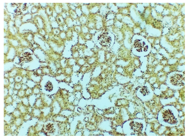 | Figure 1. Cortical parenchyma of the kidney of 3-month-old white rats "Injured with a combined injury" for 30 days, cortical material of the kidney of the research group. Balls are atrophied and not swollen. Staining: Van-Gieson. Magnification: 10x40 |
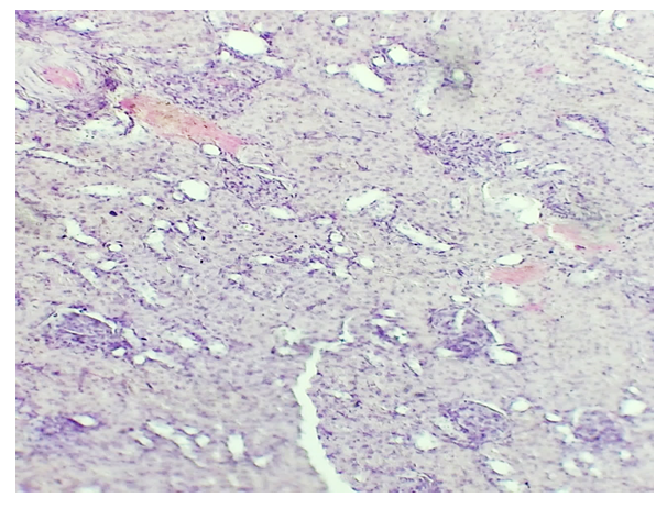 | Figure 2. Morphological changes of the kidneys of 3-month-old white rats "Combined trauma injured" for 30 days in the study group. The 1st balls are atrophied in shape, the perimeter is sharply deformed, the 2nd ball has focal sclerotic foci in the center of the capillary network, the 3rd has segmental necrosis foci in the proximal tubules. (Stained with hematoxylin-eosin. Ob: 10 x 40 |
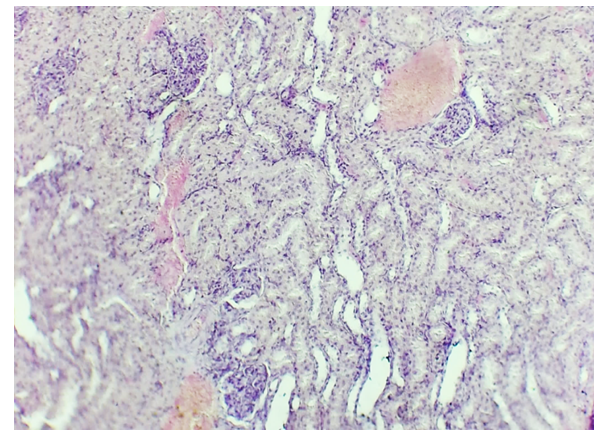 | Figure 3. Renal cortical parenchyma of 3-month-old white rats "Combined injury" for 30 days in the study group. (Stained with hematoxylin-eosin. OK 20 x OB 20. 1- focal hemorrhage zone, 2- acellular fibrotic form, 3- atrophy of straight and collecting ducts.Staining: G-E. Magnification: 10x40 |
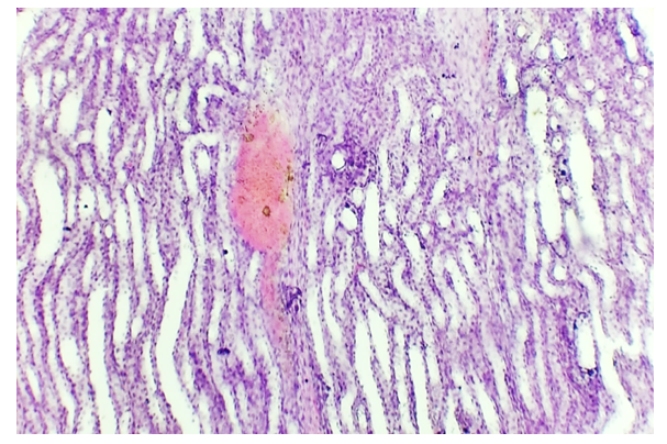 | Figure 4. Renal cortical parenchyma of 3-month-old white rats "Combined injury" for 30 days in the study group. 1- acellular fibrosis of the stroma of the renal pyramid, 2- ectasia of the collecting tubules, 3- focal hemorrhage zone, 4- atrophy of the straight and collecting tubules)Staining: G-E. Magnification: 10x40 |
4. Conclusions
- Thus, the thickness of the capsule, the size of the tubules and glomeruli, the density of cellular elements and vascular structures were studied using the morphometric method, as well as a quantitative assessment of the area of damaged zones, necrotic areas and fibrosis zones using digital microscopes and image analysis software. These data allow us to conduct a quantitative analysis of the impact of trauma and anesthesia on the morphometric characteristics of the kidneys and to assess the degree of restoration of renal tissue in various injuries.