-
Paper Information
- Next Paper
- Previous Paper
- Paper Submission
-
Journal Information
- About This Journal
- Editorial Board
- Current Issue
- Archive
- Author Guidelines
- Contact Us
American Journal of Medicine and Medical Sciences
p-ISSN: 2165-901X e-ISSN: 2165-9036
2024; 14(11): 2841-2846
doi:10.5923/j.ajmms.20241411.36
Received: Nov. 1, 2024; Accepted: Nov. 11, 2024; Published: Nov. 13, 2024

Features of Lung Morphology under the Influence of Dyes and Its Phytocorrection
 Abstract
 Reference
 Full-Text PDF
 Full-text HTML
D. A. Hasanova, Z. S. Azimova
Bukhara State Medical Institute named after Abu Ali ibn Sino, Bukhara, Uzbekistan
Copyright © 2024 The Author(s). Published by Scientific & Academic Publishing.
This work is licensed under the Creative Commons Attribution International License (CC BY).
http://creativecommons.org/licenses/by/4.0/

Food additives have become widely used for a number of reasons: to extend the shelf life of food products that must be transported over long distances; to meet the changing tastes and preferences of consumers, the desire for economy and convenience, which has led to the introduction of additives such as flavors and colors; and because of the improvement of food production technologies. So, most products in the modern world contain food additives. But not all of them pose a health risk. It's important to be aware of which supplements may be harmful and not to be afraid of eating foods labeled E without understanding their true effects. The effects of potentially harmful food additives on the body depend on their dosage and unique characteristics of the body. The safe level of intake for some is as low as a few milligrams per kilogram of body weight, while for others it can reach significantly higher thresholds.
Keywords: Food additive, Food safety, Nanomaterial, Oral exposure, Titanium dioxide, Toxicity
Cite this paper: D. A. Hasanova, Z. S. Azimova, Features of Lung Morphology under the Influence of Dyes and Its Phytocorrection, American Journal of Medicine and Medical Sciences, Vol. 14 No. 11, 2024, pp. 2841-2846. doi: 10.5923/j.ajmms.20241411.36.
1. Introduction
- The term "food additives" sounds alarming to many, although the use of some of them, such as salt, acetic acid and spices, dates back thousands of years. But over time, natural additives have given way to synthetic ones. Today, food additives are those artificial substances that are used in the production of products to provide desirable properties, such as aroma, color or shelf life, and which are not food products in themselves [1].The effects of potentially harmful food additives on the body depend on their dosage and unique characteristics of the body. The safe level of intake for some is as low as a few milligrams per kilogram of body weight, while for others it can reach significantly higher thresholds. Previously considered safe, some additives are now recognized as harmful and excluded from consumption, as shown by the examples of formaldehyde E-240 and citrus red E-121 [2].Among the various food additives, preservatives and antioxidants are considered especially dangerous due to their effect on vitamin components [5]. Some preservatives have a negative effect on nutrients: for example, sorbet acid destroys vitamin B12, and sulfur dioxide destroys vitamin B1. In addition, preservatives have the potential to be carcinogenic, contributing to the development of cancer, and can provoke allergies, asthmatic attacks, and headaches [8].At the same time, there are food additives recognized as harmless to health. However, due to a lack of consumer awareness and a unified "E" coding system, safe additives are often incorrectly associated with harmful ones. Examples of safe additives are apple pectin (E-440), citric acid (E-330) and ascorbic acid (E-300). In addition, sodium nitrite (E-250), widely used in the meat industry for smoking and curing due to its antibacterial properties, is also often misunderstood [10].Titanium dioxide (TiO2), the production of which at the global level has Zoomed significantly since the beginning of 2008, is widely used in the food industry as a white dye E-171 due to its pronounced color [1]. This color, included in food products, complies with international standards, as well as the regulations of the European Union [2]. This means that the intake of TiO2 through the human gastrointestinal tract can be quite significant.In the food industry, TiO2 is used as a colorant that gives products a white color by reducing the "gray" component in the light reflected by the product [13]. In this assessment, the possible effects of nanoparticles were not taken into account, and it was concluded that establishing safe daily levels of TiO2 intake was not necessary, mainly because of its very low solubility. However, according to the European Food Safety Authority, pigment TiO2, whose particles range in size from 0.1 to 1.0 μm, is considered safe for humans due to its low solubility in water and biological media, as well as its lack of absorption in the gastrointestinal tract [12].Thus, the available data indicate that TiO2 nanoparticles can penetrate the intestinal mucosa, but only to a limited extent. Quantification of TiO2 absorption from the gastrointestinal tract is a complex task that can be solved by detecting micro- or nanoparticles using analytical transmission electron microscopy or elemental analysis of the tissue Ti content. One of the first such studies P.U. Jani et al. identified 500 nm TiO2 microparticles in the liver, spleen, and intestinal lymphoid tissue of rats after oral administration at a dose of 12.5 mg/kg [8].In experimental studies conducted on animals, the mean lethal dose (LD50) of aluminum compounds ranged from a few hundred to 1000 mg/kg body weight. The lowest observed dose of aluminum leading to adverse reactions in rats (LOEL) was approximately 75–80 mg/kg body weight per day [1].Aluminum has a negative effect on the kidneys, causing changes such as hydronephrosis, dilation of the urinary ducts, difficulty urinating and the formation of stones. In in vitro experiments, it was found that high concentrations of aluminum can cause a genotoxic effect on bacterial cells and cells of warm-blooded animals. It has also been shown that high doses of aluminum compounds can negatively affect the reproductive system of dogs and exhibit neurotoxicity in mice and rats [8,9,10].In vitro studies conducted using the Salmonella typhimurium strain have not revealed mutagenic effects of aluminum [11]. High doses of aluminum compounds administered intragastrically demonstrated embryo toxicity in mice and rats, manifested by a decrease in fetal weight and neonatal weight, as well as delayed ossification in newborns [3].
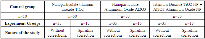
2. Materials and Methods
- For morphological analysis, the obtained tissues were fixed with 10% buffered formalin for 24 hours. Routine wiring of tissues was carried out on a carousel processor STP120, ThermoFisher, Germany, after which the samples were poured into paraffin. Sections obtained on a rotational microtome NM 325 (TFS, USA) with a thickness of 3-4 μm.
|
3. Results and Discussions
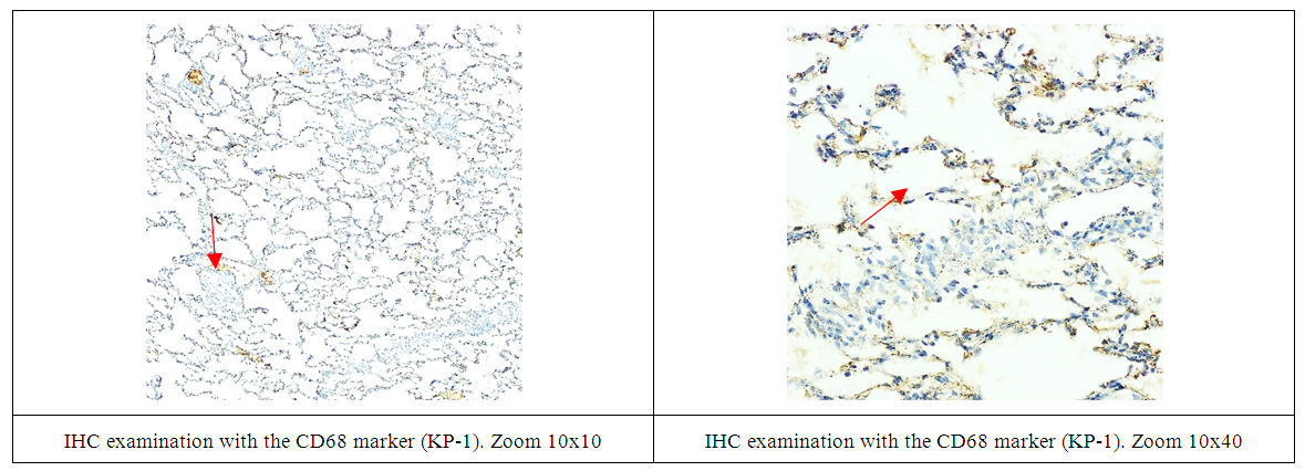
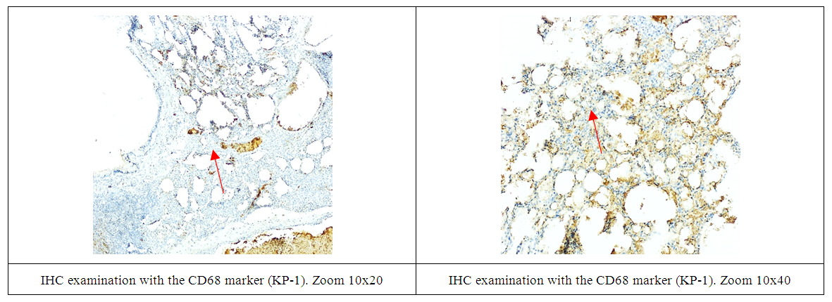
- The lung system of macrophages consists of several subpopulations that are found in different anatomical regions of the lungs, including the respiratory tract, alveolar spaces, and resident lung tissue. Alveolar macrophages make up more than 90% of the lung macrophage population. CD68 (cluster of differentiation 68, macrosialin – CD68 (clone of KP1)) is one of the specific markers of macrophages. Alveolar and interstitial macrophages are the two main subgroups, named after their location. Alveolar macrophages are responsible for clearing inhaled foreign particles of various nature. The interaction of alveolar macrophages with the removed particles through certain receptors determines the severity of the inflammatory response: from minimal to active inflammation with damage to lung tissue.Single weakly positively stained alveolar macrophages (arrow, 1-2 pieces) located in the pulmonary alveoli and intermalleolar septum in the immediate vicinity of pneumocystis’ are noted. Interstitial macrophages are not noted (Fig. 1).
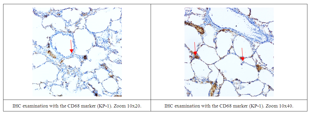 | Figure 1. Aluminum oxide control, immunohistochemical study |
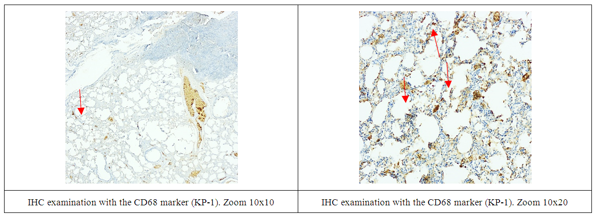
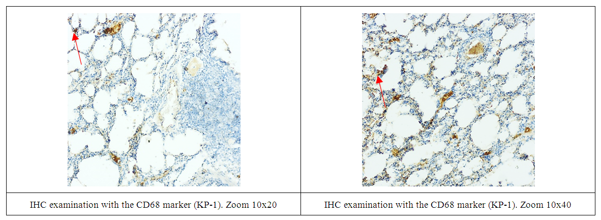
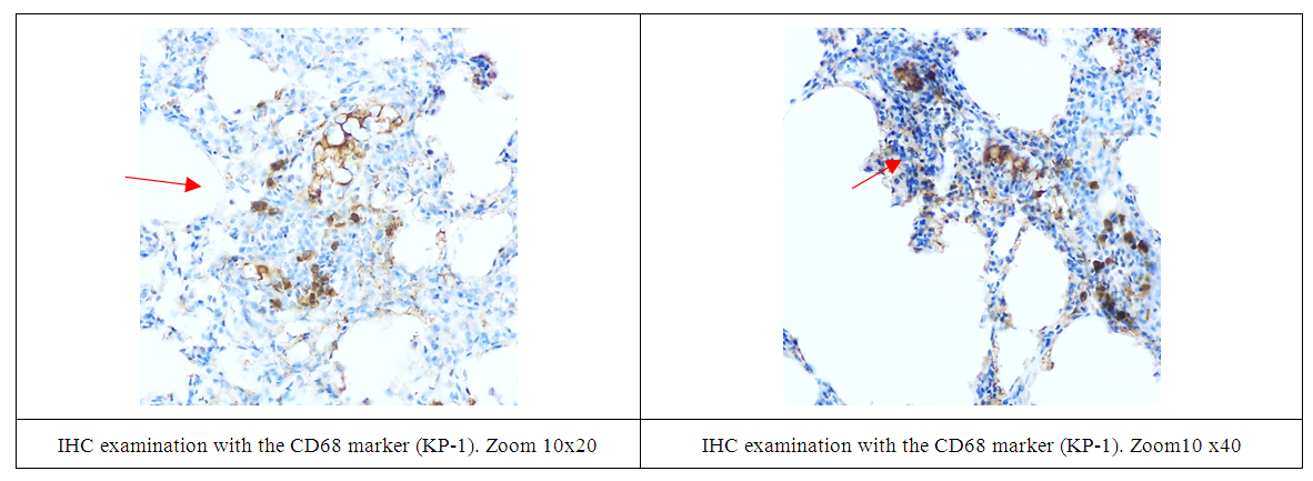 | Figure 2. Group "Aluminum oxide, experiment", immunohistochemical study |
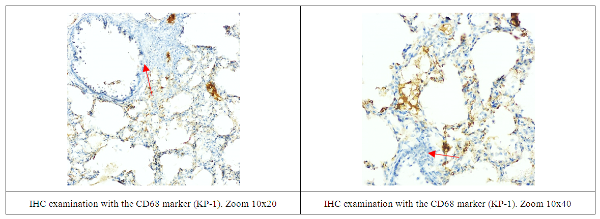
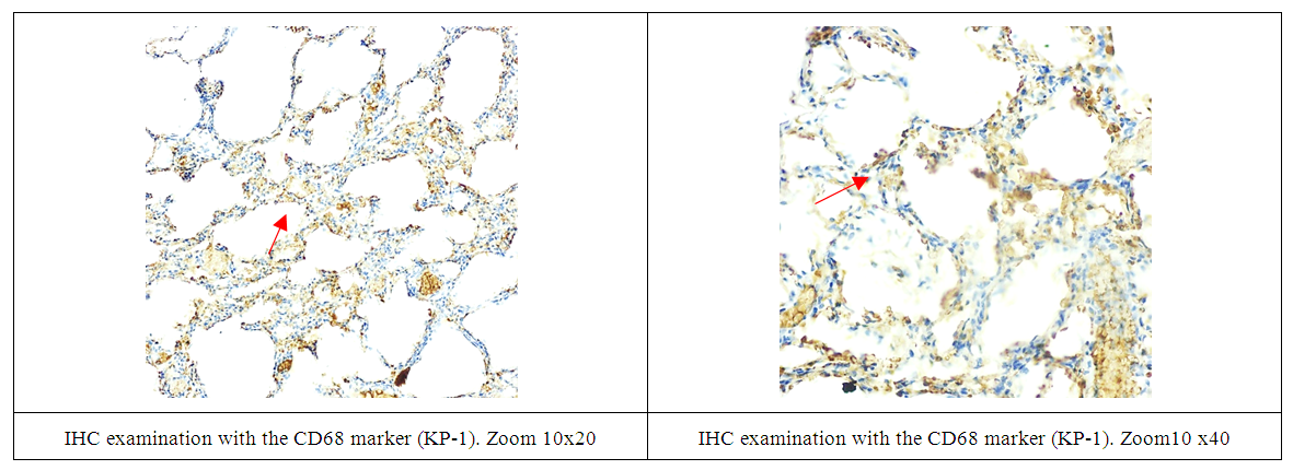 | Figure 3. Group "Aluminum oxide, correction", immunohistochemical study |
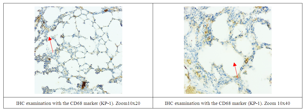
 | Figure 4. Group "Titanium dioxide, control", immunohistochemical study |
 | Figure 5. Group "Titanium, dioxide experiment", immunohistochemical study |
 | Figure 6. Titanium dioxide correction group, immunohistochemical study |
 | Figure 7. Group "E-171, E-173, control", immunohistochemical study |
 | Figure 8. Group "E-171, E173 experiment", immunohistochemical study |
 | Figure 9. Group "E-171, E-173, correction", immunohistochemical study |
4. Conclusions
- In our study, it was found that in the "experimental" groups with signs of fibrotic changes, intensive proliferation of CD68 was noted. At the same time, staining of not only alveolar and interstitial macrophages, but also histiocytic located in the formed per bronchial lymphoid groups was observed.