-
Paper Information
- Next Paper
- Previous Paper
- Paper Submission
-
Journal Information
- About This Journal
- Editorial Board
- Current Issue
- Archive
- Author Guidelines
- Contact Us
American Journal of Medicine and Medical Sciences
p-ISSN: 2165-901X e-ISSN: 2165-9036
2024; 14(11): 2830-2832
doi:10.5923/j.ajmms.20241411.33
Received: Oct. 20, 2024; Accepted: Nov. 11, 2024; Published: Nov. 13, 2024

Features of Late Morphological Changes in the Skin After Application of a Tattoo
 Abstract
 Reference
 Full-Text PDF
 Full-text HTML
Ozodov Jasur Khakimovich
Bukhara State Medical Institute, Uzbekistan
Correspondence to: Ozodov Jasur Khakimovich, Bukhara State Medical Institute, Uzbekistan.
| Email: |  |
Copyright © 2024 The Author(s). Published by Scientific & Academic Publishing.
This work is licensed under the Creative Commons Attribution International License (CC BY).
http://creativecommons.org/licenses/by/4.0/

This study investigates the late morphological changes in the skin following tattoo application. The research focuses on understanding how the skin's structure and appearance evolve over time after the introduction of tattoo ink. By analyzing skin biopsies at various intervals post-tattooing, the study aims to identify long-term alterations in the dermal and epidermal layers, including changes in collagen organization, inflammatory responses, and ink dispersion. The findings provide insights into the skin's adaptive processes and potential implications for tattoo longevity and skin health.
Keywords: Tattoo- skin morphology, Dermal changes, Epidermal changes collagen organization, Inflammatory response, Ink dispersion- long-term skin alterations
Cite this paper: Ozodov Jasur Khakimovich, Features of Late Morphological Changes in the Skin After Application of a Tattoo, American Journal of Medicine and Medical Sciences, Vol. 14 No. 11, 2024, pp. 2830-2832. doi: 10.5923/j.ajmms.20241411.33.
1. Relevance
- In the USA there are 80 million people with tattoos about statistics for 2019 in the. In Russia -14 million (10% of the population) have tattoos. Of these, 18% are men and 5% are women. The average number of tattoos among Russians is 2. 14% of people experienced complications associated with tattoos, 84% did not. An interesting study was conducted in the UK among people with tattoos. Of the 100 Englishmen with tattoos, 68% are women and only 32% are men. The average age of these people is 27 years. Of these, 88.6% are people with higher education, 24% of them with an academic degree. And only half of all respondents know about the possible risks associated with tattooing.World Health Organization (WHO) statistics “...infectious pathologies during tattooing, methicillin Staphylococcus aureus occupies a significant place...”.1 Therefore, monitoring the hygiene and sterility of paints is an urgent need. The potential carcinogenic risk of ink ingredients such as polycyclic aromatic hydrocarbons remains clinically unproven, despite tattooed individuals having been exposed to such potential risk for a century. However, research is scattered, and potential risks to the body, internal organs and fetus cannot be ruled out.Tattoos cause a wide range of clinical problems. Mild complaints, especially sun sensitivity, are very common and occur in 1/5 of cases. Predominant medical complications are allergies to tattoo pigment haptens or haptens produced in the skin, especially in red tattoos, but also in blue and green tattoos. The symptoms are severe and can be compared to severe itchy skin conditions. Allergy to tattoos and local reactions manifest as distinct clinical manifestations with plaque-like, excessive hyperkeratotic, ulcerative-necrotic, lymphopathic, neurosensory and cicatricial patterns. Reactions to black tattoos are papulo-nodular and non-allergic and are associated with agglomeration of soot nanoparticles.Purpose of the study: study of late morphological changes in the skin of white outbred rats after tattooing.
2. Materials and Methods of Research
- To conduct an experimental study, 150 white outbred rats of both sexes, weighing 200-250 grams, were selected under standard vivarium conditions. These laboratory animals received from the nursery were subjected to a mandatory veterinary examination to identify existing diseases, assess their condition and age. Accepted animals were quarantined for 21 days to prevent the introduction of infectious diseases into the vivarium.All groups were formed at the same time. The laboratory animals participating in the experiment were representative in terms of age, sex, weight, housing and feeding conditions. After 30 days of feeding, groups of laboratory animals were humanely killed and necropsies were performed. When killing and dissecting laboratory animals, biological safety rules and ethical principles for working with laboratory animals were observed. When working with laboratory animals, all rules of biological safety and ethical principles of working with laboratory animals given in the methodological manual Nuralieva N.A., Bektimirova A.M., Alimova M.G., Suvanova K.Zh. will be observed. “Rules and methods of working with laboratory animals in microbiological and immunological research”, approved by M3 of the Republic of Uzbekistan on May 25, 2016.To apply the tattoo, we used a convenient and modern tool, which is now at the peak of popularity - the Cheyenne Hawk Thunder Black tattoo pen, made in Germany.To apply the tattoo, we used ink from the most famous manufacturer - the American company World Famous Tattoo Ink (WFTI), which produces high-quality blue pigment.Morphological changes in the skin of white outbred rats were identified, the results were recorded in journals, the results were statistically processed and described in the form of a research report.Statistical processing of the obtained results was carried out using the methods of variation statistics using the application package Statistica for Windows. Digital data was processed on a personal computer using the memory of Microsoft Exell application programs. Information was considered reliable if t≥2 and p<0.05.
3. Results and Discussions
- To identify the features of late morphological changes in the skin of white outbred rats, removal from the experiment after the procedure was carried out after 30 days from the date of tattooing. We collected 15 fragments of tattooed skin, and as a control we used 10 fragments of tail skin from white outbred rats before tattooing. The material was fixed in a 10% solution of neutral formaldehyde for 24 hours, then passed through alcohols of increasing concentration: 70-80-96-100° and chloroform, embedded in paraffin, histological sections were prepared, stained with hematoxylin and eosin and Van Gieson.According to histological analysis, we found that as the time after tattooing increased (on the 30th day), the phenomena of edema and acute inflammation decreased along with the redistribution of pigment from the papillary dermis layer to the reticular layer. Thus, single small granules of pigment were observed in the papillary layer of the dermis, which were located diffusely. Vacuolar degeneration of epidermal cells was detected directly near accumulations of tattoo pigment, massive accumulation of blue pigment in the papillary layer of the dermis, and areas of hyperkeratosis were identified in the tattoo area. The fibers of the papillary layer of the dermis were located quite tightly to each other, were thickened and, when stained with hematoxylin and eosin, were represented by homogeneous eosin masses, which may indicate the focal development of hyalinosis.
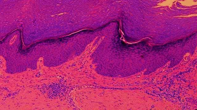 | Picture 1. Hyperecratic picture of the skin of white outbred rats during histological examination. Microscopic appearance. Hematoxylin-eosin staining. Oc 40 x 20 ob |
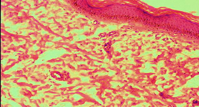 | Picture 2. Microscopic appearance. Hematoxylin-eosin staining. Oc 40 x 20 ob |
Note
- 1. https://www.who.int/ru/news/item/09-12-2020-who-reveals-leading-causes-of-death-and-disability-worldwide-2000-2019