-
Paper Information
- Next Paper
- Previous Paper
- Paper Submission
-
Journal Information
- About This Journal
- Editorial Board
- Current Issue
- Archive
- Author Guidelines
- Contact Us
American Journal of Medicine and Medical Sciences
p-ISSN: 2165-901X e-ISSN: 2165-9036
2024; 14(11): 2791-2794
doi:10.5923/j.ajmms.20241411.25
Received: Sep. 28, 2024; Accepted: Oct. 30, 2024; Published: Nov. 12, 2024

Morphofunctional Relationships of Beta– and Delta– Basophilic Cells of the Adenohypophysis During Diestrus and Estrus in Intact Animals
 Abstract
 Reference
 Full-Text PDF
 Full-text HTML
Ozоdjon Kuliev
Doctor of Philosophy (PhD) in Medical Sciences, Associate Professor, Alfraganus University, Tashkent, Uzbekistan
Correspondence to: Ozоdjon Kuliev, Doctor of Philosophy (PhD) in Medical Sciences, Associate Professor, Alfraganus University, Tashkent, Uzbekistan.
Copyright © 2024 The Author(s). Published by Scientific & Academic Publishing.
This work is licensed under the Creative Commons Attribution International License (CC BY).
http://creativecommons.org/licenses/by/4.0/

During the period of diestrus, against the background of mixed vegetative reactivity in the rat body, an appropriate relationship was ensured in the prooxidant, antioxidant system, endogenous intoxication system, and the dominance of basophilic cells with medium and low functional activity in the adenohypophysis, balanced functioning of FSH, LH, estradiol and progesterone in the blood was revealed. During the estrus period, against the background of an increase in the activity of the sympathetic nervous system 1.02 times, the functional activity of delta basophil cells of adenohypophysis, an increase in blood LH by 2.46 times, estradiol by 2.63 times, and the MDA/catalase coefficient by 1.07 times was revealed the level of MSM 254 by 1.02 times, a slight decrease in protein resistance to 1.01 times.
Keywords: Morphofunctional relationships, Diestrus, Beta basophilic cells, Estrus, Adenohypophysis, Delta basophilic cells, Intact animals, Autonomic nervous system, Catalase, Basophilic cells, MSM 254, Rat body, MSM 280, Antioxidant system, FSH, LH, Estradiol, Progesterone, Sympathetic nervous system, Protein resistance
Cite this paper: Ozоdjon Kuliev, Morphofunctional Relationships of Beta– and Delta– Basophilic Cells of the Adenohypophysis During Diestrus and Estrus in Intact Animals, American Journal of Medicine and Medical Sciences, Vol. 14 No. 11, 2024, pp. 2791-2794. doi: 10.5923/j.ajmms.20241411.25.
1. Introduction
- The day–by–day development of society in the world has its own specific manifestations, which causes an increase in extreme stressor factors of various degrees. Each stress is characterized by its origin and duration, as well as its complexity [10,21]. At the same time, it ensures the formation of adaptive and maladaptive processes of different levels and duration in the human body [5,11]. The adaptive process occurring in the human body is formed in the plane of existing systems in the body [6,7,20]. It is possible that such formation is accompanied by adaptive and maladaptive changes and causes different degrees of disease in these components [6,8,12]. Gorizontov P.D. (1983) data show the role of anabolic hormones in the last stages of the adaptive process, and the morphofunctional changes that occur in these components during the periods of clinical death and post-resuscitation disease have not been fully studied. In relation to extreme factors, the adaptive process in male and female animals is different, and it has been studied that androgen and estrogen hormones produce specific adaptive processes in this plane [1,4,13,14,16,19]. But at the same time, the morphofunctional changes in the reproductive system during diestrus and estrus in intact female animals have not been fully studied, in order to study the changes caused by extreme extreme factors in intact animals, it is appropriate to study the morphofunctional changes in the adenohypophysis in connection with the processes occurring in the autonomic nervous system, pro– and antioxidant system, endogenous intoxication level.The purpose of work: Study of morphofunctional relationships in beta and delta basophil cells of adenohypophysis in relation to autonomic nervous system, prooxidant, antioxidant systems, endogenous intoxication indicators in intact female animals during diestrus and estrus.The research of object: The research was carried out in 15 adult white female rats, weighing 150–180 g, and the morphofunctional relationships in the beta and delta basophil cells of the adenohypophysis depending on the parameters of the autonomic nervous system, prooxidant, antioxidant systems, and endogenous intoxication. was studied.
2. The Research of Methods
- Autonomic nervous system reactivity using the Hildebrant coefficient [18], morphofunctional activity in beta and delta basophil cells Polenov A.L. using the criterion [15], the amount of MDA in the blood Stalnaya I.D. using the method [17], the amount of catalase was determined using the method of Korolyuk M. A. [9], MSM254 and MSM280 using the Gabrielian method [3]. In addition, the general conditions of animals were studied. The statistical analysis of the obtained results was carried out using the Microsoft Office–Excel 2000 standard package.
3. Results and Discussion
- In the period of diestrus, the external appearance of rats in the intact group is paid attention to: the appearance is pleasant, the skin is smooth, and the movement is active. The external appearance of the penis is not observed closed separation. The number of respirations was 562.0 ± 10.4 per minute, and the heart rate was 103.6 ± 2.1 per minute. At this time, the Hildebrandt coefficient was 5.4±0.04.The amount of follicle–stimulating hormone (FSH hormone) when gonadotropin hormone was determined in the blood of rats was 157.6±1.9 mlU/ml. LH was 17.5±0.5 mlU/ml, and it was found that the amount of estradiol hormone was 88.8±1.3 pg/ml, and the amount of progesterone hormone was kept around 9.8±0.3 ng/mL Malondialdehyde (MDA) amount was 1.12±0.007 ng/mL when prooxidant activity was determined in the blood, catalase activity in the antioxidant system was 38.6±2.4 ng/mL, and the ratio of MDA/catalase was 0.029±0.002.When the indicator of endogenous intoxication was determined in rats, MSM254–0.246±0.02 unit of form, MSM280 -0.260±0.01 unit of form and it was found that the protein resistance coefficient (PRC) was equal to 1.08±0.01. At this time, the appearance of pituitary b and d basophilic cells (Figure 1):
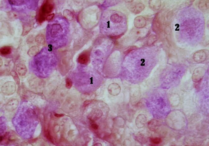 | Figure 1. In the micropreparation of the adenohypophysis of intact rats during dysterus, type I and II cells predominate among β– and d–basophilic cells. The drug is soaked with PAF and azan. Zoom: ok. 20x, ob.40x. Indicated by the number: 1. Type II delta basophilic cells; 2. Type II beta basophilic cells |
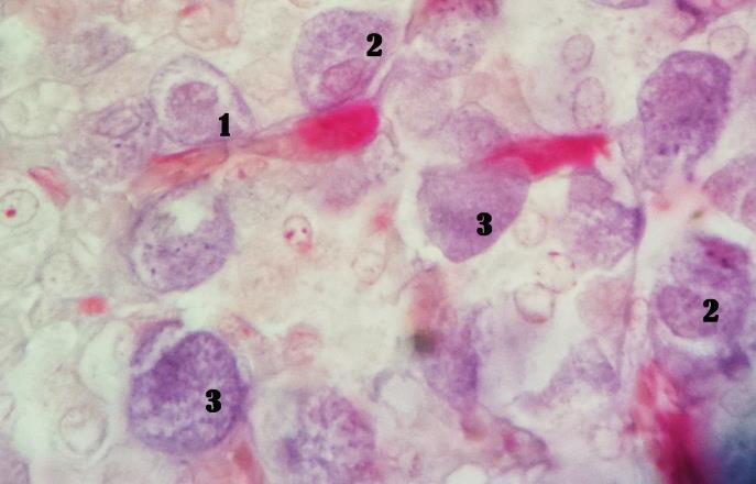 | Figure 2. In the micropreparation of the adenohypophysis of intact rats during the estrous period, type I and II cells predominate among β– and d– basophilic cells. The drug is mixed with PAF and azan. Zoom: ok. 20x, ob.40x. Indicated by number: 1. Type I neurosecretory cells; 2. Type II neurosecretory cells; 3. Type III basophilic cells compared to the index of intact animals during diestrus |
4. Conclusions
- 1. In the diestrus period, under mixed vegetative reactivity in the body of rats, a balanced state is ensured in the prooxidant, antioxidant system, endogenous intoxication system, and under the dominance of basophil cells with medium and low functional activity in the adenohypophysis, the level of FSH, LH, estradiol, progesterone hormones in the blood It was determined that it operates in a balanced state.2. During estrus, the tone of the sympathetic nervous system in rats increases by 1.02 times, the functional activity of delta basophil cells increases in the adenohypophysis, LH increases by 2.46 times, the amount of estradiol hormone increases by 2.63 times, and the MDA/catalase coefficient in the prooxidant and antioxidant systems increases. 1.07 times higher, MSM 254 was found to increase the endogenous intoxication index by 1.02 times, and the protein resistance index by 1.01 times to an insignificant level.