-
Paper Information
- Next Paper
- Paper Submission
-
Journal Information
- About This Journal
- Editorial Board
- Current Issue
- Archive
- Author Guidelines
- Contact Us
American Journal of Medicine and Medical Sciences
p-ISSN: 2165-901X e-ISSN: 2165-9036
2024; 14(11): 2689-2692
doi:10.5923/j.ajmms.20241411.01
Received: Oct. 8, 2024; Accepted: Oct. 29, 2024; Published: Nov. 7, 2024

Changes in the Histioarchitectonics of the Pancreas Associated with COVID-19 Infection
 Abstract
 Reference
 Full-Text PDF
 Full-text HTML
Sharipova Barno Ergashevna, Tursunov Khasan Ziyaevich
Assistant Lecturer, Department of Pathological Anatomy of the Tashkent Medical Academy, Uzbekistan
Copyright © 2024 The Author(s). Published by Scientific & Academic Publishing.
This work is licensed under the Creative Commons Attribution International License (CC BY).
http://creativecommons.org/licenses/by/4.0/

The peculiarity of the changes in the histioarchitectonics of the pancreas in covid-19 infection is that in covid-19 there is a sharp increase in the permeability of the vessel wall, the occurrence of interstitial edema and the blurring of all components in the gland tissue, the shape of the cells changes as a result of interstitial edema, the glands are compressed into a circle, the islets of Langerhans oval-shaped and dumbbell-shaped, at the same time, homogeneous tumors appeared around the gland ducts and blood vessels, and the sudden development of intermediate tumors in this area and the retention of gland secretions in the synthesized areas, i.e., in the acinar gland cavity, passing into the appearance of dampness, and in the general background, this picture is hypercellular. was found to have arrived.
Keywords: Pancreas, Covid-19, Necrobiosis, Pathomorphology, Pancreatitis
Cite this paper: Sharipova Barno Ergashevna, Tursunov Khasan Ziyaevich, Changes in the Histioarchitectonics of the Pancreas Associated with COVID-19 Infection, American Journal of Medicine and Medical Sciences, Vol. 14 No. 11, 2024, pp. 2689-2692. doi: 10.5923/j.ajmms.20241411.01.
Article Outline
1. Introduction
- During the covid-19 pandemic, diabetes was ignored in patients, and while the main diagnosis and treatment were focused on covid-19 infection, according to the autopsy data, it was found that hemorrhagic pancreatitis developed in many internal organs, including the pancreas [1]. According to thanotogenesis, acute hemorrhagic inflammation of the pancreas directly causes the patient's death, this information was not taken into account in lekuin covid-19. As a result, the detection of acute hemorrhagic pancreatitis during the autopsy confirmed the relevance of the problem once again. This situation in the covid-19 pandemic in the USA and European countries caused a lot of data to be lost in the period of time without autopsies of the deceased [2]. The same database was investigated after Spanish pathologists reported in a September 2020 report on the controversial autopsy data. In 23.6% of those who died from covid-19, the specificity of the pathomorphological changes of the pancreas was revealed: massive venous engorgement, acinar glands being located in a dense complex with each other, forming an edematous appearance, and massive diapedesis hemorrhages in the periacinar veins [3,4]. In this regard, the data base was not analyzed, and in the case of peragonia, parenchymatous organs were evaluated as the appearance of venous fullness. However, hyperglycemia, high level of SRB-protein, and sharp increase of AAB-protein in blood plasma were ignored in the laboratory parameters. In this research work, we have set ourselves the goal of studying specific morphological changes of the pancreas during the exudation stage of covid-19 infection and making scientific and practical recommendations based on the analysis of the obtained data.Purpose: to study specific and general morphological changes in the pancreas during covid-19 infection and to come to clear conclusions about the outcome of the obtained results.
2. Material and Methods
- Pancreatic tissue material extracted at autopsy from a total of 116 cases of covid-19 death at the Center of Pathological Anatomy of the Republic was prepared and hematoxylin and eosin dye was used to examine the morphological changes in the pancreas.
3. Research Results and Their Discussion
- The capsule of the pancreas is of different thickness, signs of fullness in the blood vessels, focal diapedesis hemorrhages around it were detected. At the same time, an edematous manza, containing lymphocytes and a small amount of neutrophils, is detected under the capsule. macrophages are rare. Although the standard histioarchitectonics of the acinar glands in the fragments located close to the subcapsular branch has not changed, they have a swollen appearance, and it is determined that dark basophilic structures remain in the cavity of the acinar glands (Fig. 1).
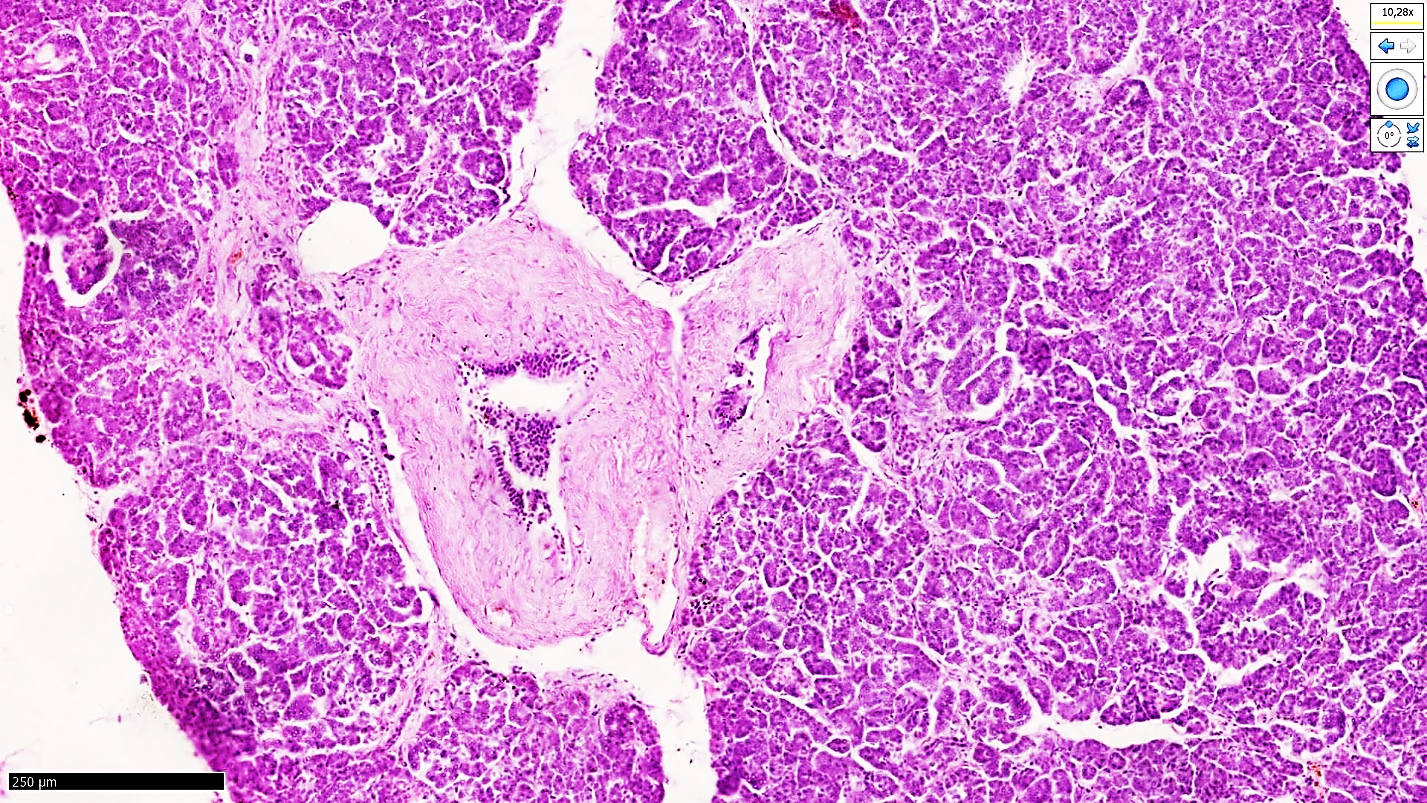 | Figure 1. Covid-19 is confirmed. Pancreas in general background. Interstitial swelling around the acinar glands developed (1), the islets of Langerhans were formed, and the cellular content decreased (2). Paint G.E. The size is 10x10 |
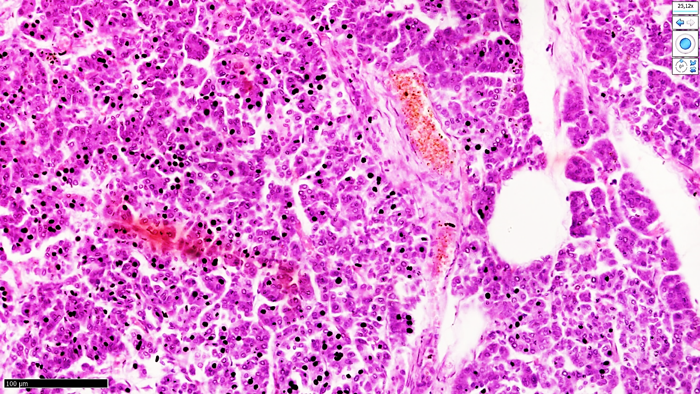 | Figure 2. Diapedesis blood flow to the pancreas islet of Langerhans and necrobiosis and interstitial edema are detected in chromophilic cells, segmental necrosis in testis and signs of fullness in pericyanar vessels are detected. Paint G.E. The size is 40x10 |
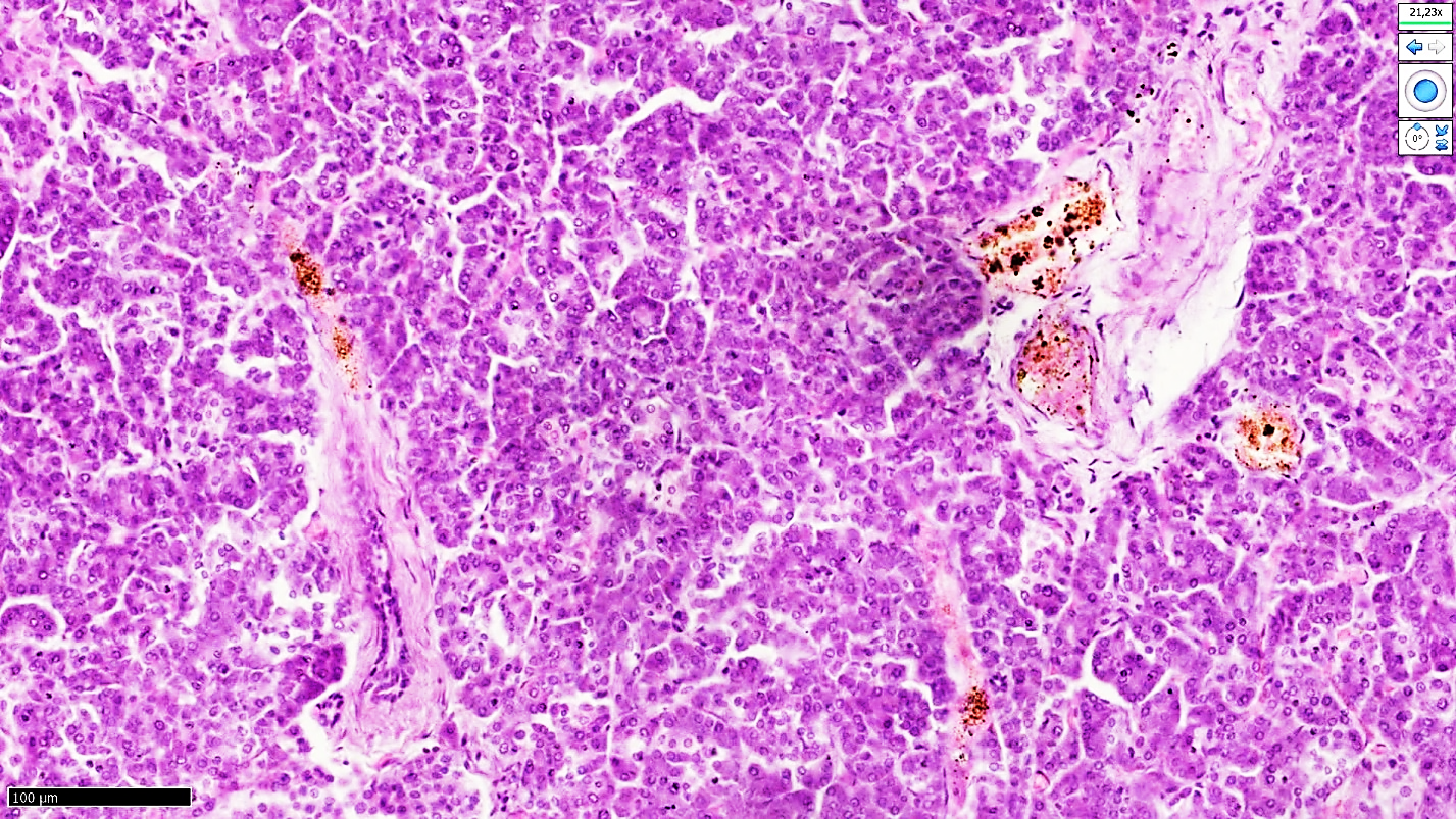 | Figure 3. Covid-19 confirmed. Minutes No. 53VI. N is 51 years old. Pancreatic acinar gland structures have multifocally developing necrosis foci in the center (1), reliefs of the glands have changed in a wave-like manner, in the form of polarization (2), the islet of Langerhans is slightly deformed (3). Paint G.E. The size is 40x10 |
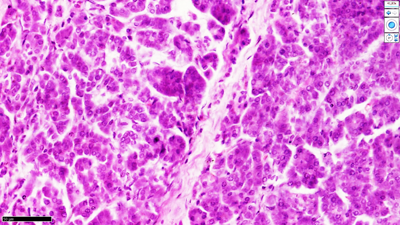 | Figure 4. Covid-19 confirmed. Minutes No. 49VI. L is a 53-year-old woman. In segmental pancreonecrosis segments with interstitial sclerosis (1), irregular proliferation of fibroblasts and a chaotic band appearance of fibrous structures (2) |
4. Conclusions
- In conclusion, it was found that in covid-19 infection, after entering the causative vessel cavity, massive dilatation and paralytic expansion of all small-caliber blood vessels and capillary walls continued with hyperemias and diapedesis hemorrhages. It is precisely this picture that develops in the same way in all parts of the pancreas, develops in the form of systemic vasculitis of vessels in covid-19, increases the plasma permeability of the basement membrane between the anatomical layers of all blood vessels, and increases the permeability of the vessel wall, including sudden hemodynamic disturbances in the pancreatic microcirculatory body. It was found that the development continued with the development of diabetes and segmental pancreonecrosis, which was first detected from a clinical morphological point of view. This confirms that in the early stages of the mechanism of thanotogenesis in covid-19, acute damage to the pancreas, polyorgan failure develops, making it difficult for the attending physician to focus on the aspects of the treatment, with the tactical help of the patient's death, and the symptomatic treatment is ineffective and the death rate is high.