-
Paper Information
- Previous Paper
- Paper Submission
-
Journal Information
- About This Journal
- Editorial Board
- Current Issue
- Archive
- Author Guidelines
- Contact Us
American Journal of Medicine and Medical Sciences
p-ISSN: 2165-901X e-ISSN: 2165-9036
2024; 14(10): 2645-2648
doi:10.5923/j.ajmms.20241410.41
Received: Oct. 2, 2024; Accepted: Oct. 21, 2024; Published: Oct. 24, 2024

Changes in Morphometric Indicators of the Placenta Depending on the Severity of Pre-Eclamsia
Rakhmanova Nodira Khodjayazovna1, Kattakhodzhaeva Maxmuda Khamdamovna2, Jumaniyazova Khulkar Atabayevna1
1Urgench Branch of the Tashkent Medical Academy, Department "Obstetrics and Gynecology", Urgench, Uzbekistan
2DSc. Professor, Tashkent State Dental Institute Department "Obstetrics and Gynecology", Tashkent, Uzbekistan
Copyright © 2024 The Author(s). Published by Scientific & Academic Publishing.
This work is licensed under the Creative Commons Attribution International License (CC BY).
http://creativecommons.org/licenses/by/4.0/

The diagnosis and management of hypertension disorders in pregnant women, along with associated consequences, get particular attention worldwide. In contemporary medicine, postpartum consequences in women with preeclampsia or eclampsia constitute a critical issue. Innovative methodologies, including the use of clinical and pathomorphological tools, are required in the study of all connections in the mother-placenta-fetus system in order to solve problems connected to maintaining the life and health of the mother and child.
Keywords: Preeclampsia, Eclampsia, Placenta, Mother-placenta-fetus system, Antenatal diagnosis
Cite this paper: Rakhmanova Nodira Khodjayazovna, Kattakhodzhaeva Maxmuda Khamdamovna, Jumaniyazova Khulkar Atabayevna, Changes in Morphometric Indicators of the Placenta Depending on the Severity of Pre-Eclamsia, American Journal of Medicine and Medical Sciences, Vol. 14 No. 10, 2024, pp. 2645-2648. doi: 10.5923/j.ajmms.20241410.41.
Article Outline
1. Topicality of the Thesis
- Preeclampsia (PE) is characterized by systemic inflammation, oxidative stress, and changes in the levels of angiogenic factors and vascular reactivity. These changes are exacerbated in normal pregnancy and are linked to a disorder of compensatory mechanisms that ultimately results in placental and vascular dysfunction. The placenta is a unique organ that develops and differentiates, affecting fetal growth and the mother's condition throughout pregnancy [1].Since PE may only occur when placental tissue is present in the body and is nearly always resolved upon removal, the placenta is crucial to the pathophysiology of the disease. When placental antiangiogenic substances reach the mother's bloodstream, they damage and disrupt the endothelium. Arterial hypertension, proteinuria, glomerular endotheliosis, HELLP syndrome, and PE may result from this [2].
2. Aim of the Study
- Using cutting-edge research techniques (scanning and atomic force electron microscopy) to examine the pathomorphological characteristics of the placenta in pregnant women with PE in order to identify strategies for reducing neonatal morbidity and mortality.
3. Materials and Methods
- The placentas of 25 women with PE (15 with moderate and 10 with severe PE) were studied. The samples were taken from the central, paracentral and peripheral parts of the organ. For subsequent analysis to implement the light-optical study, the samples were fixed in 10% neutral buffered formalin, then embedded in paraffin and sections were made from the blocks on a microtome, which were then stained with eosin and hematoxylin in the standard manner. Their photography was performed in a Topic-T Ceti microscope. Additionally, a morphometric study was carried out using the standard Video-Test Size software.For electron scanning microscopy, the samples were washed at 37°C in several portions of sodium chloride in the form of an isotonic solution. Then, at the same temperature, they were dipped into a fixation mixture consisting of glutaraldehyde (2% in phosphate buffer) and kept for two days in a refrigerator. After that, the objects were analyzed and photographed in microscopes: "FE1 Quanta 200 3D", as well as "FE1 Quanta 600 FEG".
4. Results and Discussion
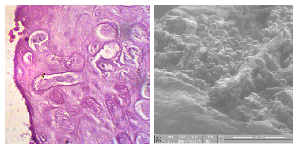
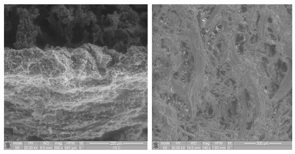
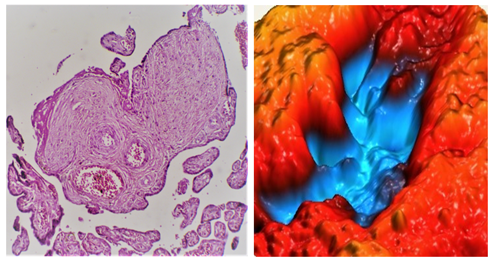
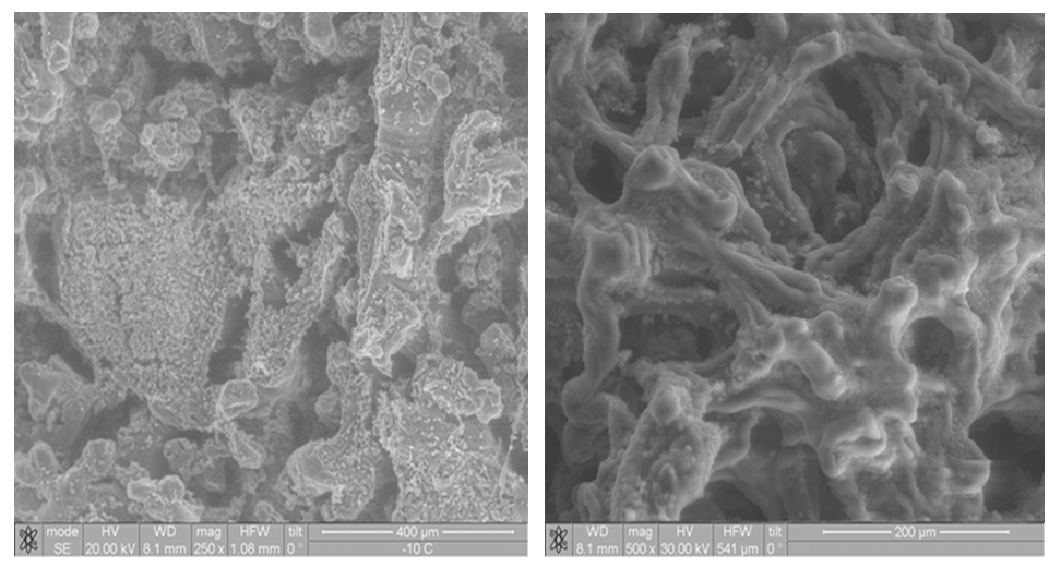
- In preeclampsia in the mother, the proportion of the placenta area occupied by large infarcts, both white (with a long history) and red, hematomas, massive intervillous thrombi, afunctional zones, caverns, calcifications (Fig. 1) increased significantly. The presence of pathologically altered fragments depended not only on the severity of the disease in the mother, but also on its duration, as well as on the options for fetal life.
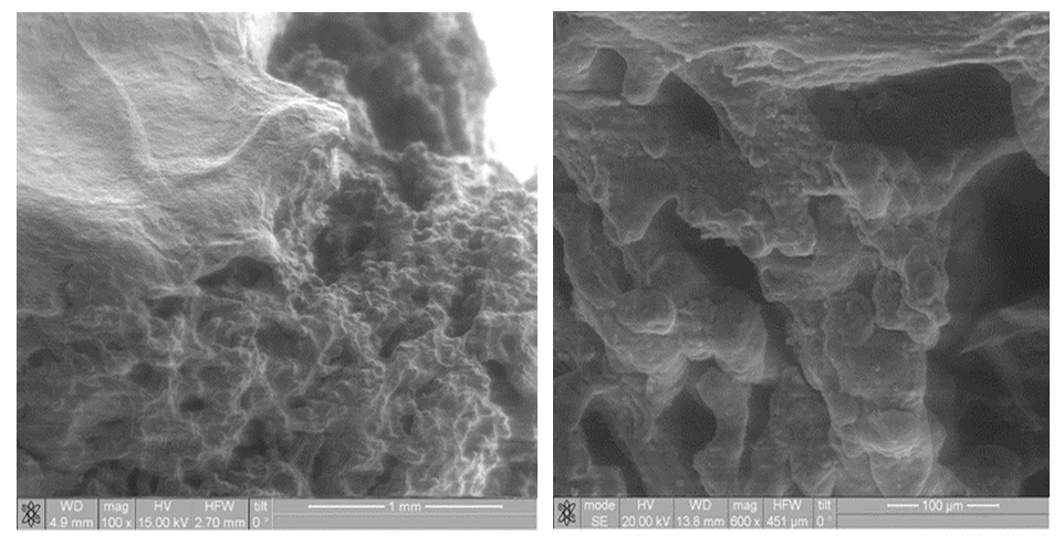 | Figure 2. Fragments of the placenta in moderate preeclampsia. The fetal surface and villous tree with a predominance of stem and intermediate villi (A, B). Fig. B (x600) fragment of Fig. A (x100) |
|
 Abstract
 Reference
 Full-Text PDF
 Full-text HTML