-
Paper Information
- Next Paper
- Paper Submission
-
Journal Information
- About This Journal
- Editorial Board
- Current Issue
- Archive
- Author Guidelines
- Contact Us
American Journal of Medicine and Medical Sciences
p-ISSN: 2165-901X e-ISSN: 2165-9036
2024; 14(10): 2641-2644
doi:10.5923/j.ajmms.20241410.40
Received: Sep. 18, 2024; Accepted: Oct. 10, 2024; Published: Oct. 24, 2024

Morphological Characteristics of the Lower Limb Muscles under Local and General Anesthesia in Mechanical Trauma of the Lower Limb
Umurov Bobirjon Fayzilloevich
Bukhara Medical Institute named after Abu Ali ibn Sino, Uzbekistan
Correspondence to: Umurov Bobirjon Fayzilloevich, Bukhara Medical Institute named after Abu Ali ibn Sino, Uzbekistan.
Copyright © 2024 The Author(s). Published by Scientific & Academic Publishing.
This work is licensed under the Creative Commons Attribution International License (CC BY).
http://creativecommons.org/licenses/by/4.0/

In this article, we studied the morphological trauma of the lower limb under anesthesia. The material was determined by morphological changes in the muscles of the lower limb of rats obtained on experimental models. Studies have shown that when using local anesthesia, less pronounced morphological changes are observed after mechanical trauma to the lower limb compared to general anesthesia. The following morphological parameters were assessed: swelling of muscle fibers, inflammatory infiltration, degenerative changes and regeneration of muscle tissue.
Keywords: Mechanical trauma, Morphology, Anesthesia, Inflammatory infiltration, Degenerative changes, Experiment
Cite this paper: Umurov Bobirjon Fayzilloevich, Morphological Characteristics of the Lower Limb Muscles under Local and General Anesthesia in Mechanical Trauma of the Lower Limb, American Journal of Medicine and Medical Sciences, Vol. 14 No. 10, 2024, pp. 2641-2644. doi: 10.5923/j.ajmms.20241410.40.
Article Outline
1. Introduction
- Muscle contractions, according to the figurative expression of I.M. Sechenov, reflect the entire diversity of external manifestations of brain activity [1]. The wide range of requirements and constantly changing conditions of the micro- and macroenvironment determine the extraordinary complexity and dynamism of the complex of mechanisms that ensure the performance of a specific function by muscle fiber. Disturbances in the delicate balance in their work, associated with changes in the parameters of cellular metabolism, can lead to the launch of a sequence of dystrophic, necrobiotic and compensatory reactions that accompany many diseases and intoxications, forming the basis of the pathological process in myopathies of various origins [2].Despite their practical and theoretical significance, the morphology and mechanisms of these changes have not been sufficiently studied. The latter is due to a number of circumstances: diseases in which damage to somatic muscles forms the basis of the pathological process (histological examination is indicated) are, as a rule, rare; muscle [4] biopsy is performed at the stage of pronounced clinical manifestations, when the histological picture reflects the complex result of the interaction of destructive and compensatory-adaptive reactions.Data on the patterns and mechanisms of damage to the muscles of the lower limb, obtained in experimental models and reflecting universal mechanisms of damage and cell death, can be extrapolated to similar processes in human pathology and used in interpreting the diversity of structural rearrangements in clinical material.
2. Research Aim
- The aim of the study was to compare morphological changes in the lower limb muscles after mechanical injury in rats receiving local and general anesthesia.
3. Materials and Methods
- The study included 48 sexually mature rats with mechanical injuries of the lower limb, divided into two groups. The first group (24 rats) received local anesthesia, the second group (24 rats) - general anesthesia. Skeletal muscle biopsies were taken from all patients 24 and 72 hours after the injury for histological analysis. The following morphological parameters were assessed: muscle fiber edema, inflammatory infiltration, degenerative changes and muscle tissue regeneration. The experiment was conducted on 48 sexually mature rats - 5-6 months old (m = 200-220 g). The animals were kept in standard conditions that comply with sanitary rules. Special marks on the body were used to identify the rats. During the experiment, the animals were healthy, without behavioral changes. In the experiment, the rats were anesthetized by intraperitoneal administration of lidocaine (350 mg / kg). In two groups, on the 1st and 3rd days after, lower limb muscles were taken during autopsy in compliance with the principles of humane treatment of animals; some animals were removed from the experiment by euthanasia, under chloroform anesthesia, by the left ventricular puncture method until complete exsanguination. The obtained biomaterial (muscle tissue) was fixed in 10% formalin solution. After fixation, muscle tissue was excised and embedded in paraffin using a standard technique. Then, 5-7 μm thick sections were made and stained with hematoxylin and eosin. Microcopying and photography were performed using an optical system consisting of a Leica microscope and an eyepiece camera at magnifications of 10x10, 10x20 and 10x40 magnification, included in the eyepiece camera delivery set. The study of the tissue structure after injury was carried out by taking autopsies of lower limb muscles that had undergone anesthesia, the following morphological abnormalities were revealed.
4. Results and Discussion
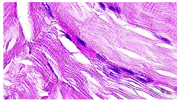
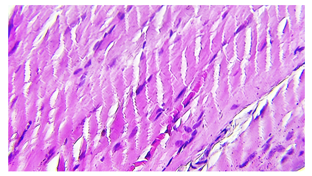
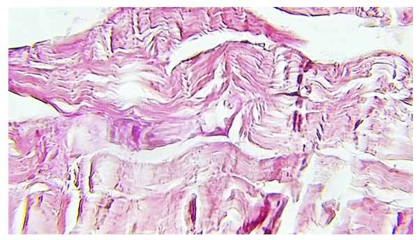
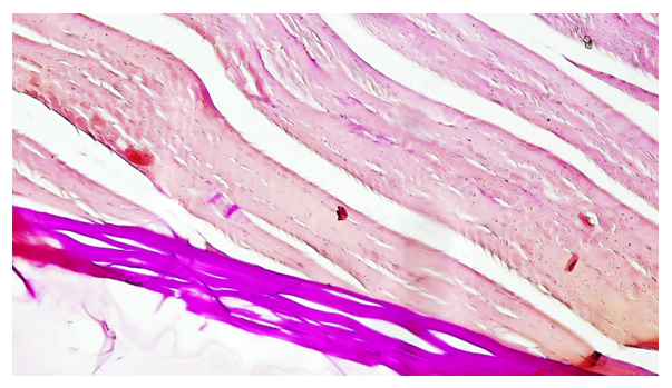
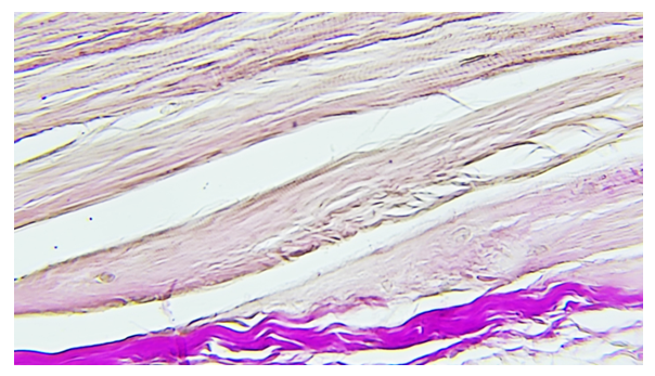
- When determining the optimal parameters for reproducing mechanical trauma, the following changes were obtained: 1) the first group of animals was given local anesthesia - the second group under general anesthesia. Rats of this group underwent microscopic [7] examination of sections of the tibialis muscle, in dynamics on the 1st and 3rd days after modeling. The study of the structure of morphological changes in skeletal muscles in intact rats that underwent anesthesia revealed morphological disorders.This is indicated by the presence of clearly defined parallel muscle fibers with transverse striation still preserved under local anesthesia (Fig. 1). Under experimental conditions on the 3rd day, the following structural changes are visualized in microphotographs, significant widened spaces are noticeable between the muscle fibers. This indicates the presence of edema, destruction of the cellular structure against the background of pathological changes under local anesthesia. Muscle fibers: muscle fibers are organized in bundles. Each fiber consists of long cylindrical cells. The nuclei of muscle cells are visible as small, dark, oval structures located on the periphery of muscle fibers. The nuclei are located in a single row along the edges of the fibers. Connective tissue: Between the muscle fibers are areas of connective tissue. This connective tissue is a lighter shade of pink and appears less dense than the muscle fibers.
5. Conclusions
- The results of the study showed that when using local anesthesia, there are less pronounced morphological changes in the muscles of the lower limb after mechanical injury to the lower limb compared to general anesthesia. Rats that received local anesthesia demonstrated less swelling, less pronounced inflammatory infiltration and degenerative changes, as well as more active regeneration of muscle tissue. These data can be used when choosing anesthesia tactics for patients with lower limb injuries, in order to minimize muscle tissue damage and accelerate recovery.
 Abstract
 Reference
 Full-Text PDF
 Full-text HTML