-
Paper Information
- Next Paper
- Previous Paper
- Paper Submission
-
Journal Information
- About This Journal
- Editorial Board
- Current Issue
- Archive
- Author Guidelines
- Contact Us
American Journal of Medicine and Medical Sciences
p-ISSN: 2165-901X e-ISSN: 2165-9036
2024; 14(10): 2624-2625
doi:10.5923/j.ajmms.20241410.37
Received: Oct. 1, 2024; Accepted: Oct. 20, 2024; Published: Oct. 24, 2024

Histological Features of the Fallopian Tube in Newborn Girls
Baqoyeva Feruza Maksudovna
Alfraganus University, Uzbekistan
Correspondence to: Baqoyeva Feruza Maksudovna, Alfraganus University, Uzbekistan.
Copyright © 2024 The Author(s). Published by Scientific & Academic Publishing.
This work is licensed under the Creative Commons Attribution International License (CC BY).
http://creativecommons.org/licenses/by/4.0/

This study presents a histological analysis of the fallopian tube in one-month-old girls. The findings reveal significant structural immaturity across the various sections of the tube. The part adjacent to the uterus is characterized by a thickened wall due to a less developed muscular layer, a narrow lumen, and poorly formed, hyperchromatic columnar epithelium. The isthmic part shows a thinner wall with disorganized smooth muscle bundles, and the epithelium is composed of cylindrical cells. The ampullary part has the thinnest wall, with only a few muscle bundles but a rich vascular network and relatively long villi. These findings underscore the importance of further research into the maturation process of the fallopian tube in early development, which could have implications for diagnosing and preventing potential reproductive system disorders later in life.
Keywords: Fallopian tube, Histological analysis, Newborn girls, Structural immaturity, Muscular layer, Epithelium, Developmental anatomy, Reproductive system maturation, Early childhood
Cite this paper: Baqoyeva Feruza Maksudovna, Histological Features of the Fallopian Tube in Newborn Girls, American Journal of Medicine and Medical Sciences, Vol. 14 No. 10, 2024, pp. 2624-2625. doi: 10.5923/j.ajmms.20241410.37.
1. Introduction
- The maturation of the fallopian tube in early childhood is an underexplored area of research, despite its critical role in the female reproductive system. Abnormal development or delayed maturation of the fallopian tube can potentially [1] lead to fertility issues or reproductive system disorders later in life. Understanding the histological features of the fallopian tube at various stages of early development is essential for identifying congenital abnormalities and for the early detection of conditions that could affect reproductive health.Research has shown that the structural immaturity of the fallopian tube in newborn females, particularly in terms of its epithelial lining and muscular layer, may have implications for future reproductive function [3,5]. The fallopian tube’s role in facilitating egg transport and supporting fertilization is dependent on the proper development of its muscular and vascular components, as well as the maturation of its epithelial cells. However, current literature lacks a comprehensive understanding of how these structures evolve during infancy and early childhood [1]. Studies have also indicated that early disturbances in the development of the fallopian tube can be associated with conditions such as ectopic pregnancies and tubal infertility if not diagnosed and managed early [4]. Therefore, it is crucial to focus on the histological maturation of the fallopian tube in newborns to better understand the potential risks and to develop preventative strategies for reproductive health issues. By filling this gap in research, we can better predict and manage potential complications related to the fallopian tube in later stages of life, contributing to overall reproductive health [2]. The fallopian tube, a crucial component of the female reproductive system, plays an essential role in the transport and maintenance of the egg. While its structure is well-developed in adult women, the histological features of the fallopian tube in newborn girls are significantly different and remain underexplored. Understanding the developmental characteristics of the fallopian tube at this early stage is important for insights into its maturation and functional [2] establishment.The aim of this article is to analyze the histological features of the fallopian tube in one-month-old girls, focusing on its different anatomical sections: the part adjacent to the uterus, the isthmic (middle) part, and the ampullary part.
2. Materials and Methods
- The study utilized sections of the fallopian tubes from one-month-old girls, stained with hematoxylin and eosin (H&E). The analysis was performed using light microscopy at a magnification of 10x10. The study focused on the structure of the fallopian tube wall, as well as the condition of the epithelial lining, mucosa, and muscular layer in its various regions.
3. Results
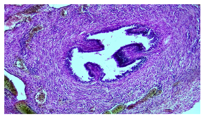
- The wall of the fallopian tube in this section was found to be thickened due to a less mature muscular layer. The luminal cavity was narrow, which is characteristic of this developmental stage. The mucosal villi were sparse and short, indicating incomplete differentiation. The lining was composed of hyperchromatic, poorly developed columnar epithelium, which exhibited signs of immaturity.In the isthmic part, the wall of the fallopian tube was relatively thinner compared to the section adjacent to the uterus. The muscular layer was composed of randomly arranged bundles of smooth muscle cells, also reflecting incomplete development. The epithelial lining consisted of cylindrical cells, which were directly positioned within the muscle bundles, a distinctive feature of this region.The ampullary part of the fallopian tube demonstrated an even thinner wall. The muscular layer was represented by only a few bundles of smooth muscle cells. This structure was rich in blood vessels, indicating its significant role in supplying blood during further development. The mucosa contained relatively long villi, but the epithelial lining remained immature, hyperchromatic, and irregularly arranged, consisting of a single layer of columnar epithelium.
 | Figure 1. 24-day-old infant, part of the fallopian tube adjacent to the uterus. The internal lumen is narrow, and the wall is thick due to the muscular layer. Staining: H&E. Magnification: 10x10 |
4. Discussion
- The findings confirm that the fallopian tube in one-month-old girls exhibits immature structural features, which undergo significant changes as it matures. The part adjacent to the uterus is characterized by a thickened wall with poorly [3] developed epithelium, reflecting its importance in the formation of functional activity. The isthmic part, with its thinner wall and disorganized muscle bundles, suggests the gradual development of the muscular layer. The ampullary part, despite having a thinner wall, displays a rich vascular network and relatively long villi, indicating its future role in capturing and transporting the egg. The hyperchromatic and disordered epithelial lining in all sections of the tube confirms the immaturity of the epithelial cells in newborn girls. This is a crucial factor that influences the future development of the reproductive system.
5. Conclusions
- The study of the histological features of the fallopian tube in one-month-old girls revealed significant differences in its structure compared to its adult state. The immaturity of the muscular layer and epithelial lining, along with pronounced vascular changes in the ampullary part, highlights the importance of further observation of the maturation processes in the early stages of life. These findings may be useful for diagnosing and preventing congenital abnormalities, as well as understanding the pathogenesis of reproductive disorders in the future.
 Abstract
 Reference
 Full-Text PDF
 Full-text HTML