-
Paper Information
- Next Paper
- Previous Paper
- Paper Submission
-
Journal Information
- About This Journal
- Editorial Board
- Current Issue
- Archive
- Author Guidelines
- Contact Us
American Journal of Medicine and Medical Sciences
p-ISSN: 2165-901X e-ISSN: 2165-9036
2024; 14(10): 2510-2513
doi:10.5923/j.ajmms.20241410.13
Received: Aug. 27, 2024; Accepted: Sep. 16, 2024; Published: Oct. 17, 2024

Arthroscopic Treatment of Diseases of the Ankle Joint
 Abstract
 Reference
 Full-Text PDF
 Full-text HTML
Kobilov Akmal Uktamovich1, Kolkhujaev Farukh Ikromovich2
1Physician-Ordinator, Samarkand Branch of the Scientific and Practical Center of Republican Specialized Traumatology and Orthopedics
2Assistant Samarkand State Medical University, Uzbekistan
Correspondence to: Kolkhujaev Farukh Ikromovich, Assistant Samarkand State Medical University, Uzbekistan.
| Email: |  |
Copyright © 2024 The Author(s). Published by Scientific & Academic Publishing.
This work is licensed under the Creative Commons Attribution International License (CC BY).
http://creativecommons.org/licenses/by/4.0/

The work is based on the experience of treating 20 patients (including 12 women and 8 men), arthroscopy of the ankle joint was performed. All patients were of working age. The average age was 38 years (from 20 to 58 years). Indications for surgery were: osteochondropathy of the talus bone in 4 (20%) patients, osteoarthritis with the presence of bone impingement in 5 (25%) patients, anterior soft tissue impingement syndrome in 4 (20%), chronic lateral instability in 2 (10%) patients, osteo-chondral fractures in 5 (25%) patients. The results obtained confirmed the high diagnostic significance of arthroscopic intervention on the ankle joint, which allows, taking into account the nature of the cartilage lesion, to determine the optimal tactics of the treatment process. The data of the functional study indicate a significant improvement in the average indicators of the ankle joint function from 31.6 points before surgery to 47.5 points at the time of the patient's repeated treatment. The pain in the ankle joint, which were the main complaints before the operation, disappeared or decreased significantly, as well as lameness disappeared, the distance that the patient can overcome and the amount of movement in the joint increased. Autografts from the tendons of the popliteal hip flexors have been successfully used to restore the The use of arthroscopic surgery, since it is a minimally invasive method, makes it possible to ensure, in comparison with open interventions, an earlier start of rehabilitation measures in the postoperative period, reduce the duration of inpatient treatment, accelerate the recovery of the patient.
Keywords: Arthroscopy of the ankle joint, Arthrosis of the ankle joint, Osteochondropathy of the talus bone
Cite this paper: Kobilov Akmal Uktamovich, Kolkhujaev Farukh Ikromovich, Arthroscopic Treatment of Diseases of the Ankle Joint, American Journal of Medicine and Medical Sciences, Vol. 14 No. 10, 2024, pp. 2510-2513. doi: 10.5923/j.ajmms.20241410.13.
1. Introduction
- Arthroscopy is an endoscopic method of diagnosis and treatment of joint diseases and injuries. For this, a special tool connected to a monitor is inserted into its cavity through a small incision above the joint. This allows the doctor to see all the changes that have occurred in the joint and perform the necessary manipulation. The history of arthroscopy begins in the 30s of the 20th century. Endoscopic examination of the hip joint was first described in 1931 by M. Burman. In 1939, K. Takagi successfully applied the experiment to cadaver joints. This study was further developed in the 70s of the 20th century, thanks to the works of the Japanese doctor M. Watanabe, using an endoscope of his own design. Detailed information on the introduction, technique and indications for hip arthroscopy. Since the mid-80s, arthroscopic interventions of the hip joint have attracted the attention of foreign and domestic surgeons more and more due to the appearance of modern arthroscope designs and the expansion of the therapeutic manipulation arsenal. In the last ten years, there have been attempts to summarize the experience gained by experts from different countries on the use of arthroscopic interventions in ankles. These data allowed us to have a clear idea about the diagnostic and therapeutic possibilities of this operation, although they did not eliminate the disagreements that exist in the literature on the justification and clinical application of this method in the pathology of the hip joint.Since the 90s, hip arthroscopy has been used in Russia. Since 2020, our clinic has been using hip arthroscopy.Anatomy. A hip joint is a complex block joint with one degree of freedom in which movements are performed around the sagittal plane. The hip joint has a range of motion of 60-90°, flexion 30-50°, extension 20-30°. The femur joint is divided into three groups depending on the location of the ligaments: 1) medial ligament complex (deltoid ligament), 2) lateral ligament complex, 3) interbarial articulation ligaments. In this case, we analyze the lateral ligament complex of the ankle joint. The lateral ligament complex consists of the anterior and posterior fibular-Taran ligaments and the calcaneal-fibular ligament. The taran-fibular ligament anterior to the lateral ligaments is the thinnest, and the sacroiliac joint is the weakest of the external ligaments.The anterior taran-fibular ligament provides stability to the anterior and lateral tibial joint. The role of the glenohumeral joint in mechanics is to limit flexion and inversion of the leg, and it also prevents rotation of the internal glenoid.The fibular ligament starts from the front surface of the lateral ankle, under the anterior Taran-fibular ligament, bends down and back, and attaches to the lateral surface of the heel bone. The more vertical direction of the ligament is an additional protection of the anterior Taran-fibular ligament, which reliably strengthens the outer part of the ankle. This ligament is extraarticular, most of its fibers are higher than the tendons of the fibular muscles.Indications for arthroscopy:• Hemarthrosis of the joint with suspicion of damage to the tendon sheath.• Chondral (osteochondral) cracks on the surface of the love bone.• Assessment of joint condition in arthritis, nonspecific synovitis.• transverse osteochondrosis.• Intraarticular bodies.• Impidgement syndrome.• The existence of free bodies.The following access methods are used for arthroscopy: front-medial, central and lateral; back - medial and lateral. Front access is of great practical importance.The anterolateral entrance is located on the lateral edge of the tendons of the long finger extensors at the level of the joint cavity. Central-m. extensor hallicis longus is located on the lateral edge at the level of the joint crack. Anteremedial entrance - the medial edge of the joint cavity along the medial edge of the tendon m. tibialis anterior (Picture 1). Posterolateral entry-at the lateral edge of the Achilles tendon at the level of the joint space. Posterior medial - on the opposite side of the Achilles tendon.
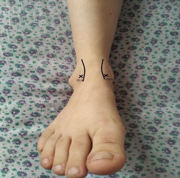 | Picture 1. Portals from the front in arthroscopy of the ankle joint |
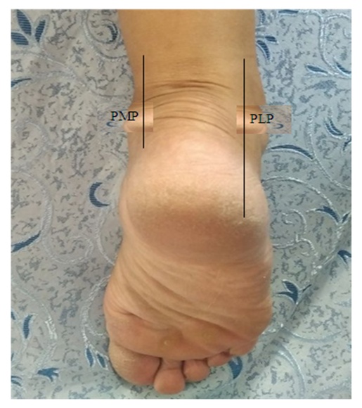 | Picture 2. Posterior portals in ankle arthroscopy |
2. Materials and Methods
- 20 patients (12 women and 8 men) underwent arthroscopy of the hip joint. All patients were able to work. The mean age was 38 years (range, 20 to 58 years). Indications for surgery: love bone osteochondropathy - 4 (20%) patients, arthrosis with presence of bone impingement - 5 (25%) patients, anterior soft impingement syndrome - 4 (20%) patients, chronic lateral instability - 2 (10%) patients, osteochondral fractures - in 5 (25%) patients.Contraindications to the operation were: local purulent inflammatory processes, severe level of arthrosis of the hip joint, i.e. III-IV level, severe level of mobility impairment, serious mental state disorders, changes that do not allow surgical intervention of internal organs.Clinical and instrumental studies were conducted to prepare patients for arthroscopy. Subjective data: patient complaints, lifestyle, previous conservative and surgical treatment (whether glucocorticosteroids are used, purulent inflammatory processes and infection, etc.), as well as additional diseases (vascular, nervous system diseases). Objective data: the patient's gait, the presence of foot axis deformity (a very important point for determining the foot's alignment during support), equinus or heel, varus or valgus deformity of the foot, evaluation of the arch of the foot (horizontal and transverse flat feet). The patient should indicate the location of the pain and the time of its occurrence. The range of motion of the hip joint was measured with a protractor. After clinical studies, standard projections-front and back and lateral x-rays and, if necessary, which allow to evaluate different levels of osteoarthritis, bone condition, limb axis, arthrosis of adjacent joints axial projections were performed. Radiography revealed bone changes, and 14 patients underwent computed tomography (CT). If soft tissue pathologies were suspected in 6 patients, a magnetic resonance imaging (MRI) examination was performed. 30 degrees, 0-2.7 mm, 1-70 mm optics, endoscopic support, arthrosheiver and ablator were used for hip arthroscopy. Since the optics were compatible, there was no need to compromise the integrity of the joint. All manipulations were usually performed under subarachnoid anesthesia with the use of a tourniquet. Operations were performed in the standard anterior and posterior ports.Two patients who underwent autoplasty of the external collateral ligament were put in a plaster cast for 2 weeks, after which they began to perform active movements of the ankle joint. Walking was allowed 3 weeks after the operation. The 6th patient who underwent microfracture was advised to walk with a stick for 2 weeks from the time of surgery.A modified ankle joint scale (Cherkes-Zade dd et al., 1999) was used to evaluate the treatment outcomes of patients. The rating scale consists of 10 characters that allow for an objective assessment of the biomechanical parameters describing the functional capabilities of the hip joint. The data of the functional study of the hip-ball joints are expressed on a five-point scale. Such indicators, complete support, the distance that the patient overcomes, trophism of paraarticular tissues, joint movement and X-ray data were studied. Each group includes three to five indicators corresponding to the evaluation system. According to this measure, the function with a total score of 45-50 is recognized as good, from 31 to 38 as satisfactory, and below 30 as unsatisfactory. Results were obtained by questionnaire before surgery and at 6-12 months.
3. Results and Discussion
- Until 2021, at the Samarkand branch of the Republican Scientific and Applied Medicine Center for Traumatology and Orthopedics, patients with sacroiliac joint pathologies were mainly operated on by an open method. Examination of patients was carried out on the basis of clinical and radiological data.In foreign literature, there are various pathologies of the ball joint, and the important role of arthroscopy in the diagnosis and treatment of such diseases is emphasized. In order to improve the diagnosis of patellar joint pathology, we began to use comprehensive research methods such as radiography, magnetic resonance imaging-MPT, multispiral computed tomography (MSCT) and arthroscopy. Radiological studies have very little information, but with its help, rupture of the intervertebral syndesmosis, intra-articular fractures, signs of deforming osteoarthritis can be determined. MSCT can be useful to determine the number and size of free bone organs, and to determine the extent of marginal bone growth [6]. Magnetic resonance imaging is the gold standard for pathologies and soft tissue injuries. MRI is the only method of radiation diagnosis, according to its results, it is possible to comprehensively evaluate the pathological condition of soft tissue and joint bone structures, damage to articular cartilage and chondromalacia. MRI allows us to rule out other pathological conditions that cause chronic pain syndrome in the ankle joint region, such as tarsal sinus syndrome, aseptic necrosis, tendon injuries, hidden and stress fractures [10]. Diagnostic arthroscopy has 100% sensitivity and specificity in the diagnosis of capsule-ligament apparatus, joint structures with soft tissue, and cartilage damage. It is also effective in the definition of osteochondral fractures and chondroma bodies.The obtained results confirmed the high diagnostic value of arthroscopic intervention of the hip joint, which allows determining the optimal tactics of the treatment process, taking into account the nature of cartilage damage. Functional study data show a significant improvement in the patient's mean ankle function scores from 31.6 points at follow-up to 47.5 points before surgery. The pain in the ankle joint, which was the main complaint before the operation, disappeared or was greatly reduced, as well as the lameness, the patient's walking distance and range of motion increased. The results of arthroscopic treatment correspond to the size and stage of chondromalacia of the hip joint surface. In the projection of N. cutaneus dorsalis intermedius associated with external access, only one patient (5%) had a complication in the form of skin sensitivity disorder.However, despite the good results, we believe that before hip arthroscopy, there should be a thorough clinical examination, radiography, MRI, and joint MSCT. In the world literature, a very large list of indications for ankle arthroscopy is given [1,10]. In addition to free intra-articular bodies, osteochondral fractures, Rheumatoid polyarthritis, anterior impingement syndrome, arthroscopic interventions of the hip joint are often used as rehabilitation in the pathology of the ligament apparatus, as well as, if necessary, in infectious arthritis. is used. The list of indications for arthroscopy is still expanding, the number of arthroscopic operations of the hip joint is increasing. The endoscopic method is used as a diagnosis, to control the repositioning of a number of intra-articular fractures and to introduce arthrodesis of the hip joint. It allows us to recommend humerus arthroscopy as the operative method of choice in the treatment of patients with osteochondropathy of the pelvis, anterior impingement syndrome, osteochondral fractures and chronic instability, because of its minimal invasiveness and high efficiency, this category of patients can be treated.The expected result in patients with arthrosis of the ankle joint is determined by the degree and size of damage, as well as the presence of pathology of the adjacent joints, which emphasizes the diagnostic value of this method. At the same time, most of the patients noted a decrease in pain syndrome and an increase in the amplitude of movements in the joints. However, this category of patients requires supportive treatment with chondroprotectors, physiotherapy and physical therapy. Information about complications varies widely in the literature. According to many researchers, arthroscopy of the knee and shoulder joints has an equally high risk of developing neurological complications. Thus, NF Sprague causes 24% of complications [9], A NC Small only 0.7%. [8] we obtained 5% (1 patient) of neurological diseases, which corresponds to the data of several other authors [4,7]. These literatures and our experience require careful preparation, careful attitude of the orthopedic traumatologist when performing arthroscopic interventional manipulations, informing the patient about possible risks and complications.
4. Conclusions
- 1. Arthroscopy of the ankle joint in post-traumatic and degenerative-dystrophic diseases is a high-tech, minimally invasive treatment and diagnosis that requires special training in traumatology and orthopedics, careful treatment of anatomical structures, and the use of modern endoscopic equipment and tools. is a method.2. Before arthroscopic intervention of the hip joint, the patient should undergo a clinical examination, including X-ray diagnostics, MRI or MSCT.3. The use of arthroscopic surgery, which is considered a minimally invasive method, ensures the beginning of rehabilitation measures, shortens the time of inpatient treatment, and accelerates the recovery of the patient compared to open interventions in the postoperative period.