American Journal of Medicine and Medical Sciences

p-ISSN: 2165-901X    e-ISSN: 2165-9036

2024;  14(9): 2432-2438

doi:10.5923/j.ajmms.20241409.66

Received: Aug. 19, 2024; Accepted: Sep. 15, 2024; Published: Sep. 30, 2024


The Role of ENT Organs in the Development of Hypoxia in Children with Secondary Temporomandibular Joint Deforming Osteoarthritis

Shaykhova H. E.1, Yakubov R. R.2, Yakubov R. K.3

1D.Sc, Professor, Tashkent Medical Academy, Uzbekistan

2Researcher, Tashkent Medical Academy, Uzbekistan

3D.Sc, Professor, Tashkent State Dental Institute, Uzbekistan

Copyright © 2024 The Author(s). Published by Scientific & Academic Publishing.

This work is licensed under the Creative Commons Attribution International License (CC BY).
http://creativecommons.org/licenses/by/4.0/

Abstract

Secondary deforming osteoarthritis of the temporomandibular joint (TMJ) in children is a multifactorial and polyetiologic disease, which is complicated by respiratory apnea with its consequences in the form of hypoxia and acidosis. The main causes and factors of endogenous hypoxia in children with TMJD in correlation with general pathology and diseases of ENT organs are insufficiently studied.

Keywords: Nasal septal deviations, Rhinitis, Pharyngeal tonsil, Hypoxia, Osteoarthritis, Osteoarthritis, Temporomandibular joint

Cite this paper: Shaykhova H. E., Yakubov R. R., Yakubov R. K., The Role of ENT Organs in the Development of Hypoxia in Children with Secondary Temporomandibular Joint Deforming Osteoarthritis, American Journal of Medicine and Medical Sciences, Vol. 14 No. 9, 2024, pp. 2432-2438. doi: 10.5923/j.ajmms.20241409.66.

1. Introduction

The problem of complex rehabilitation of children with secondary deforming osteoarthrosis of the temporomandibular joint (SDO TMJ), prediction and prevention of complications is one of the most important tasks of modern pediatrics and pediatric maxillofacial surgery. The multifactorial nature and polymorphism of this pathology are noted. Among the main causes, the authors identify trauma, inflammatory diseases of the ear, hematogenous and odontogenic osteomyelitis, as well as undifferentiated connective tissue dysplasia [8,15,27].
Yakubov R.K., Azimov M.I. (2001) note the importance of diseases of internal organs and systems. [25]. According to Khitrov N.A. (2011). Osteoarthrosis is a chronic progressive disease of the joints - not a single disease, but a whole group of diseases with similar pathomorphological processes and pathogenetic features of development. [10]. All patients with SDO TMJ require surgical and orthodontic stages of treatment: distraction osteogenesis (DO) and/or mandibular condylectomy with bone grafting, mandibular endoprosthetics in order to restore the impaired forms and functions of the TMJ. However, complications such as osteolysis, infection with rejection of the transplanted bone tissue, contracture, recurrence of deformity and respiratory problems often occur [15,22].
An important concept has recently appeared in the world literature – the «United Airways». [21] That is, the cavities of the nose, mouth, paranasal sinuses, auditory tubes, middle ear, pharynx, larynx, trachea, bronchi and lungs are actually extensions of each other and interconnected with each other.
A significant role is played by hypoxia, which stimulates the formation and action of osteoclasts, increasing bone resorption, and worsening the remodeling of bone and soft tissues [1,19,20,23].
Experimental and clinical studies have established the role of hypoxia in disrupting the processes of bone tissue remodeling, associated with the activation of inflammatory factors, angiogenesis and osteoclast formation, with the induction of osteoclastic resorption of bone tissue [3,14,16].
Manifestations of intermittent hypoxia and obstructive sleep apnea syndrome (OSA), are mainly associated with difficulty in nasal breathing, partial or complete restriction of movements of the lower jaw, one- or two-sided retro-microgenia with dislocation of the tongue, aggravated during sleep in the supine position, accompanied by severe snoring. Sleep apnea is a cessation of breathing during sleep with a decrease of up to 90% or absence of pulmonary ventilation for more than 10 seconds. It is accompanied by short-term pauses in breathing (there can be from 5 to several hundred of them during the night), snoring, anxiety, and a feeling of constant fatigue due to lack of proper sleep.
Gas exchange between the body and the environment, including the absorption of oxygen and the release of carbon dioxide, as well as the transport of these gases within the body through the system of respiratory tubes is called external respiration.
Decreased minute ventilation can occur at all levels of the respiratory system, including insufficient respiratory activity (brain stem), impaired transmission of respiratory impulses (spinal cord, peripheral nerves), as well as morphological or functional abnormalities of the musculoskeletal system, chest, scoliosis [2,7].
Clinicians recommend screening for nocturnal hypoventilation (NH) in individuals with neuromuscular diseases with a predicted <70% forced expiratory vital capacity of the lungs (FVC). <65% is predicted, especially in the presence of scoliosis. Children with nocturnal hypovetilation appear to have greater gas retention and decreased muscle strength compared to those who do not. They perceive a deterioration in their general health [9].
Researchers health-related quality of life and well-being were found to be significantly worse in pediatric patients suffering from four important otolaryngological diseases: chronic sinusitis, deviated septum, hypertrophy of the nasopharyngeal tonsil and hearing diseases [5,11].
Analysis of literature data shows that patients with SDO TMJ experience clinical symptoms. Manifestations of intermittent hypoxia and obstructive sleep apnea syndrome, the main cause of which is local pathological processes - jaw deformation, retromicrogenia, changes in the ENT organs: deformation of the nasal septum, etc. But they are the cause of structural disorders of the upper respiratory tract with narrowing of the airways and a decrease in air flow velocity. The causes of hypoxia associated with changes in other parts of the bronchopulmonary are not taken into account. Restrictive ventilation disorders are caused by processes that limit the filling of the lungs with air. The cause of such disorders can be not only changes in the lungs themselves, associated with an increase in the elasticity of the lung tissue (edema, pulmonary fibrosis, etc.), but also external causes that limit the mobility of the chest (kyphoscoliosis, trauma and deformation of the chest, etc. d.). Inspiratory disturbances in extrapulmonary diseases are caused either by weakness of the inspiratory muscles or by chest rigidity. In both cases, the effort of the inspiratory muscles is not enough to significantly expand the chest.
Detection of airway obstruction and assessment of its severity is the most common indication for spirometry. Spirometry is a non-invasive method for measuring air flows and volumes during quiescent and forced breathing maneuvers.
Narrowing of the airways leads to a drop in airflow velocity due to increased bronchial resistance, which is functionally manifested by a decrease in the speed parameters of forced exhalation. The degree of decrease in maximal expiratory flow (MEF) as you exhale from 25 to 75% forced expiratory vital capacity of the lungs (FVC) reflects the dynamics of the resistance provided by the ventilation device to breathing. Its part, corresponding to 0–25% of exhaled FVC, reflects the air patency of the large bronchi, trachea and upper respiratory tract, the section from 50 to 85% of FVC reflects the patency of the distal bronchi and bronchioles. A deflection in the descending portion of the curve in the expiratory region of 75–85% FVC indicates a decrease in the patency of small bronchi and bronchioles. These indicators are of greatest value in diagnosing initial disorders of bronchial obstruction [6,17].
Since bronchial resistance is important in the development of bronchial asthma, an increase in resistance (for example, against the background of edema of the mucous membranes of the upper respiratory tract) may be the main mechanism in the occurrence of expiratory dyspnea. Disorders of nasal breathing against the background of narrowing of the lumen of the upper respiratory tract may also be etiological factors leading to increased resistance.
The causes and factors of endogenous hypoxia in children with SDO TMJ in connection with general pathology and diseases of the ENT organs remain unstudied. Many researchers point out the need for an in-depth study of the relationship between local hypoxia factors and the development and chronicity of inflammatory diseases of the nasal cavity and paranasal sinuses.
The purpose of our research is to diagnose the pathology of the ENT organs, as a respiratory factor of endogenous hypoxia, which contributes to impaired external respiration in children with SDO TMJ, as one of the factors of the causes and/or consequences of hypoxia and contributing to the chronicity of inflammatory processes.

2. Material and Research Methods

During the period from 2018 to 2023, in order to diagnose and treat diseases of the ENT organs during their preparation for surgery under general anesthesia with endonasal intubation using a bronchoscope, 65 sick children with SDO TMJ were examined in the department of pediatric maxillofacial surgery of the Tashkent state dental institute and in the department of otorhinolaryngology at the multidisciplinary clinic of the Tashkent Medical Academy [26]. Depending on the period of development of the body, the children were divided into 3 age groups. At the age of 3-6 years, 18 patients were examined, 7-11 years-23, 12-18 years - 24 patients. The frequency of treatment in all groups was approximately the same.
The examination methods included standard methods: study of anamnesis, complaints, general examination of ENT organs, clinical and laboratory, X-ray examination (MSCT of the maxillofacial region and ENT organs). Among the factors affecting external respiration, in addition to the above-mentioned areas, the physical development of children was studied, which included: measurement of body size and weight (anthropometry by centiles), changes in the shape of the chest, physiometric indicators (such as vital capacity of the lungs) by spirometry [17].

3. Research Results

We divided all complaints presented by patients and their parents into external - and facial symmetry with violation of aesthetic proportions, limited mouth opening, difficulty chewing - in 92.3%. From the ENT organs - 76.9%. 78.4% of patients complained of the presence of neurological disorders, 53.8% of patients complained of dysfunction of the digestive organs.%. (Figure 1)
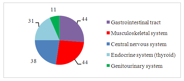
Figure 1. General incidence of children with SDO TMJ
General assessment of condition ENT organs were assessed by examining the nasal cavity and pharynx. The condition of the nasopharyngeal and palatine tonsils, nasal septum, paranasal sinuses was assessed.
Among the local causes of impaired external respiration in sick children with SDO TMJ, an important place may be occupied by difficulty in air passage due to an increase in the pharyngeal tonsils associated with inflammation and hypertrophy, curvature of the nasal septum, diseases of the nasal cavity and paranasal sinuses, as well as retroposition of the mandible and tongue.
The diagnosis of curvature of the nasal septum in its various departments, enlargement of the nasal concha and nasopharyngeal tonsils, the relationship with the degree of retroposition, was carried out by analyzing multisection tomograms in sagital, axial and coronary projections according to the Radiant DICOM program.
When studying the condition of the nasopharyngeal tonsils, out of 65 examined - 8 patients had normal tonsil sizes, 1 - degree of enlargement in 12 patients, 43 patients had 2 - degree, in two cases, 3-degree. This indicated a significant narrowing of the nasopharynx in most patients.
A deviated nasal septum significantly impairs nasal breathing by reducing air flow speed, reducing turbulence and aerodynamic filtration of inhaled particles [12,28]. (Figure 2)
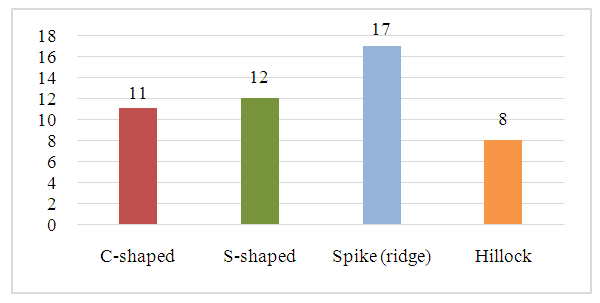
Figure 2. Frequency of occurrence and forms of deviated nasal septum
f the 65 patients examined, 48 had radiological and clinical signs of a deviated nasal septum.
The study of intrauterine development of children revealed normal centile weight-height indicators in newborns within the following range:
Weight 3334,74±109,97 gr. In centile with average indicator - 67%.
Height 51,71± 0,94 cm. In centile caverage indicator - 79%.
Weight-height index (Quetelet I), reflecting the nutritional status in the prenatal period, in the first group was within - 60.37%, which corresponded to the lower values of the norm, and in the second group - 69.55% (the norm is 60-80%).
The assessment of a child’s physical development (PD) is based on the parameters of height, body weight, proportions of development of individual parts of the body, as well as the degree of development of the functional abilities of his body (vital capacity of the lungs, muscle strength of the hands, etc.; muscle development and muscle tone, state of posture, musculoskeletal apparatus, development of the subcutaneous fat layer, tissue turgor), which depend on the differentiation and maturity of the cellular elements of organs and tissues, the functional abilities of the nervous system and the endocrine apparatus [29].
Body mass index (BMI) was determined to assess nutritional status. Calculated using the online BMI calculator for children Children BMI. com.
When determining the index in the 25-75th centile zone, “sufficient” nutrition is stated; “low” (“very low”) nutritional status is said to occur when the body mass index is below the 10th centile; about “high” (“very high”) nutritional status – in the zone above the 90-97th centile. When the index value corresponds to 10-15 or 75-90 centile intervals, we can talk about a “borderline” nutritional state (below or above average), requiring medical supervision. Based on centile estimates of body length (LB), body weight (BW), the harmony of the morphological state of the body was determined. Optimal ratios of these indicators ensure perfect functioning of the musculoskeletal system, cardiovascular, respiratory and other body systems [24]. Figure 3.
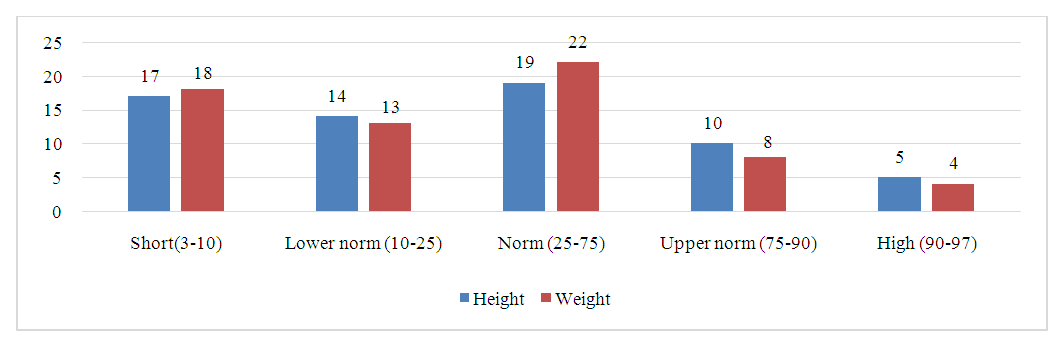
Figure 3. Results of height and weight assessment by centiles
Analysis of the results of a study of the weight and height of the body of children with SDO TMJ indicates their low indicators in 31 and 31 respectively.
Low weight was observed in 18 patients, the lower limit was observed in 13. The results obtained indicate a lag in physical development and indicate a decrease in strength indicators.
During somatoscopy of patients, the following results were obtained:
65 children examined had a cosmetic syndrome in the form of various deformities of the maxillofacial region (MFR).
39 children had various types of postural disorders and 11 had irregular chest shape.
Limb deformities were noted in 6 children, 4 children had O-shaped and 2 children had X-shaped deformities of the limbs.
These changes are associated with disruption of the structure of cartilage and delayed maturation of the epiphyseal growth zone, which is manifested by elongation of tubular bones. The basis of chest deformities is the inferiority of the costal cartilages [13]. (Figure 4)
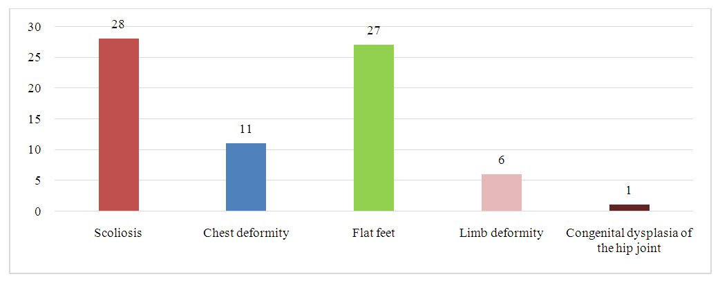
Figure 4. Combined pathology of the skeletal system in children with SDO TMJ (n=37)
Ventilation disorders were expressed in obstructive, restrictive and mixed disorders. Based on the results of spirometry, normal breathing was diagnosed in only 7 patients. The rest had various forms of disorders: restrictive in 13 patients, obstructive in 7, and mixed disorders in 16 cases. (Figure 5)
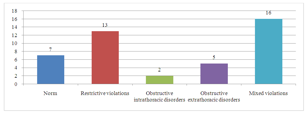
Figure 5. Detected external respiration disorders according to spirometry findings (n=43)

4. Clinical Example

Patient L., 11 years old, turned to our clinic with a diagnosis: SDO TMJ on the right, microgenia.
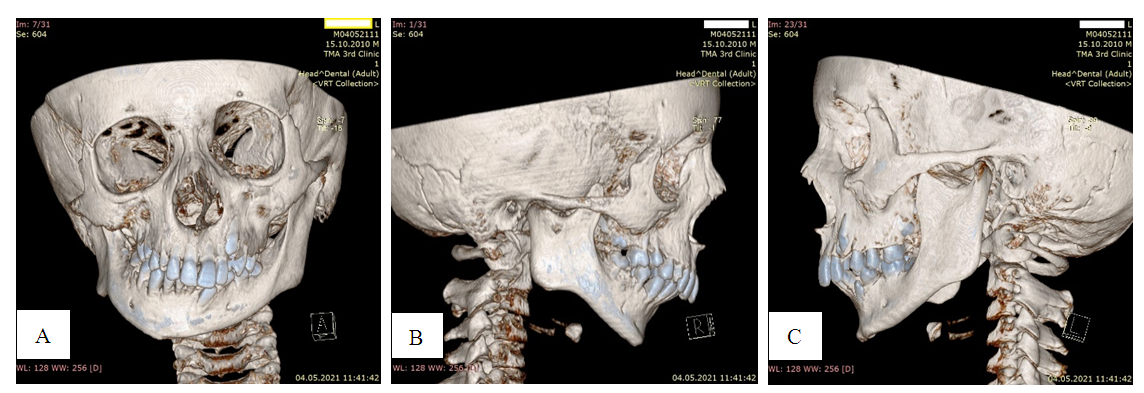
Photo 1. MSCT photographs of a patient with: A - significant deformation of the facial bones and jaws with laterognathia. B, C-microretrogeny
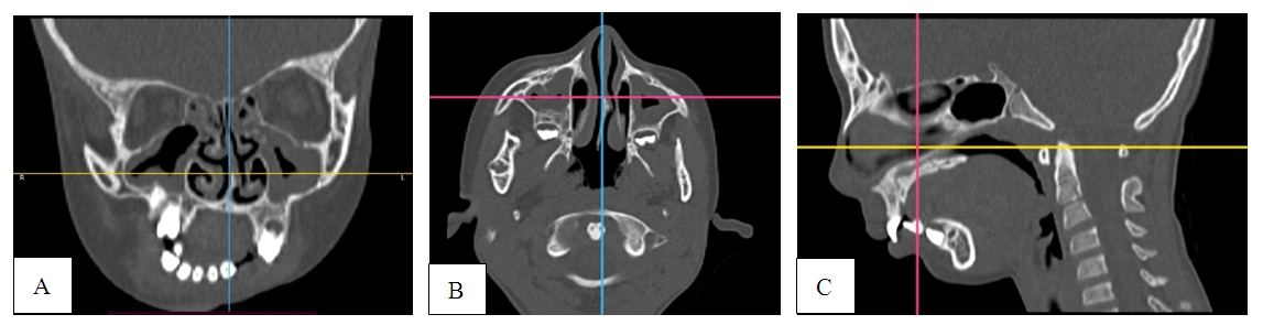
Photo 2. MSCT photograph of the same patient in coronal (A), axial (B) and sagittal projections (C). There is a deviated nasal septum, vasomotor rhinitis, grade 2 hypertrophy of the tonsils, bilateral chronic maxillary sinusitis and ethmoiditis
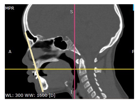
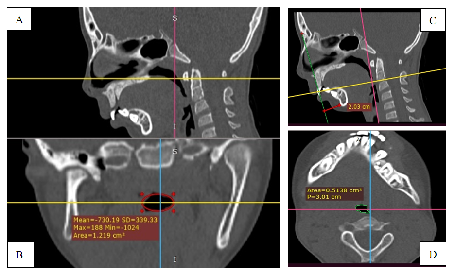
Photo 3. MSCT photograph of the same patient. The lumen of the pharynx in the area of the nasopharynx in the coronal (B), in sagittal (А) projections. In the area of the oropharynx in the axial (D), in sagittal (C) projections. Microretrogenia in sagittal projection (C)
Photo 4. MSCT of a patient with a bilateral fracture of the articular head in the stage of osteoarthritis without retromicrogenism and respiratory apnea: normal pharyngeal lumen is noted
Complaints: Facial asymmetry, nasal congestion, nosebleeds, limited movement in the temporomandibular joint, decreased appetite, bowel dysfunction (constipation), weakness, fatigue, pain in the legs, limited movement in the left elbow joint. A comprehensive examination was carried out by specialists and a conclusion was obtained.
Otorhinolaryngologist: Curvatures nasal septum, vasomotor rhinitis, grade 2-hypertrophy of the tonsils, bilateral chronic maxillary sinusitis and ethmoiditis.
Orthodontist: SDO of TMJ after osteomyelitis of the lower jaw.
Neurologist: Intoxication of the central nervous system.
Pediatrician: mild iron deficiency anemia.
Gastroenterologist: Dysfunction of the sphincter of Oddi of the biliary type, Functional intestinal disorder.
Endocrinologist: diffuse goiter, stage 1.
Orthopedist: Scoliosis of the thoracolumbar spine. Extensor contracture of the left elbow joint.
To the anamnesis: The pregnancy proceeded against a background of stress, there was a threat of miscarriage in the 1st trimester, childbirth at 38 weeks, there were no seizures. Viral hepatitis A – in 2013.
Objectively – correct physique, lagging behind in physical development (weight and height indicators correspond to 9-9.5 years).
St. localis. Deformation of the jaws. Sharply expressed microgeny, retrogeny, laterognathia, prognathia with protrusion of the front teeth, complete restriction of LF movements, breathing through the nose is difficult.
Osteoarticular system: limited movement in the left elbow joint, hypermobility of small joints. High palate, hypoplasia of tooth enamel. Poor posture: scoliotic deformity.
General blood tests: Hb – 118 g/l, RBC – 5.09*1012 l, MCV -72.1 µm3, MCН -22.8 pg, MСHC - 317 g/l, HCT - 38.8.
Blood chemistry: ALT – 17.9, AST – 9.6, total bilirubin – 10.7, Fe – 11.3 µmol/l, Ca – 1.7 mmol/l, P – 4.1 mg/dl.
Results of scatological studies on the patient: Ph-6; detritus - ++; neutral fat – none; soap - +; muscle fibers: indigestible +, digestible - +; fiber -indigestible- +; fatty acids - +++; starch- ++; iodophilic flora - +; Fungi - +.
Results of bacteriological studies: from the nose: Staph. Pyogenus-105CFU, from the throat: Str. Pyogenus -104CFU. Tank culture of stool for dysbacteriosis - Reduced: Total number of anaerobes - 109 CFU, Bifidobacteria - 104 CFU, Lactobacilli - 106 CFU, E. coli lactose-positive - 105 CFU. Increased: Total number of aerobes - 109 CFU, E. Coli negative - 106 CFU, Candida albicans - 105 CFU.
Urine screening tests: Sulkowicz test: +, Lactic acid: ++.
Ultrasound examination: the gallbladder is bent in the body area, the walls are not changed. The content is homogeneous. S1- 8.4 cm2; after a yolk breakfast S2 - 6.7 cm2; The liver is normal, not enlarged, the edges are smooth. RV is within normal limits. Conclusion: Dysfunction of the biliary tract of hypomotor type, deformation of the gallbladder.
ECG: respiratory arrhythmia heart rate 75-88 bpm. Electrical axis of the Heart- rightogram, electrical heart position- vertical. Slow intraventricular conduction.
Spirometry: the ratio of changes in the MEF of the beginning and the second half of the FVC indicates the presence of stenosis of the extrathoracic airways.
Dopplerography of brachiocephalic vessels: a decrease in linear blood flow speed is determined in all carotid arteries and in the supratrochlear arteries. Blood flow is increased through the vertebral arteries. The contours of the Dopplerogram are unclear in the vertebral arteries on the left.
EEG – Periodic pathological pointed waves are recorded in the central-parietal-occipital leads, more on the left.

5. Conclusions

In children with retarded physical development, diseases of both the gastrointestinal tract, nervous, endocrine, and musculoskeletal systems were observed, manifested by poor posture, deformities of the chest, changes in muscle tone involved in ventilation of the lungs, masticatory muscles and other parts of the body.
Diseases of the ENT organs that occur with respiratory disorders negatively affect the child’s health and lead to the development or progression of somatic pathology.
Somatic pathology in children with SDO TMJ leads to delayed physical development and bronchopulmonary syndrome with impairment of external respiration in the form of obstructive, restrictive and mixed forms respiratory disorders, which in many cases are the trigger for the development of hypoxic conditions.
Disturbances in the area of the upper and lower respiratory tract negatively affect the child’s body, in the development of age-related dynamics during periods of morphogenesis, neurohumoral restructuring, sexual and somatic development. Of course, this is important in the preparation and conduct of surgical treatment and rehabilitation of patients with SDO TMJ.

References

[1]  Arnett, T.R.; Gibbons, D.C.; Utting, J.C.; Orriss, I.R.; Hoebertz, A.; Rosendaal, M.; Meghji, S. Hypoxia is a major stimulator of osteoclast formation and bone resorption. J. Cell. Physiol. 2003, 196, 2–8.
[2]  Böing S, Randerath WJ. Chronic hypoventilation syndromes and sleep-related hypoventilation. J Thorac Dis. 2015 Aug; 7(8): 1273-85. doi: 10.3978/j.issn.2072-1439.2015.06.10. PMID: 26380756.
[3]  Burns PA, Wilson DJ. Angiogenesis mediated by metabolites is dependent on vascular endothelial growth factor (VEGF). Angiogenesis. 2003; 6(1): 73-7. doi: 10.1023/a:1025862731653. PMID: 14517407.
[4]  Chesnokova N. P., Brill G. E., Polutova N. V., Bizenkova M. N. Lecture 10 hypoxia: types, etiology, pathogenesis. Medical sciences. - 2017. - No. 2. - pp. 53-54; (In Russ.).
[5]  Chmielik, L. P., Mielnik-Niedzielska, G., Kasprzyk, A., & Niedzielski, A. (2023). A review of health-related quality of life issues in children suffering from certain key otolaryngological illnesses. Frontiersinpediatrics, 10, 1077198. https://doi.org/10.3389/fped.2022.1077198.
[6]  Chuchalin A. G., Aisanov Z. R., Chikina S. Yu., Chernyak A.V., Kalmanova E. N. Federal clinical guidelines of the Russian Respiratory Society on the use of spirometry. Pulmonology. 2014; (6): 11-24. https://doi.org/10.18093/0869-0189-2014-0-6-11-24 (In Russ.).
[7]  Fahey PJ, Hyde RW. “Won’t breathe” vs “can’t breathe”. Detection of depressed ventilatory drive in patients with obstructive pulmonary disease. Chest 1983; 84:19-25.
[8]  Kasparova N. N., Kolesov A. A., Vorob'ev Yu. I. Temporal-mandibular joint disease in children and adolescents, Moscow: Meditsina Publ., 1981. (In Russ.).
[9]  Katz SL, Gaboury I, Keilty K, et al. Nocturnal hypoventilation: predictors and outcomes in childhood progressive neuromuscular disease. Arch Dis Child 2010; 95: 998-1003.
[10]  Khitrov N. A. "Osteoarthrosis" Medical Council, No. 11-12, 2011, pp. 132-139. (In Russ.).
[11]  Khodzhaeva K. A. Yakubov R. K. Sharipova A. U. Assessment of the condition of the ear, throat and nose of children with jaw deformities. Stomatologiya. - Sredneaziatskiy nauchno-prakticheskiy zhurnal, Tashkent, 2005, no 1-2, pp. 23-25. (In Russ.).
[12]  Khodzitskaya V. K., Khodzitskaya S. V. Nasal obstruction: some aspects of morphology, etiopathogenesis, clinic and treatment. Chasopis. 2012; 1 (87): P-111. (In Russ.).
[13]  Klemenov A.V. Extracardial manifestations of undifferentiated connective tissue dysplasia // Clinical medicine. - 2003. - T. 81, No. 10. - p. 4-7. (In Russ.).
[14]  Murray B, Wilson DJ. A study of metabolites as intermediate effectors in angiogenesis. Angiogenesis. 2001; 4(1): 71-7. doi: 10.1023/a:1016792319207. PMID: 11824381.
[15]  Qudah MA, Qudeimat MA, Al-Maaita J. Treatment of TMJ ankylosis in Jordanian children - a comparison of two surgical techniques. J Craniomaxillofac Surg. 2005 Feb; 33(1): 30-6. doi: 10.1016/j.jcms.2004.07.005. Epub 2005 Jan 11. PMID: 15694147.
[16]  Semenza GL. Regulation of physiological responses to continuous and intermittent hypoxia by hypoxia-inducible factor 1. Exp Physiol. 2006 Sep; 91(5): 803-6. doi: 10.1113/expphysiol.2006.033498. Epub 2006 Jun 1. PMID: 16740642.
[17]  Shtanenko, N.I., Buinevich I.V., Kenya A. I., Respiratory system: textbook. -method. handbook for students of all faculties of medical universities, clinical residents, post-graduate students, trainee doctors / edited by N. I. Shtanenko, I. V. Buinevich. Gomel: GomSMU Publ., 2015, 15-30 p. (In Russ.).
[18]  Тam C.S., Wong M, McBain R, Bailey S, Waters KA. Inflammatory measures in children with obstructive sleep apnoea. J Paediatr Child Health. 2006 May; 42(5): 277-82. doi: 10.1111/j.1440-1754.2006.00854.x. PMID: 16712558.
[19]  Tan, J.K.; Mohamad Hazir,N.S.; Alias, E. Impacts of Hypoxia on Osteoclast Formation and Activity: Systematic Review. Int. J. Mol. Sci. 2021, 22, 10146. https://doi.org/10.3390/ijms221810146.
[20]  Tang, Y.; Zhu, J.; Huang, D.; Hu, X.; Cai, Y.; Song, X.; Song, Z.; Hong, C.; Feng, Z.; Kang, F. Mandibular osteotomy-induced hypoxia enhances osteoclast activation and acid secretion by increasing glycolysis. J. Cell. Physiol. 2019, 234, 11165–11175.
[21]  Tarasova G.D.: Adenoids: cause, effect or...? // RMJ. 2016. No. 6. pp. 391–394. (In Russ.).
[22]  Ukwas A, Elshik M, Elbialy M. TMJ Ankylosis in Children: A Case Report and Literature Review. Case Rep Dent. 2023 Jan 5; 2023:6474478. doi: 10.1155/2023/6474478. PMID: 36643593.
[23]  Utting, J.C.; Flanagan, A.M.; Brandao-Burch, A.; Orriss, I.R.; Arnett, T.R. Hypoxia stimulates osteoclast formation from human peripheral blood. Cell Biochem. Funct. 2010, 28, 374–380.
[24]  Vasilyeva E. I., Physical development of children: educational and methodical manual for foreign students. - State Budgetary Educational Institution of Higher Professional Education IGMU of the Ministry of Health of the Russian Federation. Irkutsk: IGMU Publ., 2013, 32p. (In Russ.).
[25]  Yakubov R. K., Azimov M. I. Results of a comprehensive examination of children with secondary deforming TMJ osteoarthritis. - Stomatologiya, Moscow, 2001, no. 5, pp. 37-39. (In Russ.).
[26]  Yakubov R. K., Azimov M. I., Marufkhanov Kh.M. Yusupova B. Yu. Surgical treatment of jaw deformities in children taking into account the operational and anesthetic risk. –Alma-Ata, 2001. №4 (14). –P. 28-30. (In Russ.).
[27]  Yakubov R. K., Usmankhodzhayeva A. A., Vysogortseva O. N., Yakubova N. A., Fayziev B. R. Application of therapeutic gymnastics in children with congenital dysplastic syndrome Proceeding of European Conference on Biology and Medical Sciences (March 28, 2015). «East West» Association for Advances Stadies and Higher Education Gmb H. Vienna. 2015. 232 Р. 169-175. (In Russ.).
[28]  Yunusov A.S. Deformation of the nasal septum. Practical recommendations, Moscow 2014; P.2-5. (In Russ.)
[29]  Yuryev V. V., Simakhodsky A. S., Voronovich N. N., Khomich M. M. Growth and development of the child: for students of medical universities and pediatricians, Editions. 3rd. St. Petersburg: Peter, 2007. 260 p. (In Russ.).