-
Paper Information
- Next Paper
- Previous Paper
- Paper Submission
-
Journal Information
- About This Journal
- Editorial Board
- Current Issue
- Archive
- Author Guidelines
- Contact Us
American Journal of Medicine and Medical Sciences
p-ISSN: 2165-901X e-ISSN: 2165-9036
2024; 14(9): 2382-2387
doi:10.5923/j.ajmms.20241409.55
Received: Aug. 22, 2024; Accepted: Sep. 16, 2024; Published: Sep. 30, 2024

Assessment of the Functional Balance of the Mastical Apparatus in Patients with Disocclusion During Post-Orthodontic Treatment by Electromyography
 Abstract
 Reference
 Full-Text PDF
 Full-text HTML
Khabilov B. H.1, Vakhobova M. B.1, Khamrayev Kh. Kh.2
1Tashkent State Dental Institute, Tashkent, Uzbekistan
2Samarkand State Medical University, Tashkent, Uzbekistan
Copyright © 2024 The Author(s). Published by Scientific & Academic Publishing.
This work is licensed under the Creative Commons Attribution International License (CC BY).
http://creativecommons.org/licenses/by/4.0/

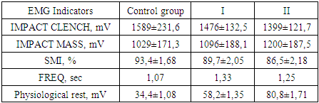
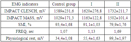
Introduction. Changes in occlusal contacts between teeth, leading to disturbances in antagonism, can provoke destabilization of the dentofacial system and cause symptoms of occlusal trauma, including a dysfunctional state of the temporomandibular joint. These changes can manifest themselves in the form of pain and discomfort when chewing, tooth sensitivity, as well as restrictions in mouth opening, creaking or noise when moving the jaws, which requires timely intervention by the dentist to prevent further complications. The article presents the results and discussions of the study of IMPACT CLENCH - the total EMG impulse of muscles during compression, IMPACT MASS - dynamic potential during chewing, SMI - index of symmetry (efficiency) of chewing, FREQ - frequency of chewing movements, and electromyograms were recorded in the state relative physiological rest in the position of central occlusion. Purpose of the study. To study the change in the electrical potential of pairs of the masticatory and temporal muscles in patients with disocclusion during post-orthodontic treatment. Materials and methods. The object of the study were 60 patients who applied to the clinic of the Faculty of Orthopedic Dentistry of the Tashkent State Dental Institute after previously undergoing orthodontic treatment. We studied the statistical and physical parameters of the bioelectrical activity of the masticatory muscles according to the method of Ferrario (2001) using the “Freely” apparatus (DeGotzen, Italy). Results. An electromyographic study conducted 1 year after orthopedic treatment indicated an improvement in some indicators of changes in the amplitude of the electrical potential of the masticatory muscles in group I of patients who were treated for disocclusion with emax inserts. The frequency of chewing movements during the study period after 2 years in group II increased by almost 50% relative to the main group of patients, which is associated with mechanical abrasion of the contact occlusal surfaces of the teeth restored with light-curing composite material and the physical property of shrinkage of this material. The studied indicator of the electrical potential of pairs of masticatory muscles during physiological rest in patients of group I indicated the similarity of the results obtained in the control group, however, in patients of group II we observed the development of compensatory efforts after 2 years of orthopedic rehabilitation, which is confirmed by an increase in the value of the studied indicator relative to the control and the main group by 65.4% and 44.2%, respectively. Conclusion. In the oral cavity, the process of high-quality formation of the occlusal surface of a direct composite restoration is difficult, which can cause neuromuscular disorders of the masticatory apparatus and complicate the process of holding the lower jaw in a central position, resulting in excessive load on the temporomandibular joint. This study complements the opinion of a number of scientists about the disadvantages of direct dental restoration.
Keywords: Electromyography, Disocclusion, Post-orthodontic treatment, Dental inlays
Cite this paper: Khabilov B. H., Vakhobova M. B., Khamrayev Kh. Kh., Assessment of the Functional Balance of the Mastical Apparatus in Patients with Disocclusion During Post-Orthodontic Treatment by Electromyography, American Journal of Medicine and Medical Sciences, Vol. 14 No. 9, 2024, pp. 2382-2387. doi: 10.5923/j.ajmms.20241409.55.
1. Introduction
- The study of functional balance and occlusal relationships in school-age children allows not only to assess the current state of their dental system, but also to take measures for proper correction and prevention of potential problems in the future. This approach helps maintain dental health and ensures optimal functioning of the chewing apparatus throughout life. There are also disagreements among authors regarding the optimal time to identify problems associated with occlusion and anomalies of the masticatory apparatus, but despite the disagreements, all authors express a unanimous opinion on the advisability of conducting such studies in order to prevent dental anomalies, deformations and subsequent relapses after orthodontic treatment. Early identification and assessment of the functional balance of the masticatory apparatus and occlusal relationships in children is important to prevent potential problems with the development of the dental system [4,6,15].Research in this area makes it possible to identify and correct imbalances of the masticatory apparatus and occlusion, eliminate developmental anomalies and prevent possible relapses after orthodontic treatment. This approach helps achieve optimal results and long-term dental health, which is an important aspect in the field of dentistry. [2,8,11] It is worth paying special attention to diagnosing the functional state of the articular apparatus of the masticatory system - the temporomandibular joints, as well as assessing the occlusal relationships of the teeth. The discovery of dysfunction in the joints is often associated with violations of the occlusal contacts of the teeth, and conversely, improper restoration of the occlusal relationships can become a source of problems in the functioning of the joint. [9,10] Assessing the occlusion and functional state of the temporomandibular joints allows us to identify possible factors influencing dysfunction, which can lead to pain symptoms, limited mobility and other negative consequences. Such studies contribute to more accurate diagnosis and determination of treatment strategies, including correction of occlusion and necessary joint treatment [1,13,14].Maintaining a balance between proper occlusion and joint function is an essential aspect of maintaining dental health and preventing the development of dysfunction. Therefore, research in this area plays a key role in optimizing treatment and preventing complications associated with the functioning of the temporomandibular joints and occlusion [12].The study of the electrical potential of the chewing pairs of muscles of the maxillofacial area allows us to determine neuromuscular problems of the dentofacial system, and also allows us to determine changes in the functional state of the muscles under physical loads to monitor the quality of orthodontic treatment objectively assess the functionality of the occlusion [3].
2. Materials and Methods
- The object of the study were 60 patients who applied to the clinic of the Faculty of Orthopedic Dentistry of the Tashkent State Dental Institute after previously undergoing orthodontic treatment. Of these, 23 were men and 37 were women; the average age of patients admitted to the clinical department was 34 years. In order to optimize diagnostic methods and methods for correcting destabilization of occlusal relationships in patients after orthodontic treatment, we formed the following groups of subjects: The main study group consisted of 30 patients with post-orthodontic disclusion who required restoration of defects in the hard tissues of the chewing teeth, namely, replacement of failed composite restorations and production of ceramic onlays (inlays), according to the IROPZ classification from 0.3 to 0.6.The comparison group consisted of 30 patients with post-orthodontic disclusion who required the restoration of defects in the hard tissues of the chewing teeth, namely, the replacement of failed composite restorations with new ones, and/or the manufacture of onlays (inlays) from a composite light-curing material, according to the IROPS classification up to 0.3.For the control group, 30 people without violations of the occlusal relationships of the jaws and with intact dentition were selected.We studied: IMPACT CLENCH - total EMG impulse of muscles during compression, IMPACT MASS- - dynamic potential during chewing, SMI - index of symmetry (efficiency) of chewing, FREQ - frequency of chewing movements, and recorded electromyograms in a state of relative physiological rest in position central occlusion. Almonds were used as food (stimulant). An electromyographic study in patients in groups with disocclusion during post-orthodontic treatment was carried out at the following times: before treatment, 1-2 weeks after treatment, 1 year after treatment, 2 years after treatment.
3. Results and Its Discussion
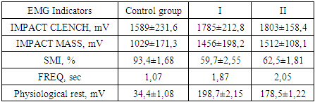
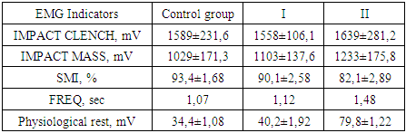
- When studying the electrical potential of pairs of the actual masticatory and temporal muscles in patients in the control group, results were obtained that did not go beyond normal values. Thus, the average value of the maximum tension during jaw compression was 1589±231.6 mV, and the average total value of the dynamic chewing tension was 1029±171.3 mV. The percentage of the chewing efficiency index in individuals in the control group, without occlusion pathology, was quite high and amounted to 93.4 ± 1.68%, which corresponds to the results of the chewing test with almonds, which also indicates the high coordination of the studied muscles during chewing movements. The frequency of chewing movements averaged 1.07 seconds. We used the obtained values of the studied EMG parameters in the control group throughout the entire period of the experimental study as material for a comparative assessment of the results obtained in the study of muscle electrical potential in patients of the working groups.
|
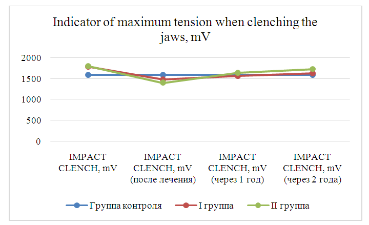 | Diagram 1. Values of the maximum tension during jaw compression in patients in the comparison and control groups, obtained before and after treatment, over time, mV |
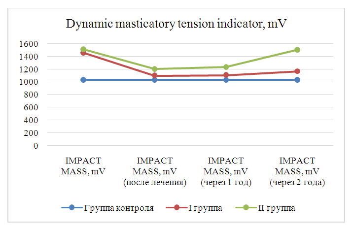 | Diagram 2. Average total value of the dynamic masticatory tension indicator in patients in the comparison and control groups, obtained before and after treatment, over time, mV |
|
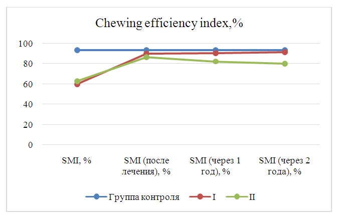 | Diagram 3. Average total value of the chewing efficiency index in patients in the comparison and control groups, obtained before and after treatment, over time, % |
|
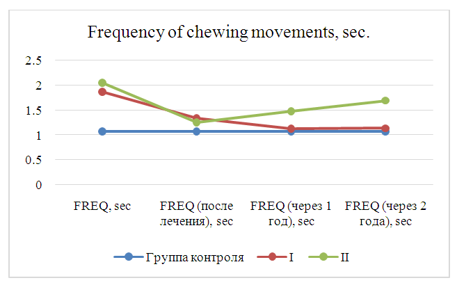 | Diagram 4. Average total value of the frequency of chewing movements in patients in the comparison and control groups, obtained before and after treatment, over time, sec |
|
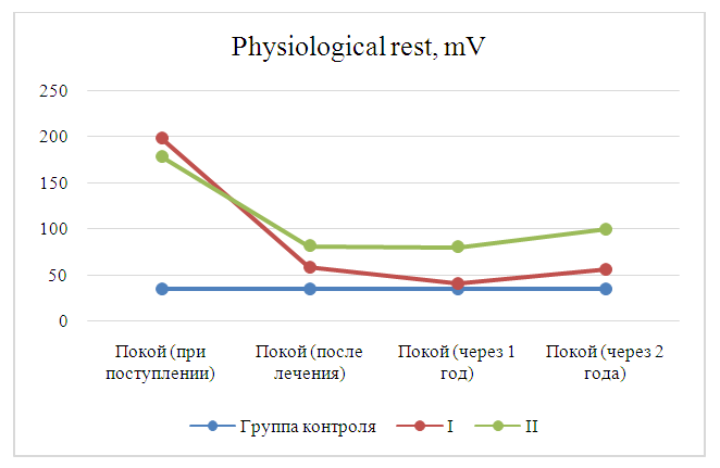 | Diagram 5. Average total value of the electrical potential of pairs of masticatory muscles at physiological rest in patients in the comparison and control groups, obtained before and after treatment, over time, sec |
4. Conclusions
- Data from clinical and sociological studies have confirmed the feasibility of wider use of myographic research as a method of additional diagnosis of disorders of the dental system in patients, in order to increase the effectiveness of orthopedic treatment and prevent functional complications of the masticatory apparatus. Assessing the functional balance of the masticatory apparatus allows us to identify potential problems with occlusion, abnormalities in the development of the jaws and teeth, which is important for early detection and prevention of possible further complications.