American Journal of Medicine and Medical Sciences

p-ISSN: 2165-901X    e-ISSN: 2165-9036

2024;  14(9): 2333-2336

doi:10.5923/j.ajmms.20241409.44

Received: Sep. 2, 2024; Accepted: Sep. 20, 2024; Published: Sep. 21, 2024


Indicators of Biochemical and Immunological Profile in Premature Discharge of Amniotic Fluid

Kayumova Guzal Mukhtorovna

Bukhara State Medical Institute named after Abu Ali ibn Sina, Bukhara, Uzbekistan

Correspondence to: Kayumova Guzal Mukhtorovna, Bukhara State Medical Institute named after Abu Ali ibn Sina, Bukhara, Uzbekistan.

Email:

Copyright © 2024 The Author(s). Published by Scientific & Academic Publishing.

This work is licensed under the Creative Commons Attribution International License (CC BY).
http://creativecommons.org/licenses/by/4.0/

Abstract

Complication of birth due to premature rupture of the umbilical cord is one of the most important problems of modern obstetrics. According to the duration of the pregnancy, ectopic pregnancy causes the onset of labor in 8-92% of cases. Premature discharge of amniotic fluid is closely related to infection, which in turn increases the risk of purulent-septic complications in a pregnant woman 10 times. Chorioamnionitis is a major risk for a pregnant woman that worsens the delivery process and results.

Keywords: Premature birth, Premature discharge of amniotic fluid, Chorionic amnionitis, Femoflor, Progesterone

Cite this paper: Kayumova Guzal Mukhtorovna, Indicators of Biochemical and Immunological Profile in Premature Discharge of Amniotic Fluid, American Journal of Medicine and Medical Sciences, Vol. 14 No. 9, 2024, pp. 2333-2336. doi: 10.5923/j.ajmms.20241409.44.

1. Introduction

One of the most serious complications of preterm birth is premature amniocentesis. In full-term pregnancy, PDOAF is the most important risk factor for pregnancy complications that threaten the health of the fetus and mother, determines the high level of perinatal and infant morbidity and mortality [1,3,8]. Premature amniocentesis in preterm labor is the most important risk factor for the fetus and mother, as it determines the high risk of morbidity and mortality of perinatal babies. Therefore, complications of premature birth due to rupture of the membranes are one of the most important problems of modern obstetrics [12,18].
PDOAF is a complication of pregnancy and is characterized by a violation of the integrity of the fetal membrane and the release of amniotic fluid (before the onset of labor). The frequency of this pathology, according to different authors, varies widely and ranges from 1.0% to 19.8%. According to the World Health Organization, every year in the world 15 million children are born prematurely [16].
The problem of premature birth remains one of the most urgent problems in obstetrics and perinatology, because of the polyetiological nature of this pathology and the lack of a single tactic in the delivery process. Despite the emergence of many drugs used for the risk of preterm birth on the pharmaceutical market, the frequency of PB in different countries has remained stable from 5% to 12% over the past 30 years. In the period from 22 to 28 weeks of pregnancy, the frequency of PB is 5-7% of all cases of this pathology, from 29 to 34 weeks - 33-42%, from 34 to 37 weeks - 50-60% [4,17].

2. Materials and Methods

Premature and perinatal amniocentesis occupies one of the first places among modern obstetric problems, because it determines the high level of perinatal and infant morbidity and mortality. Premature discharge of amniotic fluid during premature and full-term pregnancy is the most important risk factor for the fetus and the mother [5,7,15].
Premature amniocentesis is an urgent problem of modern obstetrics, causing complications for the mother and the fetus possible. Therefore, delivery complicated by premature amniocentesis should be carried out in a maternity hospital based on scientific examination [6,11]. Premature amniocentesis is a common obstetric pathology, occurring in 10-19% of cases in term and in 25-54% of premature births. Childbirth with SCI is often accompanied by various anomalies of uterine contraction activity, in particular, weakness of labor activity (4.9-23%) or discoordination of labor activity (8.3-41.8%) [9,10].
Premature rupture of the placenta occurs in 3-10% of all pregnancies. It is determined by the violation of the integrity of the fetal membranes and the release of amniotic fluid before the onset of labor, regardless of the period of pregnancy [13,19].
Premature discharge of amniotic fluid in premature pregnancy is one of the urgent problems. This problem should be considered not only medically, but also socially and economically. Premature discharge of amniotic fluid accompanies every third case of premature birth [2,14].
PB complicates up to 8% of all pregnancies and is the main reason for the development of spontaneous PB in 30-51%. When preterm labor begins before the 26th week of pregnancy, the percentage of SCC reaches 90-92%.
Complication of birth due to premature rupture of the umbilical cord is one of the most important problems of modern obstetrics. According to the duration of the pregnancy, PDOAF causes the onset of labor in 8-92% of cases [13,20].

3. Goal of Study

A differential approach to pregnancy and childbirth based on changes in the immunological status of the mother-placental-fetus system during premature amniocentesis.
In 2022, 177 pregnant women with vaginal discharge who were under control at the perinatal center of Bukhara region were prospectively analyzed. The examined women were divided into 2 groups: 1 group included 107 women who had premature rupture of membranes and gave birth; 30 pregnant women with amniocentesis were divided into 2 groups; 40 relatively healthy pregnant women were selected for the control group.

4. Result and Discussion

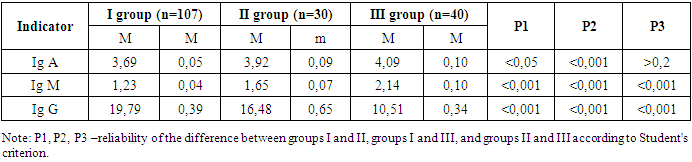
As part of the study, a comparative analysis of the level of immunoglobulin was conducted in three groups of women. Analysis showed statistically significant changes in immunoglobulin levels between groups. The average value of IgA increased by 3.69 mg/L in the third group compared to the first group, and reached 4.09 mg/L, with the most significant increase between the first and second groups (p<0.001). This indicates an activation of the immune system in response to premature ejaculation, as IgA plays an important role in local immunity and mucosal protection. The amount of IgM was also studied in detail, from 1.23 mg/l in the first group to 2.14 mg/l in the third group. it was found that it increased up to, a high statistical index of changes was noted between all groups (p<0.001). The primary response of the IgM immune system to infection may be due to inflammation associated with potential agents or premature ejaculation. The greatest change was observed in the amount of IgG, which decreased from 19.79 mg/l in the first group to 10.51 mg/l in the third group, the difference between all groups being statistically significant (p<0.001).
A decrease in IgG, the main component of humoral immunity, may reflect a long-term load on the immune system or a violation of its regulatory mechanisms under conditions of stress due to premature dehydration (Table 1).
Table 1. Comparative analysis of immunoglobulin levels in three studied groups n=177
     
Quantitative analysis of IgG antibody against infectious agents such as Toxoplasma gondii, Herpes simplex virus type I and II, as well as Ureaplasma urealyticum is important in assessing the risk of premature ejaculation. Immunoglobulins of class G (IgG) are the main indicators of the adaptive immune response and provide long-term protection against infections, which is important in maintaining the normal course of pregnancy.
A positive IgG antibody to Toxoplasma gondii is indicative of previously acquired infections, which is important for pregnant women because primary infection during pregnancy can lead to severe birth defects or premature ejaculation. Herpes simplex virus types I and II, IgG detection indicates prior exposure to these viruses, which may react during pregnancy and cause an inflammatory process that potentially affects the amniotic membrane and causes premature discharge. Ureaplasma urealyticum, which is often associated with urogenital infections, is also of clinical importance, as infections caused by these microorganisms can cause inflammation and disruption of the integrity of the membranes surrounding the fetus, which increases the risk of premature ejaculation.
Thus, the quantitative analysis of IgG is important for obstetricians and gynecologists to assess the immune status and possible risks associated with infectious agents during pregnancy. This allows timely and clinically informed management decisions to be made, avoiding potential risks to maternal and fetal health, while minimizing the risk of preterm delivery.
We performed a quantitative analysis of IgG to infectious agents, including Toxoplasma gondii, Herpes simplex virus types I and II, as well as Ureaplasma urealyticum. The obtained results are shown in the table below.
Table 2. Quantitative analysis of IgG antibodies to infectious agents
     
The average indicator for Toxoplasma gondii antibody was 0.24 ME/ml in the first group, 0.01 ME/ml in the second group, and 0.21 ME/ml in the third group. The difference between the first and second, as well as the first and third groups was statistically significant (p<0.05 and p<0.001, respectively), which indicates a higher immune response in the first group compared to the other groups.
Antibody to Herpes simplex virus type I was higher in the first group (0.24 ME/ml) compared to the second group (0.01 ME/ml) and the third group (0.17 ME/ml), a higher statistical value was noted between the first and second groups (p< 0.001), but the difference between the first and third groups is not significant (p>0.2).
The value of Herpes simplex virus type II was 0.19 ME/ml in the first group, 0.01 ME/ml in the second group and 0.18 ME/ml in the third group. There was no significant difference between the first and second groups (p>0.2), but there was a significant difference between the second and third groups (p<0.05).
Antibody values for Ureaplasma urealyticum were higher in the first group (0.30 ME/ml) compared to the second group (0.01 ME/ml) and the third group (0.18 ME/ml), the difference between all groups was highly statistically significant (p<0.001).
For the results of the prospective study, we used the algorithm for the assessment and management of the PDOAF. This algorithm is based on the use of binary logistic regression, which significantly increases the forecast accuracy. The algorithm includes assessment of cervical microbiota, hormone levels, and presence of antibodies to infectious agents.
The risk of preterm discharge is calculated as a sum of binary variables, each representing a higher or lower risk for different parameters, such as the percentage of specific bacterial species, hormone levels, and the presence of antibodies. The overall risk score ranges from 0 to 5, and pregnancy management recommendations are made based on this score. This algorithm is described in detail in the next paragraph of the study. The level of IgA increases with premature rupture of membranes, because this immunoglobulin protects the mucous membranes. Increased IgA levels are associated with increased inflammation (positive correlation r = 0.65). The level of IgM also increases with PDOAF, because it is the main response against infection, and it is associated with the presence of an infectious process (positive correlation r = 0.78). IgG levels decrease in PDOAF as this immunoglobulin provides long-term protection, and the decrease in levels is associated with increased inflammatory stress (negative correlation r = -0.72). Quantitative analysis of IgG antibodies to infectious agents shows that a high level of Toxoplasma gondii IgG indicates a stronger immune response, and a decrease in its level is associated with the risk of PDOAF (negative correlation r = -0.58). High levels of Herpes simplex I IgG indicate past infection, and a decreased level correlates with the risk of PDOAF (negative correlation r = -0.62). Similar to Herpes simplex I, a decrease in Herpes simplex II IgG level is associated with the risk of PDOAF (negative correlation r = -0.59). A high level of Ureaplasma urealyticum IgG indicates an immune response to urogenital infections, and a decrease in levels is associated with the risk of UTI (negative correlation r = -0.67). IgA and IgM are elevated in PDOAF indicating an active immune response against infection and inflammation.

5. Conclusions

In order to determine the risk factors for the development of the pathology of premature discharge, it is necessary to analyze the patients' catamnestic and anamnestic data during outpatient or inpatient follow-up. According to the results of the dissertation research, the most common risk factors of pregnancy that cause PDOAF are: hypoxic conditions, pre-pregnancy or related somatic diseases, inflammatory diseases of the genital system. shows support for stress and protection. How it responds to this unaltered state in immunoglobulin levels has an important bearing on the diagnosis and treatment of PDOAF.

ACKNOWLEDGMENTS

The authors are grateful for the support and helpful comments provided by the department of Obstetrics and gynecology in Bukhara State Medical Institute, as well as other experts consulted as part of the process. We would also like to thank Bukhara city maternity complex for her assistance in creating the figure for our literature search.

References

[1]  Kayumova, G. M., & Dustova, N. K. (2023). ASSESSMENT OF THE STATE OF THE GENITAL TRACT MICROBIOCENOSIS IN PREGNANT WOMEN WITH PREMATURE RUPTURE OF THE MEMBRANES USING THE FEMOFLOR TEST. Modern Scientific Research International Scientific Journal, 1(1), 70-72.
[2]  Valeryevna, S. L., Mukhtorovna, K. G., & Kobylovna, E. S. (2019). Premature Birth In A Modern Aspect. International Journal of Bio-Science and Bio-Technology, 11(10), 31-37.
[3]  Ikhtiyarova, G. A., Dustova, N. K., & Qayumova, G. (2017). Diagnostic characteristics of pregnancy in women with antenatal fetal death. European Journal of Research, (5), 5.
[4]  Kayumova, G. M. (2023). To Determine the Features Of Pregnancy and Children During Antenature Ruption Of Ambient Fluid. American Journal of Pediatric Medicine and Health Sciences (2993-2149), 1(9), 66-72.
[5]  Kayumova, G. M. (2023). Features of the Hormonal Background During Premature Relation of Ambitional Fluid. American Journal of Pediatric Medicine and Health Sciences (2993-2149), 1(9), 73-79.
[6]  Kayumova, G. M. (2023). The Significance Of Anti-Esterogen And Progesterone Antibodies As A Risk Factor In Premature Rupture Of Amniotic Fluid. American Journal of Pediatric Medicine and Health Sciences (2993-2149), 1(9), 58-65.
[7]  Galaz, J., Romero, R., Xu, Y., Miller, D., Slutsky, R., Levenson, D., ... & Gomez-Lopez, N. (2020). Cellular immune responses in amniotic fluid of women with preterm clinical chorioamnionitis. Inflammation Research, 69(2), 203-216.
[8]  Romero, R., Yoon, B. H., Kenney, J. S., Gomez, R., Allison, A. C., & Sehgal, P. B. (1993). Amniotic fluid interleukin-6 determinations are of diagnostic and prognostic value in preterm labor. American journal of reproductive immunology, 30(2-3), 167-183.
[9]  Khamroyev Kh.N. «The morphofunctional changes in internal organs during alcohol intoxication». European journal of modern medicine and practice vol. 2 no. 2 (2022) ejmmp issn: 2795-921x.
[10]  Hamroev Khudoyshukur Nutfilloevich, Khasanova Dilnoza Akhrorovna. «Сomparative classification of liver morphometric parameters in the liver and in experimental chronic alcoholism». International Journal of Cognitive Neuroscience and Psychology IJCNP, Volume 1, Issue 1, 2023.
[11]  Rodríguez-Coutiño S.L., Ramos-González R., Hernández-Herrera R.J. Rodríguez-Coutiño S.I., Risk factorsofprematurity. A case control study. Ginecol. Obstet. Mex. - 2013. - Vol. 81, № 9. - P. 499-503.
[12]  Rodriguez-Trujillo A., Cobo T., Vives I. et al. Gestational age is more important for short-term neonatal outcome than microbial invasion of the amniotic cavity or intra-amniotic inflammation in preterm prelabor rupture of membranes. Acta Obstet. Gynecol. Scand. - 2016. - Vol. 95, № 8. - P. 926-933.
[13]  Sadeh-Mestechkin D., Samara N., Wiser A.et al. Premature rupture of the membranes at term: time to reevaluate the management Arch. Gynecol. Obstet. - 2016. - Vol. 294, № 6. - P. 1203-1207.
[14]  Shook L.L., Buhimschi C.S., Dulay A. T. et al. Calciprotein particles as potential etiologic agents of idiopathic preterm birth. Sci. Transl. Med. - 2016. - Vol. 8, № 364. - P. 154.
[15]  Smith A., Allen V.M., Walsh J. et al. Is preterm premature rupture of membranes latency influenced by single versus multiple agent antibiotic prophylaxis in group B streptococcus positive women delivering preterm? J. Obstet. Gynaecol. Can. - 2015. - Vol. 37, № 9. - P. 777-783.
[16]  Souza A.S., Patriota A.F., Guerra G.V., Melo B.C. Evaluation of perinatal outcomes in pregnant women with preterm premature rupture of membranes. Rev. Assoc. Med. Bras. - 2016. - Vol. 62, № 3. - P. 269-275.
[17]  Soydinc H.E., Sak M.E., Evliyaoglu O. et al. Prolidase, matrix metalloproteinases1 and 13 activity, oxidative-antioxidative status as a marker of preterm premature rupture of membranes and chorioamnionitis in maternal vaginal washing fluids. Int. J. Med. Sci. - 2013. - Vol. 10, № 10. - P. 1344-1351.
[18]  Stepan M., Cobo T., Maly J. et al. Neonatal outcomes in subgroups of women with preterm prelabor rupture of membranes before 34 weeks J. Matern. Fetal Neonatal Med. - 2016. - Vol. 29, № 14. - P. 2373-2377.
[19]  Wagner P., Sonek J., Mayr S. et al. Outcome of pregnancies with spontaneous PPROM before24+0 weeks' gestation Eur. J. Obstet. Gynecol. Reprod. Biol. - 2016. - Vol. 203. - P. 121-126.
[20]  Wolf M.F., Miron D., Peleg D. et al. Reconsidering the current preterm premature rupture of membranes antibiotic prophylactic protocol. Am. J. Perinatol. - 2015. - Vol. 32, № 13. - P. 1247-1250.