American Journal of Medicine and Medical Sciences

p-ISSN: 2165-901X    e-ISSN: 2165-9036

2024;  14(9): 2287-2294

doi:10.5923/j.ajmms.20241409.35

Received: Aug. 25, 2024; Accepted: Sep. 16, 2024; Published: Sep. 20, 2024


Metabolic Status of the Brain in Children and Adolescents with Diabetes, Taking into Account the Presence of or the Absence of Cognitive Deficits

Alidjanova D. A., Madjidova Ya. N.

Tashkent Pediatric Medical Institute, Tashkent, Uzbekistan

Copyright © 2024 The Author(s). Published by Scientific & Academic Publishing.

This work is licensed under the Creative Commons Attribution International License (CC BY).
http://creativecommons.org/licenses/by/4.0/

Abstract

This publication will present an analysis of the results obtained from a study of the metabolic status of the brain using multivoxel proton magnetic resonance spectroscopy in children and adolescents with cognitive dysfunction in type 1 diabetes. According to the results obtained, correlations between the main metabolites of the brain – N-acetylaspartate (NAA), creatine (Cr), choline (Cho) and the results of the MoSA test will be demonstrated. The biochemical shifts discovered in the course of the study may be the primary links in the pathogenetic mechanisms of the formation of cognitive disorders in children and adolescents with type 1 diabetes.

Keywords: Cognitive dysfunction, Children and adolescents, Diabetes mellitus, Proton magnetic resonance spectroscopy, Brain metabolites

Cite this paper: Alidjanova D. A., Madjidova Ya. N., Metabolic Status of the Brain in Children and Adolescents with Diabetes, Taking into Account the Presence of or the Absence of Cognitive Deficits, American Journal of Medicine and Medical Sciences, Vol. 14 No. 9, 2024, pp. 2287-2294. doi: 10.5923/j.ajmms.20241409.35.

1. Introduction

Diabetes mellitus (DM) refers to autoimmune pathologies of the endocrine system. The formation of diabetes occurs as a result of an absolute or relative lack of insulin induced by the disintegration of beta cells of the pancreas [1]. To date, diabetes is the most serious problem of the entire global medical community, as it does not lose its leading position in the deterioration of the quality of life and the formation of early disability due to the occurrence of long-term complications.
According to the International Diabetes Federation, the expected increase in the number of diabetic patients in the world by 2045 is up to 628.6 million people. At the same time, there has been a sharp increase in type 1 diabetes in children and adolescents in recent years. Unfortunately, the Republic of Uzbekistan is no exception to this statistic, so at the end of 2023, more than 260 thousand people were registered with diabetes mellitus in our country, of which almost 3,500 thousand children, more than 1,000 adolescents.
Among the entire range of variability of complications of type 1 diabetes in children and adolescents, deviations from the brain occupy a primary place, the fundamental manifestations of which are changes in the processes of cognitive activity. In recent years, many scientific studies have been conducted and published confirming both direct and indirect effects on the occurrence of structural and functional disorders of the brain in diabetes mellitus [2]. Such close attention to cerebral, namely cognitive abnormalities in diabetes is associated not only with the normal daily functioning and social adaptation of the child, but also with regular and clear self-control of glycemia, which has a direct impact on the overall dynamics of the underlying disease.
According to epidemiological studies, all kinds of changes on the part of higher brain functions in type 1 diabetes can begin to form within the first 2-8 years after the onset of the disease. And their wide range of manifestations (10-75%) is almost always associated with such factors as the age of onset of the disease, duration of diabetes mellitus, reference values of glycemia, diagnostic methods used, etc. [3,4].
The pathogenetic mechanisms of the formation of cognitive deficits in type 1 diabetes mellitus, for the most part, remain uncertain and poorly understood. To date, hypo- and hyperglycemia, insulin deficiency, malfunction of pro- and antioxidant activity systems, activation of the polyol pathway of glucose metabolism, intensification of non-enzymatic glycation of proteins, hypoxic changes in brain tissues, induction of cell apoptosis, hemodynamic and hemorheological changes are distinguished among the pathogenetic components. In addition, in some research papers, the demonstrated correlations between cognitive dysfunctions and the degree of hyperglycemia gave the authors reason to assert that this main metabolic change may be the cause of cognitive disorders in DM [5,6]. Thus, in the process of cerebral disorders, there is an interest of various pathogenetic links, which may explain the polymorphism of the clinical manifestations of these disorders in type 1 diabetes. However, it is worth noting here that if the picture is more or less clear with the formulation of various pathogenetic factors, then the question of determining the main primary pathogenetic mechanism for the development of cognitive dysfunction in type 1 diabetes in children and adolescents remains open.
In order to diagnose cognitive deficits in a variety of pathological processes, researchers widely use methods of neuropsychological testing of patients. Among these numerous methods, the Montreal Cognitive Assessment Scale – the Montreal Cognitive Assessment (MoSA test), developed for screening disorders of higher brain functions in a fairly short time, is the most popular. This technique is characterized by ease of use, reliability and is considered the most rational method of analyzing cognitive activity, the test is characterized by a fairly large range of study areas of the cognitive sphere [7]. The specificity of this method is 90% [8]. However, even with all the positive assessments of this method, it is not necessary to exclude the possible presence of a subjective assessment of indicators, which entails a significant percentage of both false positive and false negative results of neuropsychological testing. In this regard, and in order to maximize the objectification of indicators, it is necessary, in addition to neuropsychological testing, to conduct neuroimaging diagnostic methods, which at the present stage have found their wide application. For example, the proton magnetic resonance spectroscopy (PMRS) method is a lifetime, non-invasive neuroimaging method, which in a short time by means of metabolic state analysis makes it possible to determine biochemical shifts in any pathology in brain tissues, while at the earliest stages of the disease, when there are no structural changes yet. In addition, PMRS allows you to track both the dynamics of these metabolic changes and shows the effectiveness and expediency of prescribed therapy [9,10]. During the application of MR spectroscopy, it is possible to obtain data, that is, to visualize the following main metabolites of the brain: N-acetylaspartate (NAA); choline (Cho); creatine (Cr)/creatine phosphate (PCr); lactate (Lac); glutamate (Glu). If we consider each of the metabolites in more detail, then, for example, N-acetylaspartate can be determined only in nervous tissue and at the same time it is considered a marker of neuronal and axonal integrity. A decrease in its concentration indicates the loss or damage of neurons, which can occur with various brain injuries [11,12]. The level of choline provides information and is a marker of the functioning of cell membranes. An increase in its concentration can be observed with an increase in the number of cells, an increase in membrane synthesis, or with increased membrane destruction [13]. Creatine is a marker of the metabolic state of brain cells and has the greatest stability among other metabolites, however, an increase in its concentration occurs in a hyperosmolar response to any damage, and a decrease indicates degenerative changes in brain tissues [14]. Thus, cognitive disorders, along with type 1 diabetes itself in children, are among the primary problems of the modern medical community. The reasons for this circumstance are the high prevalence of both DM and cognitive dysfunctions with it, as well as their medical, social and socio-economic significance. In the future, the method of MR spectroscopy may make it possible to analyze the dynamics of the metabolic status of children and adolescents with type 1 diabetes, while starting from the earliest stages of the disease, demonstrating the progression of pathology, as well as the effectiveness of selected therapy to correct abnormalities of higher cortical activity.
The purpose of the study. Determination of the metabolic status of the brain in children and adolescents with type 1 diabetes, taking into account the presence or absence of cognitive impairment.

2. Material and Methods

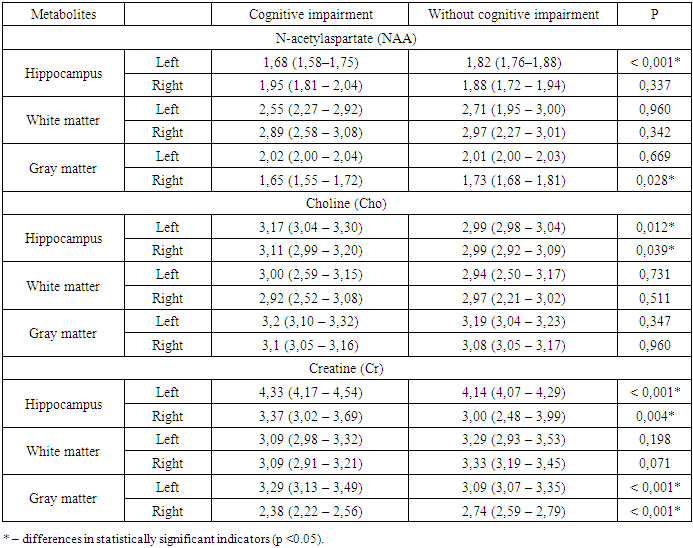
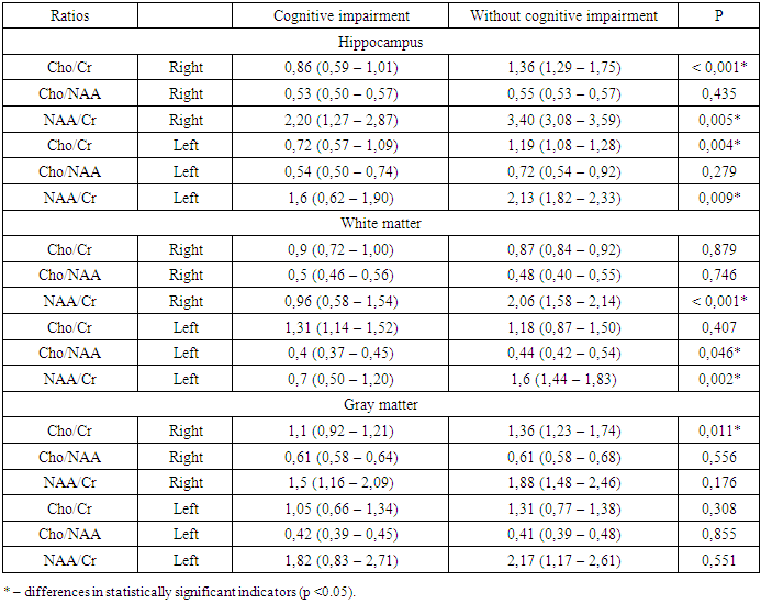
Our study involved 205 children in the age range from 7 to 18 years old suffering from type 1 diabetes. Children from 7 to 11 years old (average age – 9.0±1.6 years) accounted for 81 patients –39.5%), 124 (60.5%) patients from 12 to 18 years old were examined, the average age of which was 14.7±1.8 years. In accordance with the conditions of the study, all patients underwent an objective clinical and neurological examination with a detailed examination of personal data and clarification of anamnesis. Changes in higher brain activity were determined using the Montreal Cognitive Function Assessment Scale (MoSA test) [15]. The level of glucose concentration in the blood was determined using a quantitative method on a Hitachi 912 biochemical tester (Germany). On the DS5 tester (Netherlands), the concentration of glycated hemoglobin (HbA1c) in capillary blood was determined by liquid chromatography. MR spectroscopy of the main metabolites of the brain was performed immediately after the introduction of routine MRI, while not changing either the apparatus or the position of the child's body, with a relaxation time of TE = 135 ms, the volume of one voxel was 1.5 cm3. [16]. The survey was carried out in a multi-pixel mode, which provides an option for placing 64 voxels on one slice simultaneously. Peaks of the main spectra of N-acetylaspartate, choline, and creatine, as well as their ratios, were recorded in the pre-selected areas of greatest interest. The choice of mathematical methods was determined taking into account the tasks set in each specific case and according to the criteria for processing medical data.

3. The Results and Their Discussion

When evaluating the total score of the MoCA test, it was found that in 156 (76.09%) patients with type 1 diabetes, the result was less than 26 points. Of these, 2 (1.3%) children had 22 points; 44 (28.2%) patients had 23 points; 29 (18.5%) children had 24 points and 81 (51.9%) patients had 25 points. At the same time, the performance of testing tasks by patients was significantly (p < 0.001) worse than in healthy children (Figure 1).
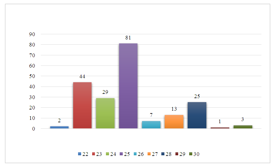
Figure 1. The average score on the MoCA scale, depending on the presence of diabetes
Out of 49 patients who scored 26 or more points, they coped with all the test tasks in full, i.e. they scored the most points for the test – 30 points, only 3 (6.1%) patients with type 1 diabetes. These indicators demonstrate the presence of moderate and mild abnormalities in higher cortical activity in this category of patients. Therefore, the need to provide neuropsychological examination to patients with type 1 diabetes for earlier detection of cognitive dysfunctions, the presence of which, in turn, as evidenced by literature data, lead to a decrease in self-control and have a negative impact on the general course of the disease, must be considered reasoned and appropriate.
During the analysis of individual tasks, it was determined that patients with type 1 diabetes performed significantly worse with the tasks "clock" (p<0.001), "attention" (p<0.001), "phrase repetition" (p<0.001) and "delayed reproduction" (p<0.001) in relation to the control group (Figure 2).
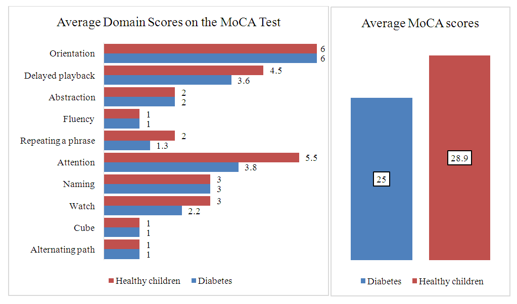
Figure 2. The average score for the domains of the MoCA scale, depending on the presence of SD
Thus, the analysis of the results of neuropsychological testing determined the presence of mild and moderate cognitive deficits in 76.09% of children and adolescents with type 1 diabetes, with the most pronounced deviations in memory and attention processes. There was also a difference in the task characterizing opto-spatial activity. Both boys and girls coped with the tasks to the same extent, that is, there were no gender differences in this category of patients. In our opinion, the initial manifestations of a decrease in cognitive functions in patients already at the early stages of the disease (up to 3 years of experience) are associated with the peculiarities of the psychological state and pronounced stress of the mechanisms of psychological adaptation. Both boys and girls coped with the tasks to the same extent, that is, there were no gender differences in this category of patients. However, it is worth noting that there are a number of studies in the scientific literature that determine the influence of gender on higher cortical functions. For example, Schoenle et al. (2002) presented results on a simultaneous decrease in verbal intelligence in boys with type 1 diabetes aged 7-16 years, whereas no similar changes were observed in girls of the same age [17]. Nevertheless, most research papers indicate the unconditional absence of differences in the assessment of cognitive activity by gender. As for the initial manifestations of cognitive decline in patients already in the early stages of the disease (up to 3 years of experience), in our opinion, this is associated with the peculiarities of the psychological state and with a significant strain on the mechanisms of psychological adaptation. In addition, according to Bremner J.D. (1997), the negative impact of stress associated with the underlying disease on the activity of the hippocampus responsible for the organization of cognitive processes is not excluded [18]. According to A.R. Luria (2002), it is in the hippocampus and the limbic sections directly related to them (amygdala, olfactory bulb, olfactory tract and tubercle, anterior nuclei of the thalamus, cingulate gyrus, reticular formation, areas of the frontal, temporal and parietal lobes of the large brain) that a significant number of neurons are contained that respond to each transformation of stimuli, and thus are considered equally to be neurons of attention and neurons of memory [19].
Further, out of 156 children with cognitive changes of varying severity identified during neuropsychological testing, we selected 59 patients to involve them in the PMRS study. The control group consisted of children with type 1 diabetes, but without clinical, neurological and cognitive changes (Table 1).
Table 1. Analysis of metabolites in children and adolescents with type 1 diabetes depending on the presence of cognitive impairment
     
Table 1 shows that the content of N-acetylaspartate in the hippocampus on the left in patients with CH – 1.68 mmol/l was significantly lower (p < 0.001) than in children with DM, but without cognitive abnormalities – 1.82 mmol/L. In addition, in the group of children with CN, there was a statistically significant (p=0.028*) decrease in N-acetylaspartate in the gray matter on the right – 1.65 mmol/l. compared with patients without cognitive impairment – 1.73 mmol/l. According to other parameters, namely the content of NAA in the hippocampus on the right, the content of NAA in the white matter on the left, the content of NAA in the white matter on the right, the content of NAA in the gray matter on the left, depending on cognitive impairment, statistically significant differences could not be detected (p = 0.337, p = 0.960, p = 0.342, p = 0.669 accordingly). A decrease in the content of N–acetylaspartate in the hippocampus and gray matter may indicate changes in the integrity of neurons with the presence of abnormal neuronal activity and transfer of oxidative energy, in addition, its decrease in the gray matter of the cerebral cortex on the right indicates the appearance of early signs of atrophy of this area, which corresponds to the preclinical period of impaired memory and attention processes. In addition, one of the tasks of NAA is to maintain, by combining the metabolism of axons with the metabolism of oligodendrocytes (glial cells), myelination processes, and according to N.K. Singhal et al., (2017), a decrease in NAA synthesis in neurons and catabolism in oligodendrocytes is the reason for the vulnerability of axons to the formation of demyelination due to changes in the expression of genes in oligodendrocytes and the composition of myelin lipids [20]. Thus, NAA is directly related to both the ability of nerve cells and their processes to perform their direct functions (neuronal integrity, information transmission, regulation of cell energy supply, due to the connection with ATP metabolism) and pathogenetic mechanisms of demyelination, this indicates a high degree of informativeness of this metabolite in assessing the functional ability of neurons and axons, and also when assessing the severity of the degree of myelin damage in the studied areas of the brain. The noted NAA shifts can lead to significant deviations in the dynamic processes of neuronal interaction, negatively affecting the dissemination of information in neuronal networks.
The assessment of choline content (Cho) showed that in the group with cognitive deficits, its significant increases were determined in the hippocampus on the left – 3.17 mmol/l (p=0.012*) and on the right – 3.11 mmol/l (p=0.039*), compared with the group without cognitive impairment – 2.99 mmol/L. and – 2.99 mmol/l. accordingly. According to Khomenko Yu.G. (2016), an increase in the peak of choline is associated with accelerated metabolism in cell membranes. And its possible decrease in patients with poorer cognitive performance is associated with depletion of compensatory mechanisms associated with the production of acetylcholine from the breakdown products of membrane phospholipids [21]. In addition, in the scientific works of P.A. Narayana (2005) and M. S. van der Knaap (2005), it was proved that increased concentrations of peak Cho indicate an increase in membrane metabolism, which occurs during the processes of demyelination, remyelination, inflammation and gliosis [22,23]. According to other indicators, in particular, the content of choline in the white matter on the left, the content of choline in the white matter on the right, the content of choline in the gray matter on the left, the content of choline in the gray matter on the right, depending on cognitive changes, statistically significant differences could not be detected (p = 0.731, p = 0.511, p = 0.347, p = 0.960, respectively). Thus, an increase in the level of choline (Cho), which is part of the structure of the complex phosphorus-containing compound phosphotidylcholine and is a structural component of cell membranes, may reflect the formation of destructive abnormalities in the membranes of cells of the central nervous system and be an indicator of a violation of their activity. In addition, these disorders can reverse the signs of functional disorders of the cholinergic neurotransmitter system of the brain.
The study of creatine content, which reflects the energy processes and metabolism of cells, and an increase in its concentration means increased oxidative stress, mitochondrial dysfunction in both neurons and glial cells [24], demonstrated its statistically significant increases in the group of patients with CN were detected in the hippocampus on the left – 4.33 mmol/L. (p < 0.001*), and on the right – 3.37 mmol/L. (p= 0.004*), in addition, in the gray matter on the left – 3.29 mmol/L. (p < 0.001*), whereas in the group of children with diabetes, but without CN, similar values were equal – 4.14 mmol/L., – 3.00 mmol/L., – 3.09 mmol/l. accordingly. In addition, in patients with CN, a significant decrease in creatine (p < 0.001*) was noted in the gray matter on the right – 2.38 mmol/L. in relation to persons without CN – 2.74 mmol/L. High creatine levels in the hippocampus on both sides, as well as in the gray matter on the left in patients with type 1 diabetes, can be associated with compensatory reactions occurring in these areas of the brain. An increase in energy metabolic processes in cells indicates that for optimal activity, the hippocampus requires high energy costs, which in the future may cause the formation of pathology at the anatomical level. As for the low creatine content in the gray matter of the cerebral cortex on the right in this category of patients, this indicates the appearance of initial signs of atrophy of this area due to depletion of energy reserves. It is well known that creatine plays a significant role in ensuring the energy metabolism of cells of the nervous system. Its concentration in the brain is quite high. With a decrease in the level of this metabolite, a deficiency of macroergs occurs, as a result of which the entire set of processes that are the basis of the functional activity of the nervous tissue is disrupted. One of the negative features of this process is the formation of an uncontrolled increase in the concentration of Ca2+ ions in the cytoplasm, which causes secondary damage to mitochondrial membranes and an even greater increase in energy deficiency. The chain of these circumstances plays an essential role in triggering the processes of neurodegeneration and ultimately determines the death of cells in the studied areas.
According to the results of the study, we established both direct and inverse correlations, which testified to the relationship between metabolic changes and cognitive deficits. Thus, a noticeable close relationship (p <0.001*) was established between the content of NAA in the hippocampus on the left and the scores on the MoCA test; moderate crowding feedback (p= 0.018*) NAA in white matter on the left and weak crowding feedback (p= 0.039*) on the right; moderate crowding direct relationship of NAA content in gray matter on the right (p= 0.003) and weak crowding feedback (p= 0.196) on the left; noticeable crowding feedback (p <0.001*) creatine in the hippocampus on the left; moderate tightness direct connection (p= 0.004*) of creatine in the gray matter on the right and weak tightness direct connection (p=0.360) of creatine in the gray matter on the left and the MoHS index of the test. Thus, all of the above metabolic shifts lead to energy imbalance, changes in neurotransmission, acceleration of neurodegeneration and demyelination, and also cause atrophic changes in the brain in patients of this category.
The evaluation of the ratio parameters in patients with type 1 diabetes, depending on the presence of cognitive deficits, demonstrated a significant decrease in the choline to creatine (Cho/Cr) ratios in hippocampal cells on the right - 0.86 mmol/l (p < 0.001*) and on the left – 0.72 mmol/l (p= 0.004*) compared with the group without KN is 1.36 mmol/l and 1.19 mmol/l. accordingly. In addition, patients with CN in the hippocampus on the right and left showed a significant decrease (p= 0.005*, p= 0.009*) in the ratio of N-acetylaspartate to creatine (NAA/Cr) – 2.20 mmol/l and 1.6 mmol/l, compared with diabetic patients, but without CN – 3.40 mmol/l. and 2.13 mmol/l, respectively. These changes (due to the involvement of creatine) demonstrate the formation of an energy imbalance (Cho/Cr and NAA/Cr) in the early stages of development, that is, the process occurs at the cellular level (Cho/NAA), not yet spreading to nerve tissues. In an earlier study by Sarac K., Akinci A., (2005) involving patients aged 8 to 19 years with type 1 diabetes, similar changes in the NAA/Cr and Cho/Cr ratios were detected in the bridge and in the left posterior parietal region of the white matter. At that time, the author concluded that the detected changes can be detected at a stage when there are no structural lesions yet [25] (Table 2).
Table 2. Analysis of the ratios of metabolites in children and adolescents with type 1 diabetes depending on cognitive impairment
     
The following changes in the ratios of metabolites were detected in the white matter of the brain (subcortical structures) in patients with cognitive dysfunction with type 1 diabetes: statistically significant (p < 0.001*) decreases in NAA/Cr on the right – 0.96 mmol/L.; Cho/NAA on the left (p= 0.046*) – 0.4 mmol/L.; NAA/Cr on the left (p= 0.002*) – 0.7 mmol/L., compared with patients without cognitive impairment – 2.06 mmol/L., – 0.44 mmol/L. and – 1.6, respectively.
The evaluation of the gray matter ratios showed a significant decrease (p= 0.011*) in patients with choline to creatine (Cho/Cr) content on the right – 1.1 mmol/L. Compared with children without CH – 1.36 mmol/L., in addition, there was a decrease in Cho/Cr on the left, NAA/Cr on the left and NAA/Cr on the right, however, no statistical differences were found according to these ratios (p= 0.308; p= 0.551; p= 0.176, respectively, the method used: Mann–Whitney U–test). However, there was a certain pattern of NAA/Cr ratios on both sides, since their reduced level in the early stages of diabetes decreased with the duration of the disease and was more pronounced in patients with more than six years of experience.
According to the results of the study, the coefficients of the ratios of the studied metabolites, taking into account the presence of cognitive deficits, we established the following correlations: moderate crowding direct relationship of Cho/Cr in the hippocampus on the right and MoCA scores (p <0.001*); weak crowding direct relationship of Cho/NAA in the hippocampus on the right and MoCA scores (p= 0.328); weak crowding direct association of NAA/Cr in the hippocampus on the right and MoCA scores (p= 0.296); moderate crowding direct association of Cho/Cr in the hippocampus on the left and MoCA scores (p= 0.011*); weak crowding direct association of NAA/Cr in the hippocampus on the left (p= 0.046*); noticeable tightness direct association of NAA/Cr in white matter on the right and MoCA scores (p <0.001*); moderate tightness direct association of Cho/NAA in white matter on the left and MoCA scores (p= 0.012*); moderate tightness direct association of NAA/Cr in white matter on the left and MoCA scores (p <0.001*); weak crowding direct connection of Cho/Cr in gray matter on the right and MoCA scores (p= 0.170); weak crowding direct connection of Cho/Cr in gray matter on the left and MoCA scores (p= 0.410); weak crowding direct connection of NAA/Cr in gray matter on the left and MoCA scores (p= 0.200). In his work, Y. G. Khomenko (2016) provides data on a significant decrease in the content of NAA in patients with moderate cognitive impairment in the white and gray matter of supraventricular regions and indicates that a more pronounced decrease in Cho/Cr and NAA/Cr and correlations with the results of cognitive tests indicate that the ratio of Cho/Cr and NAA/Cr may have diagnostic value reflecting the severity of cognitive deficits [21]. At the same time, the author only assumes the presence of this value, whereas according to the results of our extensive research, we deny any assumptions and state with absolute probability that these metabolite ratios have enormous diagnostic and prognostic value.

4. Conclusions

Thus, when analyzing MR spectroscopy in children and adolescents with cerebral changes in type 1 diabetes, the main abnormalities of metabolites were determined in areas directly related to the processes of cognitive activity, that is, in the structures of the limbic-reticular complex. The changes we found revealed a multi-vector nature of biochemical abnormalities that occur during the formation of cerebral disorders, which were largely interrelated, complemented and enhanced the pathological effect of each other. At the same time, as mentioned above, one of the key factors was the energy deficit of nerve cells, which causes the activation of catabolic processes, which in turn contributes to the disruption of the synaptic apparatus, leading to a decrease in the efficiency of the information dissemination process in neurons. In addition, changes in the cerebral metabolic status are manifested by a decrease in the neuronal activity of the hippocampus, as well as the white and gray matter of the brain, and with an increase in the length of the disease and depletion of energy resources, these disorders tend only to progress with the occurrence of neuronal degenerations and demyelination processes in these areas of the brain. Given the importance of this circumstance for the optimal functioning of higher cortical activity, the detected biochemical shifts should be considered within the framework of the main links in the pathogenesis of cognitive disorders in children and adolescents with type 1 diabetes. And the use of PMRS as a method of the earliest diagnosis of cerebral changes in patients with type 1 diabetes is considered appropriate and justified. The PMRS study, in addition to being able to localize the areas of the brain responsible for cognitive decline in children and adolescents with type 1 diabetes, provided more accurate information about metabolic changes in these areas of the brain. In general, this method allows you to determine the dynamics of the condition of patients with type 1 diabetes mellitus, the presence of cerebral changes in them, while at a stage when there are no clinical manifestations yet, in addition, to monitor the possible progression of cognitive impairment, as well as monitor the adequacy of selected therapy to correct abnormalities of higher cortical activity, in addition PMRS it makes it possible to detect not only metabolic changes in the brain at the early stages of the formation of cognitive deficits, but even to predict the development of atrophic changes in the cerebral cortex, taking into account the length of the disease, since the level of NAA, Cr and Cho decreases 3.2 times faster than the normalized volume of the brain, which indicates the preceding loss of neurons and axons to the formation of atrophic changes.

References

[1]  Matveeva M.V., Samoilova Yu.G., Zhukova N.G., Tolmachov I.V., Brazovskiy K.S., Leiman O.P., Fimushkina N.Yu., Rotkank M.A. Neuroimaging methods for assessing the brain in diabetes mellitus (literature review). Bulletin of Siberian Medicine. 2020; 19 (2): 189–194.
[2]  Li W., Huang E., Gao S. Type 1 Diabetes Mellitus and Cognitive Impairments: A Systematic Review. J. Alzheimers Dis. 2017; 57(1): 29– 36.
[3]  Ghetti S., Kuppermann N., Rewers A., Myers S.R., Schunk S.E., Stoner M.J., Garro A., Quayle K.S., Brown K.M., Trainor J.L., … Pediatric Emergency Care Applied Research Network (PECARN) DKA FLUID Study Group. Cognitive function following diabetic ketoacidosis in children with new-onset or previously diagnosed type 1 diabetes. Diabetes Care. 2020; 43: 2768–2775.
[4]  Chen H.J., Lee Y.J., Huang C.C., Lin Y.F., Li S.T. Serum brain-derived neurotrophic factor and neurocognitive function in children with type 1 diabetes. J. Formos. Med. Assoc. 2021; 120(1 Pt 1): 157–164.
[5]  Bykov Yu.V., Baturin V.A. Cognitive impairment in type 1 diabetes mellitus. Siberian Scientific Medical Journal. 2023; 43(1): 4–12.
[6]  Wolf V., Abdul Y., Ergul A. Novel targets and interventions for cognitive complications of diabetes. Front. Physiol. 2022; 12.
[7]  Zakharov V.V. Evolution of cognitive deficit: mild and moderate cognitive impairment / Zakharov V.V. // Neurology, neuropsychiatry, psychosomatics. - 2012. - No. 2. - P. 16-21.
[8]  MoCA: Montreal Cognitive Assessment. – URL: www.mocatest.org (access date: 06.06.2018).
[9]  Samoilova Yu.G., Rotkank M.A., Zhukova N.G., Matveeva M.V., Tolmachev I.V., Tonkih I.V., Kudlay D.A. Application of magnetic resonance methods of brain research in patients with type 1 diabetes mellitus and cognitive dysfunction. Nevrologicheskij zhurnal. 2018; 23(2): 86-92.
[10]  Aulich J, Cho YH, Januszewski AS, Craig ME, Selvadurai H, Wiegand S, Jenkins AJ, Donaghue KC. Associations between circulating inflammatory markers, diabetes type and complications in youth. Pediatr Diabetes. 2019 Aug 28.
[11]  Gujar S.K., Maheshwari S., Bjorkman-Burtscher I., Sundgren P.C. Magnetic Resonance Spectroscopy. J. Neuroophthalmol. 2005; 25(3): 217–26.
[12]  Tyurina, A. N. Proton 3D MR spectroscopy of gray and white matter of the brain. Study of 15 volunteers / A. N. Tyurina, I. N. Pronin, N. E. Zakharova, et al. // Issues of Neurosurgery named after N. N. Burdenko. - 2018. - No. 4. - 4 p.
[13]  Zhao X, Han Q, Lv Y, Sun L, Gang X, Wang G. Biomarkers for cognitive decline in patients with diabetes mellitus: evidence from clinical studies. Oncotarget. 2017; 9(7): 7710-7726.
[14]  Sergeev AM, Pozdnyakov AP, Ivanov DO, Atamanova EE, Olshansky OV, Pozdnyakova OF, Suf El-Zhil LR, Grechanyi SV. The results of a study of the brain in children with autism spectrum disorders using proton magnetic resonance spectroscopy. Luchevaya diagnostika i terapiya. 2020; 11(2): 66-75.
[15]  Samoilova Yu.G., Novoselova M.V., Zhukova N.G., Tonkikh O.S. Analysis of the role of neurospecific proteins in the diagnosis of cognitive dysfunction in patients with type 1 diabetes mellitus // Diabetes mellitus. 2014. No. 2. P. 83–90.
[16]  Grechany S.V., Suf El-Zhil L.R., Ivanov D.O., Pozdnyakova O.F., Sergeev A.M., Atamanova E.E., Pozdnyakov A.V. Proton magnetic resonance spectroscopy in children with atypical autism combined with psychomotor disinhibition syndrome. Korsakov Journal of Neurology and Psychiatry. 2021; 121(11 issue 2): 12–18.
[17]  Schoenle E.J. Impaired intellectual development in children with type I diabetes: association with HbA(1c), age at diagnosis and sex / E.J. Schoenle, D. Schoenle, L. Molinari, R.H. Largo // Diabetologia. – 2002. – Vol. 45. – P. 108–114.
[18]  Bremner J.D. MRI-based measurement of hippocampal volume in posttraumatic stress disorder // Biological Phychiatry. 1997. Vol. 41. Р. 23-32.
[19]  Luria A.R. Fundamentals of Neuropsychology / A.R. Luria. - M.: Publishing Center "Academy", 2002. - 384 p.
[20]  Singhal, N.K. The neuronal metabolite NAA regulates histone H3 methylation in oligodendrocytes and myelin lipid composition. N.K. Singhal, H. Huang, S. Li, R. Clements, J. Gadd, A. Daniels, E.E. Kooijman, P. Bannerman, T. Burns, F. Guo, D. Pleasure, E. Freeman, L. Shriver, J. McDonough. // Exp Brain Res. – 2017, Jan. – Vol. 235, №1. – P. 279-292.
[21]  Khomenko Yu.G., Bogdan A.A., Kataeva G.V., Chernysheva E.M. \\ Using Multivoxel Magnetic Resonance Spectroscopy in Examining Patients with Cognitive Disorders \\ Bulletin of St. Petersburg State University. Ser. 4. Vol. 3 (61). 2016. Issue 1.
[22]  Narayana, P.A. Magnetic resonance spectroscopy in the monitoring of multiple sclerosis. / P.A. Narayana // J Neuroimaging. – 2005. – Vol.15, № 4. – P.46–57.
[23]  Van der Knaap, M. S. Magnetic resonance spectroscopy: basic principles and application in white matter disorders. / M. S. van der Knaap, P. J. W. Pouwels. // Magnetic Resonance of Myelination and Myelin Disorders. Berlin: Springer. – 2005. – P.859-880.
[24]  Ross, B. Magnetic resonance spectroscopy of the human brain / B. Ross, S. Bluml // Anat. Rec. – 2001. – Vol. 265, No. 2. – P. 54–84.
[25]  Sarac K., Akinci A., Alkan A., Aslan M., Baysal T., et al. Brain metabolite changes on proton magnetic resonance spectroscopy in children with poorly controlled type 1 diabetes mellitus. Neuroradiology. 2005; 47: 562–532.