-
Paper Information
- Next Paper
- Previous Paper
- Paper Submission
-
Journal Information
- About This Journal
- Editorial Board
- Current Issue
- Archive
- Author Guidelines
- Contact Us
American Journal of Medicine and Medical Sciences
p-ISSN: 2165-901X e-ISSN: 2165-9036
2024; 14(9): 2248-2252
doi:10.5923/j.ajmms.20241409.26
Received: Aug. 10, 2024; Accepted: Sep. 6, 2024; Published: Sep. 18, 2024

Use of Arthroscopy in the Treatment of Early Fractures of the Greater Tibial Tuberosity
 Abstract
 Reference
 Full-Text PDF
 Full-text HTML
Irismetov M. E., Tukhtayev M. K.
Ministry of Health of the Republic of Uzbekistan, Republican Center of Specialized Traumatology and Orthopedic Scientific Applied Medicine, Uzbekistan
Copyright © 2024 The Author(s). Published by Scientific & Academic Publishing.
This work is licensed under the Creative Commons Attribution International License (CC BY).
http://creativecommons.org/licenses/by/4.0/

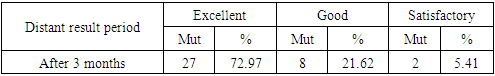
The aim of the study was to improve the results and efficiency of treatment by using arthroscopy in the treatment of patients diagnosed with early fractures of large tibial masses. It was found that in the treatment of fractures of the big tibia, it was shown that performing an operation under the control of an arthroscope allows you to fully see the changes in bone fragments and soft tissues, the state of the joint, the accuracy of repositioning can be maximally controlled, and it can be performed in a minimally invasive way. When observing the results of arthroscopic treatment, a high result in 72.97% of cases, a good result in 21.62% of cases, and a satisfactory result in 5.41% of cases indicated the high efficiency of the long-term results of this method of treatment.
Keywords: Early fractures of the greater tibial tuberosity, Arthroscopic treatment, Long-term results of treatment
Cite this paper: Irismetov M. E., Tukhtayev M. K., Use of Arthroscopy in the Treatment of Early Fractures of the Greater Tibial Tuberosity, American Journal of Medicine and Medical Sciences, Vol. 14 No. 9, 2024, pp. 2248-2252. doi: 10.5923/j.ajmms.20241409.26.
Article Outline
1. Introduction
- Last in years The frequency of intra-articular fractures of proximal metaepiphysis of the large tibia is not decreasing, these fractures make up to 10% of intra-articular fractures in the musculoskeletal system, from 2% to 5% of total fractures, and up to 30% of leg injuries [1].In injuries of the bones forming the knee joint, these fractures make up 29.4%, femur fractures - 5.2%, and knee cap fractures - 65.4%. Also, fractures of the external condyle of the greater tibia occur in 55-70% of cases, and fractures of the internal and both condyles occur in 10-30% of cases. The peculiarity of these fractures is that the fracture surfaces are located inside the joint, and in most cases it is the joining of soft tissue elements of the joint [2].Damage to the integrity of the surface of the knee joint is the reason for the severity of the injury and the long recovery process. It has been shown that fractures of the knee joint are overgrown with regenerating tissue, which in turn causes damage to the surface integrity of the patient's joint and changes in the shape of the joint, due to which it has been shown that patients often have limited movement in the injured joint and deforming arthrosis [3].It should be noted that the accumulation of blood in the fractured joint causes the formation of scars and adhesions, and even permanent contractures of the joint are unlikely. The complexity of knee fractures is also based on the difficulty of repositioning bone fragments and their stable fixation. Incomplete diagnosis of these fractures leads to incorrect selection of treatment tactics and, as a result, unsatisfactory results, today these cases are observed in 6-39%, and disability in them is observed in 6-15% [4].It is known that the diagnosis of fractures of the greater tibial tubercle in older patients is difficult, and in the first days of the injury, a complete diagnosis is often not made. Initially, the patient's knee joint is x-rayed in standard projections, and multispiral computer tomography (MSCT) and magnetic resonance imaging (MRT) examinations clearly show the direction and displacement of intra-articular fractures, as well as the degree of injury to the intra-articular soft tissues (menisci, iliac ligaments) [5].Currently, the use of high-tech minimally invasive methods is required in the treatment of large tibial tumors, and wide arthrotomy is still used in many clinics. It was found that in such practices, the crushing of the soft tissues of the joint increases, which, in turn, causes the increase of purulent complications, the appearance of contractures, the lengthening of the rehabilitation period, the inability to work, and the increase of disability [6]. Arthroscopic treatment of such fractures is still not widely practiced.Based on the above, the aim of this research work was to improve the results and increase the effectiveness of treatment by using arthroscopy in the treatment of patients diagnosed with early fractures of the large tibia.In the treatment with the help of the recommended method, restoring the surface integrity and shape of the knee joint in patients, preventing post-operative scars, preventing joint contractures and arthrosis, shortening the rehabilitation period, and returning patients to work by ensuring pain-free walking were among the priority tasks.
2. Materials and Methods of Research
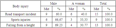
- In order to achieve the set goal, 37 patients who were treated in 2019-2023 in the Department of Sports Traumatology and Orthopedic Scientific Applied Medicine Center of the Republic of Uzbekistan and the Department of Traumatology, Orthopedics and Neurosurgery of the Multidisciplinary Medical Center of Navoi region were involved in the research.All patients (n= 37) had a diagnosis of early fracture of the tibia, they were between 25 and 56 years old, 28 were men (75.68%) and 9 were women (24.32%). Table 1 shows the results of distribution of patients according to the causes of injuries.
|
3. The Obtained Results and Their Discussion
- All studied patients (n= 37) complained of knee swelling, pain, deformation, limitation of movement, difficulty in walking (100.0%) within 14 days after the injury.In 17 patients (45.95%) meniscal injuries were detected between bone fragments, in 2 cases (5.41%) lateral meniscus anterior horn tear was detected and "P" sutures were placed, and in the remaining cases (48.65%, n=18) underwent a partial meniscectomy.All cases were repositioned and osteosynthesized with cancellous screws under arthroscopic control. The patients were put in a plaster cast with a tutor for 14 days, after which the patients began to perform flexion-writing exercises in the knee joint.Below, as a clinical example, we found it necessary to present the medical history of one of the adult patients involved in the treatment.Patient D. 37 years old, (medical history No. 8514) 8 days before the winter of applying to the clinic, he received an injury to his left leg as a result of a fall from a height. placed However, the patient referred to the clinic because of persistent pain, and additionally underwent MSCT (Fig. 1) and MRI (Fig. 2).
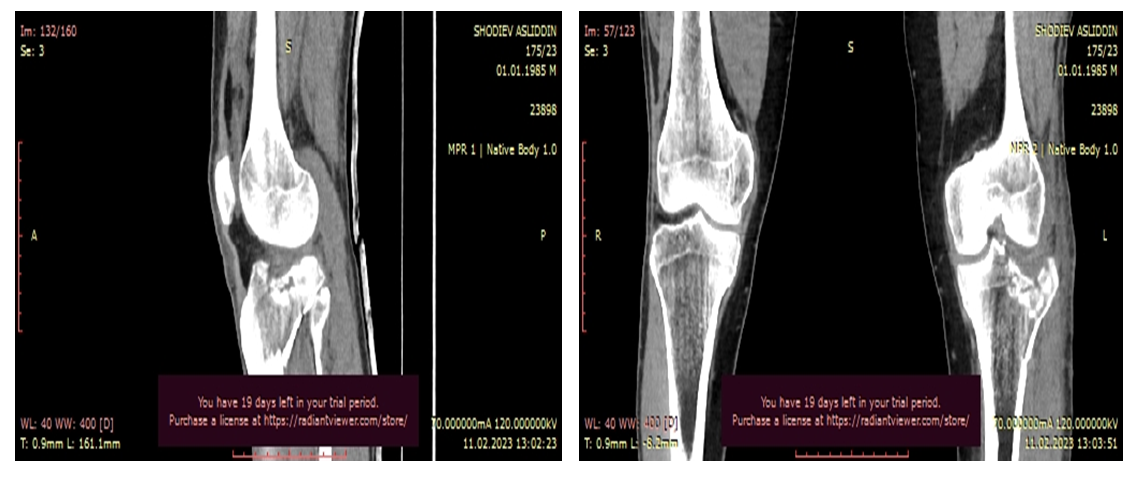 | Figure 1. MSCT view of a patient with a diagnosis of fracture and displacement of the outer condyle of the left tibia (patient D. 37 years old, (case history No. 8514) |
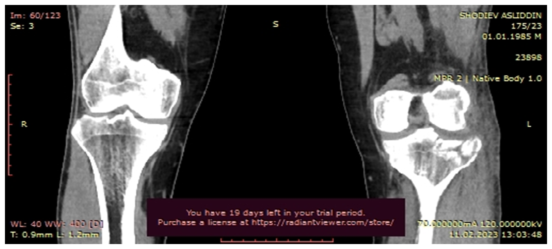 | Figure 2. MRI image of a patient with a diagnosis of fracture and displacement of the outer tubercle of the left tibia, partial tear of the anterior horn of the lateral meniscus (patient D. 37 years old, (case history #8514) |
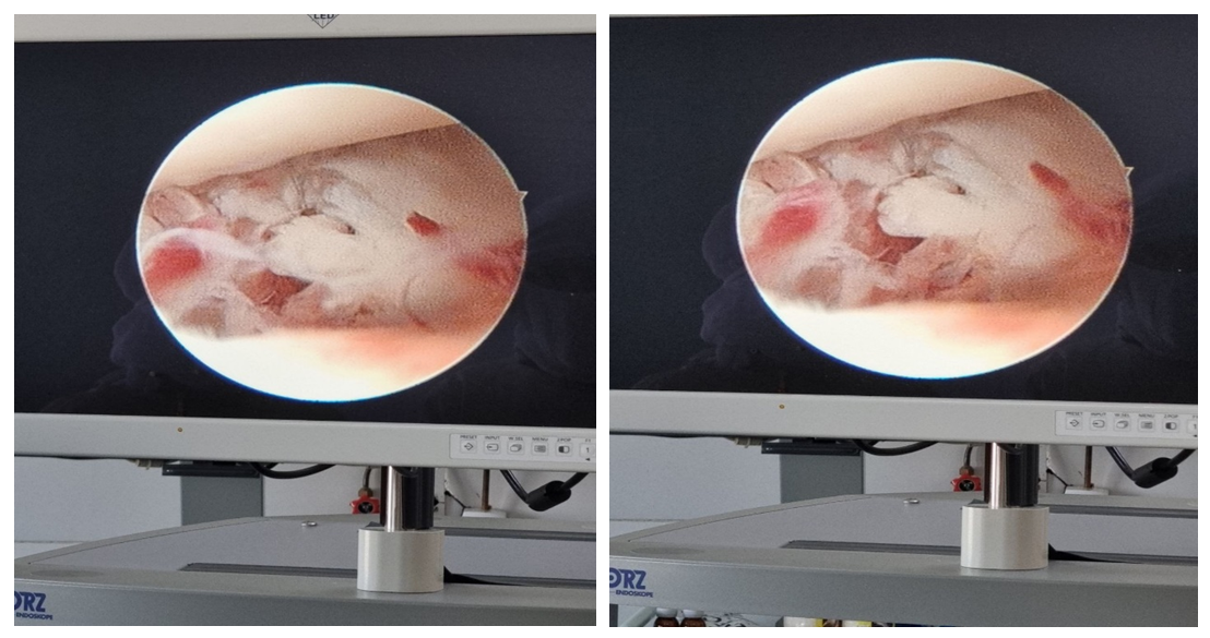 | Figure 3. Arthroscopic view of a patient diagnosed with a fracture and displacement of the outer condyle of the left tibia, a partial tear of the anterior horn of the lateral meniscus (patient D. 37 years old, case history No. 8514) |
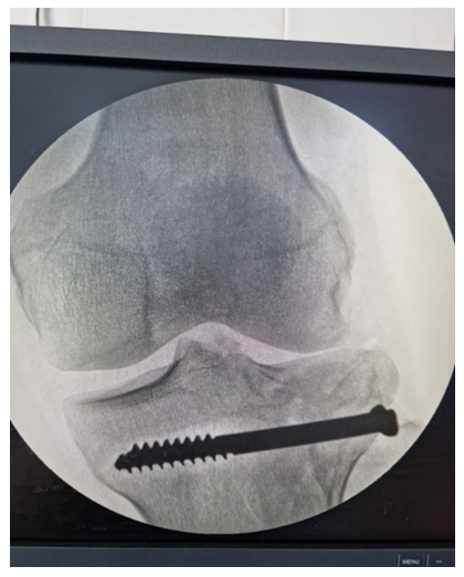 | Figure 4. Radiographic view after arthroscopic surgery of a patient diagnosed with a fracture and displacement of the outer condyle of the left tibia, a partial tear of the anterior horn of the lateral meniscus (patient D. 37 years old, (case history # 8514) |
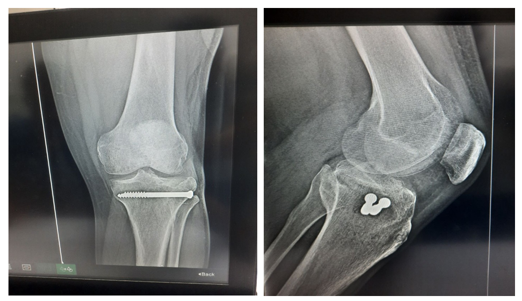 | Figure 5. Radiographic view of a patient diagnosed with a fracture and displacement of the outer condyle of the left tibia, a partial tear of the anterior horn of the lateral meniscus, 3 months after arthroscopic treatment (patient D. 37 years old, (case history No. 8514) |
|
4. Conclusions
- 1. It has been shown that in the treatment of fractures of the large tibial bone, performing an operation under the control of an arthroscope allows you to fully see the changes in bone fragments and soft tissues, the state of the joint, the accuracy of repositioning can be maximally controlled, and it can be performed in a minimally invasive way.2. The advantages of arthroscopy include the absence of scars on the skin after the procedure, the reduction of the risk of purulent complications, the absence of long-term use of plaster bandages, and the absence of contractures limiting movement in the knee joint.3. When observing the results of arthroscopic treatment, a high result in 72.97% of cases, a good result in 21.62% of cases, and a satisfactory result in 5.41% of cases indicated the high efficiency of the long-term results of this method of treatment. This made it possible for early rehabilitation of patients and their return to work in a short period of time.