American Journal of Medicine and Medical Sciences

p-ISSN: 2165-901X    e-ISSN: 2165-9036

2024;  14(9): 2183-2190

doi:10.5923/j.ajmms.20241409.12

Received: Aug. 5, 2024; Accepted: Sep. 7, 2024; Published: Sep. 10, 2024


Dynamics of Cellular Immunity Indices in Kidney Damage in Patients Carried SARS-CoV2

Xursandov Ilyos Axmedovich1, Кhamdamov Bakhtiyor Zarifovich2, Khamdamov Ilkhomjon Bakhtiyorovich2, Кhamdamova Muhayyoxon To`xtasinovna2

1Multidisciplinary Medical Center “Sultan Hospital", Termez, Uzbekistan

2Bukhara State Medical Institute, Bukhara, Uzbekistan

Correspondence to: Кhamdamov Bakhtiyor Zarifovich, Bukhara State Medical Institute, Bukhara, Uzbekistan.

Email:

Copyright © 2024 The Author(s). Published by Scientific & Academic Publishing.

This work is licensed under the Creative Commons Attribution International License (CC BY).
http://creativecommons.org/licenses/by/4.0/

Abstract

Relevance. The SARS-CoV-2 coronavirus has evolved into a life-threatening pandemic disease, Covid-19. Acute respiratory distress syndrome (ARDS) and diffuse alveolar involvement are known to be the main manifestations of this disease. Purpose of the study: To study the indices of cellular immunity in renal injury in SARS-CoV2 patients. Material and methods of research: The paper presents data on the comprehensive examination and treatment of 62 patients with kidney injury who underwent SARS-CoV-2. The distribution of patients was based on a prospective targeted open randomized study. The period of research and collection of clinical material started in the second quarter of 2020 and ended in December 2023. Conclusion. Thus, acute kidney damage in patients after SARS-CoV2 occurs against the background of a low number and activity of CD4+ cells and a high value of CD8+, CD19+ cells, which ultimately leads to impaired regeneration of individual parts of damaged nephrons, stimulating the growth and accumulation of circulating immune complexes and changing their antigenic structure.

Keywords: Cellular immunity, Kidney damage SARS-COV2, Regeneration

Cite this paper: Xursandov Ilyos Axmedovich, Кhamdamov Bakhtiyor Zarifovich, Khamdamov Ilkhomjon Bakhtiyorovich, Кhamdamova Muhayyoxon To`xtasinovna, Dynamics of Cellular Immunity Indices in Kidney Damage in Patients Carried SARS-CoV2, American Journal of Medicine and Medical Sciences, Vol. 14 No. 9, 2024, pp. 2183-2190. doi: 10.5923/j.ajmms.20241409.12.

1. Relevance

The SARS-CoV-2 coronavirus has evolved into a life-threatening pandemic disease, Covid-19. Acute respiratory distress syndrome (ARDS) and diffuse alveolar involvement are known to be the main manifestations of this disease [1,3,5,7,9,11,13,15].
Although the respiratory system is the primary target of SARS-CoV-2, other organs in the body can be affected by the virus via the circulatory system. Initially, information on kidney involvement (KI) was sparse. Publications regarding PP in SARS-CoV-2 early in the pandemic were not systematic and were characterized by isolated clinical cases ranging from mild proteinuria to progressive acute renal failure (ARF) [2,4,6,8,10,12,14,16,18,20].
During SARS-CoV-2 infection, decreased CD4+ and CD8+ cell counts and increased cytokine levels cause inflammation [17,19]. Regarding the specificity of SARS-CoV-2 virus, different T cell subsets are activated [18]. The overproduction of cytokines that leads to ARDS is associated with the outcome and severity of SARS-CoV-2. The enhanced inflammatory response induced by SARS-CoV-2 plays a key role in the severity of infection, development of PP and death [3].

2. Purpose of the Study

To study the indices of cellular immunity in renal injury in SARS-CoV2 patients.

3. Material and Methods of Research

The paper presents data on the comprehensive examination and treatment of 62 patients with kidney injury who underwent SARS-CoV-2. The distribution of patients was based on a prospective targeted open randomized study. The period of research and collection of clinical material started in the second quarter of 2020 and ended in December 2023. At the same time for the period from April to August 2020 the clinic functioned still as a specialized covid center, with involvement of specialists of all directions according to quarantine requirements. All patients were combined into one main group.
Criteria for inclusion of patients in the main group were: age of patients not younger than 20 and not older than 75 years; presence in the history of coronavirus infection disease, with severe course, with signs of kidney damage during the treatment period; persistence of signs of kidney disease (proteinuria, albuminuria, micro- or macrohematuria, decreased rate of glomerular filtration, high values of creatinine and urea in blood, etc.); presence of negative results of PCR in the clinic.); negative PCR test result for SARS-CoV-2 during hospitalization in our clinic; voluntary informed consent of the patient to participate in the clinical trial.
Due to the fact that the patients of the main group came to the clinic with different duration of the anamnestic period of the disease development and the time of SARS-CoV-2 before us, there was a need to randomize the main group itself. Based on the principles of evidence-based medicine the main group of patients was subdivided by us into two subgroups:
The first subgroup - 34 (54.8%) patients who were transferred from a specialized covid infectious disease clinic after achieving a negative PCR test result for SARS-CoV-2 with signs of kidney damage and the need for renal replacement therapy.
The second subgroup - 28 (45.2%) patients who were transferred from other therapeutic clinics or hospitalized as in the primary treatment for the underlying disease. All of them had a history of SARS-CoV-2 and were treated as inpatients in a specialized covid infectious disease clinic. During treatment, they were diagnosed with acute kidney injury, but remission of complications was achieved by the time of hospital discharge. However, later, after 1 to 3 months, the patients came to our clinic with the need for renal replacement therapy.
The chronology of hospitalization of patients with kidney injury after SARS-CoV-2 showed that patients of the first subgroup were hospitalized in our clinic predominantly (70.6%) during the peak of the first and second waves of the COVID-19 pandemic, whereas patients of the second subgroup sought specialized care predominantly after the passage of the COVID-19 pandemic waves for the period 2021-2023 (92.9%).
In the comparative evaluation of clinical and immunological changes, the data of 20 healthy individuals recognized by the medical commission as absolutely healthy were used. All of them were combined into a control (reference) group.
The criteria for inclusion of volunteers in the control group were: age of patients from 20 to 50 years; absence of any chronic somatic or psychiatric diseases; obligatory conclusion of the medical commission about healthy condition during the last 3 months before the study; absence of diseases from the urinary system in the anamnesis; absence of SARS-CoV-2 in the anamnesis; negative result of PCR-test for SARS-CoV-2; voluntary informed consent for participation in the clinical trial.
The diagnosis of kidney damage was established according to the recommendations of the Global Foundation for the Improvement of Kidney Diseases (KDIGO) in the presence of one of three signs of pathological process manifestation in the form of: increase of serum creatinine level at 26.5 μmol/l and higher within 48 hours; increase of serum creatinine above 1.5 times compared to the baseline level during the previous 7 days; decrease of urine output less than 0.5 ml/kg/hour within 6 hours.
All patients in the main group were subdivided according to the stages of kidney damage after SARS-CoV-2, which were also recommended by KDIGO. As our data showed, the third stage of renal damage prevailed among the patients of the main group (62.9%), in which the level of creatinine in the blood of patients increased 3 times from the baseline or the beginning of renal replacement therapy. In the remaining 37.1% of cases, patients had stage 1 and 2 kidney damage after SARS-CoV-2. From 17.7% of cases the first stage of kidney damage was diagnosed, when blood creatinine level increased 1.5-1.9 times from baseline, and in 19.4% of cases - 2 - 2.9 times under the same conditions. Distribution of patients by age showed predominance of patients in the age category of 51 and older (67.8%). At the same time between patients in the age category from 51 to 60 years and from 61 to 75 years the distribution of the number of patients was the same (21 patients or 33.9% each). The second place was occupied by patients aged 41-50 years (24.2%). Patients in young age were only in 8.1% of cases. It should be noted that among the patients of the first subgroup prevailed in the age category from 51 to 60 years, and in the second subgroup - from 61-75 years.
Thus, renal damage was predominantly characteristic of mature and elderly patients, while acute kidney injury after SARS-CoV-2 was characteristic of younger age.
Male patients predominated (67.7%), both among patients of the first and second subgroups.
When performing renal replacement therapy, we used a capillary dialyzer.
Special multi-stage purified water was used for dialysis according to the approved standards of the Ministry of Health of the Republic of Uzbekistan. Devices for renal replacement therapy contained a set of hydraulic pumps for mixing solutions containing electrolytes Na+, K+, Ca++, Mg++, Cl-. General clinical laboratory, biochemical, bacteriological, molecular genetic (PCR) studies were performed in a centralized clinical diagnostic laboratory. The calculation of the glomerular filtration rate (ml/hour/kg) was performed according to the formula:
Club filtration rate (ml/hour/kg) = [Urine volume (ml) x urine creatinine (mg/mL)] / [time (hour) x serum creatinine (mg/mL) x patient weight (kg)].
All patients underwent ultrasound examination of the kidneys in a functional diagnostics room. To determine morphostructural changes in the kidneys, all patients of the main group underwent percutaneous puncture biopsy of the kidneys in the surgical department under aseptic conditions. The biopsy material was investigated by standardized histological technique with staining under light in the conditions of the central research laboratory.
Immunologic studies were performed in the Bukhara branch of the Institute of Human Immunology and Genomics. Peripheral blood served as the object of study. A 6 ml sample of venous blood was collected at one time into tubes with heparin (20 U/mL) and 5 ml into a dry tube for subsequent serum separation. The immune status and cytokine profile of the patients were assessed twice - at the initial visit of the patients to us, which usually occurred on the 1-2 day of inpatient treatment and after the treatment before discharge from the hospital, which usually occurred on average on 14-21 days of treatment.
When assessing the immunologic status of the study indicators of cellular (leukocytes, neutrophils, eosinophils, monocytes, lymphocytes (CD3+), T-helpers (CD4+), T-killers (CD8+), B-lymphocytes (CD19+), phagocytic number and phagocytic index, circulating immune complexes) and humoral (immunoglobulins A, M, G and cytokines IL-1β, TNF-α, IL-8, IL-10 and IFN-γ) immunity. Quantitative determination of cytokines IL-1β, TNF-α, IL-8, IL-10 and IFN-γ was performed in blood serum using ELISA test systems of Cytokin LLC (St. Petersburg) on an automatic analyzer for enzyme-linked immunosorbent assay.
Mononuclear cells were isolated on a density gradient of ficollaverographin. To identify lymphocytes and their subpopulations, immunophenotyping of mononuclear cells was performed by direct immunofluorescence using monoclonal antibodies CD3+, CD4+, CD8+, CD19+.
Determination of the concentration of immunoglobulins IgM, IgG, IgA in blood serum was carried out by immunoturbometric method using an automatic biochemical analyzer.
Determination of the concentration of circulating immune complexes in serum was carried out by polyethylene glycol-6000 precipitation method according to the method of Y.A. Grinevich and A.N. Alferov, 1981. Assessment of phagocytic activity included the use of leukocyte suspension, which was incubated with standard latex particles with a diameter of 1.35 μm (Immunoscreen, Moscow) for 30 min, swabbed, stained with Azur-eosin and counted the number of neutrophils that captured latex particles. The percentage of phagocytizing neutrophils was denoted as phagocytic index. The phagocytic number was determined, which corresponded to the number of particles taken up on average by one phagocytic neutrophil.
In order to assess the prognostic significance of the studied parameters of immunologic disorders in patients with kidney damage after SARS-CoV-2 we used the method of calculating the dependence between multivariate variables and the model created by the statistical model for predicting the disorder. This analysis was performed by constructing linear regression analysis. Factor analysis was used to examine the mutual relationship of the various traits studied, which determined the variance of specific factors in the form of a variance cloud. Statistically significant linear combinations of a number of factors allowed us to reduce the dimensionality of the trait graphical cloud that can be analyzed.
The linear plotting of the data allowed us to compare specific factors against each other. This in turn was the key to examining the actions on the final outcome of the factors analyzed in each variance data cloud. Using this method of statistical analysis allowed us to quantify the observed signs of change. The studied factors and their constituent immunologic data were presented in quantitative form; the observed traits influenced by the studied factors were the outcome traits.

4. Results and Their Discussion

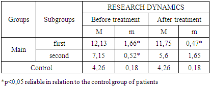
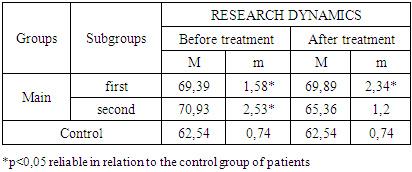
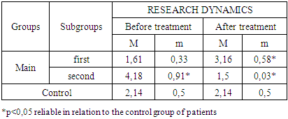
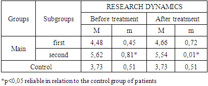
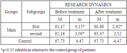
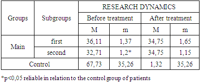
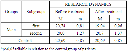
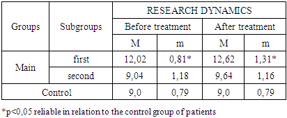
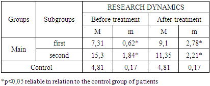
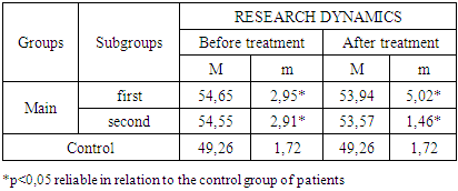
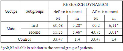
The study of the state of cellular immunity in patients with kidney damage after SARS-CoV2 was mainly aimed at assessing their activity against viruses. At the first stage we studied the dynamics of leukocyte changes in blood (Table 1).
Table 1. Nature of changes in the dynamics of leukocyte count (x109/l) in peripheral blood in patients with kidney injury after SARS-CoV2
     
The general dynamics of leukocyte level among patients with kidney injury after SARS-CoV2 was characterized by the presence of leukocytosis. Thus, the mean value of leukocytes in blood during the whole period of the study in patients with kidney damage after SARS-CoV2 was 9.16±1.08 x10⁹/l [CI:8.08; 10.08]. The maximum level of leukocytosis was observed among the patients of the first subgroup before treatment [CI:10.47; 13.79], which was 2.9 times higher than reference values (p<0.05). Even after treatment in patients of the first subgroup, the leukocytosis rate was 2.75 times higher [CI:11.28; 12.22] than the reference value [CI:4.08; 4.44].
In patients of the second subgroup at the time of referral to our clinic, the leukocyte level corresponded to the range of physiologic values [CI:6.63; 7.67]. By the end of the treatment, this index decreased 1.3 times compared to the time of patients' admission to our clinic [CI:3.93; 7.25].
The number of neutrophils in patients with kidney damage after SARS-CoV2 was characterized by the presence of high values, although in the dynamics of the conducted treatment there was a relative decline (Table 2).
Table 2. Nature of changes in the dynamics of neutrophil counts (%) in peripheral blood in patients with kidney injury after SARS-CoV2
     
The average level of neutrophil count in patients during the whole period of the study was equal to 68,89±1,91% (p<0,05 in relation to reference values). At the same time in patients before treatment its level was equal to 70,16±2,01% (p<0,05 in relation to reference values), and after treatment it decreased to 67,62±1,77% (p<0,05 in relation to reference values). The ranges of the confidence interval in the dynamics of treatment ranged between [CI: 68.11; 72.22] before treatment and between [CI: 65.86; 69.4] after treatment. The mean level of this index was [CI: 66.98; 70.81].
Separate analysis of the dynamics of the neutrophil count change in blood depending on the terms of treatment showed that in patients of the first subgroup the difference between the neutrophil count before treatment [CI: 67,81; 70,97] and after treatment [CI: 67,55; 72,23] was not reliable, whereas in patients of the second subgroup there were significant changes in this indicator before [CI: 68,4; 73,46] and after [CI: 64,16; 66,56] treatment (p<0,05). The mean neutrophil count among the patients of the first subgroup was 69.64±1.96%, and it was lower among the patients of the second subgroup - 68.15±1.87%.
The number of eosinophils during the whole period of the study in patients with kidney damage after SARS-CoV2 averaged 2.61±0.89% [CI: 1.72; 3.51], exceeding the reference value 1.2 times [CI: 1.64; 2.64] - Table 3. At the same time, the number of eosinophils in the patients of the main group during the period of treatment in the clinic was characterized by the increase of this index 2,9±0,62% [CI: 2,28; 3,52] in comparison with the reference values (2,14±0,5%; p<0,05).
Table 3. Nature of changes in the dynamics of the number of eosinophils (%) in peripheral blood in patients with kidney damage after SARS-CoV2
     
At separate analysis of changes in blood eosinophils level in patients of different subgroups we noted relative Eosinophilosis after the treatment course among patients of the first subgroup [CI: 1,61; 0,33]. It should be noted that the initial value of blood eosinophils in patients of the first subgroup on admission to our clinic was 1.3 times less than the reference [CI: 1.64; 2.64] values (p<0.05). After the treatment, in patients of the first subgroup, there was an increase in the number of eosinophils [CI: 2,58; 3,74], which exceeded the reference values 1,5 times. At the same time, the growth (2-fold) of eosinophils after the treatment was also noted in relation to the period before the treatment [CI: 1.28; 1.94] in the patients of the first subgroup (p<0.05). Thus, the mean value of eosinophils among the patients of the first subgroup for the whole period of the study equaled 2.39±0.46% [CI: 1.93; 2.84], which was lower than the overall mean value [CI: 1.72; 3.5].
In patients of the second subgroup the mean value of eosinophils, which amounted to 2.84±0.47% [CI: 2.37; 3.31], was 1.2-fold [CI: 1.93; 2.84] and 1.3-fold [CI: 1.64; 2.64] higher than that of patients of the first subgroup. When patients applied to the clinic, before the beginning of treatment measures, high values of this index [CI: 3.27; 5.09] were detected, which exceeded the reference values on average 1.9 times (p<0.05), values of the first subgroup patients before the treatment 2.6 times (p<0.05) and values of the first subgroup patients after the treatment 1.3 times (p<0.05). We intentionally emphasize this fact, because the level of eosinophils in blood of the second subgroup patients before the treatment exceeded (1,4 times; p<0,05) the average value of this period of all patients with kidney damage, after SARS-CoV2.
After the conducted treatment in patients of the second subgroup the mirror opposite picture in change of eosinophils quantity in peripheral blood is noted. The number of eosinophils in patients of the second subgroup after treatment [CI: 1.47; 1.53] decreased 2.8 times (p<0.05), compared to the previous study period. In comparison with the same period of the study of the patients of the first subgroup a 2.1-fold decrease in the number of eosinophils was also noted (p<0.05). The specific weight of this index in the share of the total average value of the patients of the main group after the treatment amounted to 64.4%, and among the patients of the second group - 52.8%.
Thus, the growth of eosinophils in chronic kidney injury after SARS-CoV2 is characterized by a decrease below the reference value after treatment, whereas in patients with acute kidney injury after SARS-CoV2 there is an opposite picture, characterized initially by a low number of these cells with a gradual increase after treatment.
The mean number of monocytes in peripheral blood of patients with kidney damage after SARS-CoV2 was 5.08±1.25% [CI: 3.83; 6.32], which was 1.4 times higher than the reference value (3.73±0.51% [CI: 3.22; 4.24]) (p<0.05) - Table 4.
Table 4. Nature of changes in the dynamics of the number of monocytes (%) in the peripheral blood in patients with kidney damage after SARS-CoV2
     
The mean value of monocyte count in patients with kidney injury after SARS-CoV2 at initial visit to our clinic equaled 5.05±0.63% [CI: 4.42; 5.68], and after treatment this index was almost unchanged and amounted to 5.10±1.86% [CI: 3.24; 6.96].
In patients with acute kidney injury after SARS-CoV2 the mean monocyte content in peripheral blood equaled 4.57±0.59% [CI: 3.99; 5.16], which was 1.22 times higher than reference values. When the patients were admitted to the clinic, the number of monocytes in peripheral blood [CI: 4.03; 4.93] was 1.24 times higher than the reference values. The number of monocytes in the peripheral blood of patients of the first subgroup at admission to the clinic was 1.25 times lower than in patients of the second subgroup during the given period of the study. The share of the average number of monocytes of patients before the beginning of treatment measures amounted to 44.4%. The same level was observed in relation to the total average value of peripheral blood monocytes of the whole main group of patients.
After the treatment the number of monocytes in the peripheral blood slightly increased [CI: 3,94; 5,38] in comparison with the previous term of the study [CI: 4,03; 4,93], while among the patients of the second subgroup there was an inverse pattern of changes dynamics (decrease in the number of monocytes from 5,62±0,81% to 5,54±3,0%).
Thus, the general dynamics of changes in the number of monocytes in peripheral blood was characterized by an unreliable increase after the treatment. At that, the similar tendency of changes was noted among patients with acute kidney injury after SARS-CoV2, which characterizes the pathological process as a determining mechanism of changes in leukocyte structure.
The character of lymphocyte changes by CD3+ cells was almost identical in patients with different forms of kidney damage, after SARS-CoV2 - Table 5. It was characterized by a decrease in values in patients compared to control data. Thus, the mean value of the total number of CD3+ T-lymphocytes amounted to 61.87±3.0% [CI: 58.88; 64.87], which was less than the reference values by 5.86±1.97% [CI: 62.76; 72.7]. At the same time, the number of CD3+ T-lymphocytes in patients before treatment was lower (61.68±3.62%; [CI: 58.06; 65.3]) than in patients after treatment (62.07±2.37%; [CI: 59.7; 64.44]), which indicates the positive effect of methods of kidney damage correction in patients who underwent SARS-CoV2 in the form of CD3+ cells expression.
Table 5. Nature of changes in the dynamics of the number of CD3+ T-lymphocytes (%) in peripheral blood in patients with kidney damage after SARS-CoV2 transfer
     
Separate analysis depending on the timing of treatment in our clinic of patients with kidney damage after SARS-CoV2, we can note low mean lymphocyte counts in patients with acute pathologic process (60.92±3.39%; [CI: 57.53; 64.30]) than in patients with chronic pathologic process (62.83±2.61%; [CI: 60.22; 65.43]).
In patients of the first subgroup, the number of CD3+ cells in peripheral blood was higher before treatment [CI: 57.02; 65.32] than after replacement therapy [CI: 58.04; 63.28]. In both cases, these values were lower than reference values by 6.56±1.32% and 7.07±2.85%, respectively. At the same time, in patients of the second subgroup the trend of CD3+ cells number changes corresponded to the general average value, was higher than in patients with acute kidney injury after SARS-CoV2 and was characterized by CD3+ cells growth ([CI: 59,09; 65,27] before treatment and [CI: 61,35; 65,59] after treatment).
We obtained relatively similar dynamics of changes when we studied the number of T-helper lymphocyte subpopulations by CD4+ cells (Table 6).
Table 6. Nature of changes in the dynamics of the number of CD4+ T-helper cells (%) in peripheral blood in patients with kidney damage after SARS-CoV2
     
The average level of CD4+ T helper cells among patients with kidney damage after SARS-CoV2 was lower (34.58±1.34%) than the reference values (35.26±1.32%), although the confidence interval range did not have such a high difference ([CI: 33.94; 36.58] in the control group and [CI: 33.24; 35.92] the average level of the main group of patients). At the same time, between patients with kidney damage after SARS, the average value was before (34.41±1.29%; [CI: 33.13; 35.7]) and after (34.75±1.4%; [CI: 33.35; 36,15]) treatment did not have a significant difference, which was due to different phases of the pathological process (p>0.05).
The average value of CD4+ T-helper cells among patients with acute kidney injury after undergoing SARS-CoV2 was 35.43±1.51% [CI: 33.92; 36.94], whereas among patients with Over a longer period of the pathological process, this indicator was lower and amounted to 33.73±1.18% [CI: 32.56; 34.91].
When conducting a separate analysis, it can be noted that in acute kidney injury, in patients who underwent SARS-CoV2, the number of CD4+ T helper cells increased [CI: 34.74; 37.48], which was the maximum value compared with the general main group [CI: 33.13; 35.7]. A decrease in CD4+T helper cells was characteristic of patients with kidney damage after undergoing SARS-CoV2 [CI: 31.51; 33.91], which acquired a chronic course.
Renal replacement therapy in patients of the first subgroup led to a decrease in the number of CD4+ T helper cells in peripheral blood [CI: 33.1; 36.4], and hemodialysis in the second subgroup of patients ended with an increase in the number of CD4+ T helper cells [CI: 33.6; 35.9]. In other words, the acute inflammatory process occurred against the background of suppression of T-helpers, whereas the chronic one was stimulated. However, in both cases, the data obtained indicated that the reference values were not reached, which was typical for kidney damage after SARS-CoV2. After the treatment, the level of CD4+ cells in both subgroups becomes the same.
The average number of cytotoxic T lymphocytes CD8+ cells in patients with kidney damage after SARS-CoV2 was almost at the level of reference values, which was determined by the similarity of fluctuations in the confidence interval ([CI: 19.84; 21.54] reference values and [CI: 19.02; 21.22] in the main group of patients) - Table 7.
Table 7. The nature of changes in the dynamics of the number of CD8+ T-killers (%) in peripheral blood in patients with kidney damage after SARS-CoV2
     
The average number of CD8+ cells in the control group was 20.69±0.85%, whereas in patients of the main group it was 20.12±1.1%.
In patients of the main group, the number of CD8+ cells when contacting our clinic was higher than 20.37±1.04% [CI: 19.33; 21.41] than in subsequent periods of therapy 19.87±1.17% [CI: 18.71; 21.04], that is, against the background of replacement therapy, the number of cytotoxic T lymphocytes CD8+ cells it decreased, reaching a level below the reference values.
We noted higher values of CD8+ T cells among patients with acute kidney injury after undergoing SARS-CoV2 [CI: 19.93; 21.55]. This value was higher than the reference value [CI: 19.84; 21.54] and the values of patients in the second subgroup [CI: 18.73; 21.27]. After the therapy, the number of CD8+ T cells in patients of the first subgroup decreased [CI: 18.08; 20.0], whereas in patients of the second subgroup it increased [CI: 19.33; 23.07].
The average number of CD19+ B lymphocytes in patients with kidney damage after SARS-CoV2 was 10.83±1.12% [CI: 9.72; 11.95], which was higher than the reference values (9.0±0.79% [CI: 8.21; 9.79]). The variance of B lymphocyte values among patients with kidney damage was low at the time of admission to the clinic (10.53±1.0% [CI: 9.54; 11.53]), followed by an increase to 11.13±1.24% [CI: 8.21; 9.79] as a result of substitution therapy. In general, patients with kidney damage after SARS-CoV2 were characterized by an increased value of CD19+ cells (Table 8).
Table 8. The nature of changes in the dynamics of the number of CD19+ B lymphocytes (%) in peripheral blood in patients with kidney damage after SARS-CoV2
     
In patients with acute kidney injury after undergoing SARS-CoV 2, the level of CD19+ cell elevation was higher than among patients with a chronic pathological process. The number of B lymphocytes increased after the treatment.
The phagocytic number in patients with kidney damage after undergoing SARS-CoV2, both when contacting the clinic and during treatment, was 2.35 times higher than the reference values (p<0.05) and 2.13 times (p<0.05), respectively (Table 9).
Table 9. The nature of changes in the dynamics of phagocytic number (cu) in peripheral blood in patients with kidney damage after undergoing SARS-CoV2
     
In patients of the second subgroup, this difference was more pronounced upon admission to the clinic (3.18 times; p<0.05) and tended to decrease after therapy. In patients of the first subgroup, changes in the phagocytic number were of a mirror–like nature, that is, after treatment, they increased.
The phagocytic index in patients of the main group, which was 54.18±3.09 CU [CI: 51.09; 57.26], was higher than the reference values (Table 10).
Table 10. The nature of changes in the dynamics of the phagocytic index (cu) in peripheral blood in patients with kidney damage after undergoing SARS-CoV2
     
At the same time, in the dispersion value, the reserve capabilities of phagocytes were relatively high in patients before replacement therapy and amounted to 54.6±2.93 CU [CI: 51.67; 57.53]. After treatment, the phagocytic index decreased to 53.76±3.24 CU [CI: 50.52; 57.0].
On the day of hospitalization of patients to the clinic, relatively high values of the phagocytic index were detected by us in patients of the first subgroup. The same value was noted by us among patients of the second subgroup. After treatment in patients of both the first and second subgroups, the number of phagocytic index decreased in an unreliable value.
The average number of phagocytic index in patients of the first subgroup was 54.3±3.99 cu [CI: 50.31; 58.28], and in patients of the second subgroup - 54.06±2.19 CU [CI: 51.88; 56.25].
The reference values of circulating immune complexes were 33.47±1.4 cu [CI 32.07; 34.87], whereas in patients of the main group they increased to 57.25±4.96 CU [CI: 52.28; 62.21] – Table 11.
Table 11. The nature of changes in the dynamics of circulating immune complexes (CU) in peripheral blood in patients with kidney damage after undergoing SARS-CoV2
     
The prevalence of circulating immune complexes was noted among patients before the treatment - 62.52±5.37 CU [CI: 57.15; 67.89]. After replacement therapy, the level of circulating immune complexes in the blood decreased to 51.98±4.56 CU [CI: 47.42; 56.53], although it remained 1.55 times higher than the reference values (p<0.05).
The highest values were found among patients with acute kidney injury after SARS-CoV2 up to 64.94±5.7 CU [CI: 59.25; 70.64], which was 1.94 times higher than the reference values (p<0.05). As a result of the treatment, the decrease in the concentration of circulating immune complexes was insignificant – by 9.48 ± 1.17 cu. We deliberately focus on this fact, since in patients of the second subgroup, the relatively low values of circulating immune complexes in the blood at admission to the clinic, after treatment, decreased by 11.60±2.46 cu.

5. Conclusions

Thus, acute kidney damage in patients after SARS-CoV2 occurs against the background of a low number and activity of CD4+ cells and a high value of CD8+, CD19+ cells, which ultimately leads to impaired regeneration of individual parts of damaged nephrons, stimulating the growth and accumulation of circulating immune complexes and changing their antigenic structure. High values of phagocytic number and index may indicate the continued destruction of the basement membranes of nephrons, which ultimately leads to a decrease in the functional volume of the kidneys with the development of chronic renal failure.

References

[1]  Khamdamov B.Z., Eshbekov M.A., Khamdamov А.B. Сharacteristics of the thyroid profile in various clinical forms of glomerulonephritis and correlations with the cytokine system // American Journal of Medicine and Medical Sciences 2024, 14(2): 458-465.
[2]  Khamdamov B. Z., Akhmedov R. M., Khamdamov A. B. (2019) The use of laser photodynamic therapy in the prevention of purulent-necrotic complications after high amputations of the lower limbs at the level of the lower leg in patients with diabetes mellitus. International Journal of Pharmaceutical Research. Vol 11. Issue 3. – Р. 1193-1196. DOI: 10.31838/ijpr/2019.11.03.089.
[3]  Khamdamov, B.Z., Teshayev, S., Khamdamov, I.B. (2020). Improved method of amputation at shin level in severe forms of diabetic foot syndrome. Russian Journal of Operative Surgery and Clinical Anatomy. 4(2), P. 37–40.
[4]  COVID-19-related acute kidney injury; incidence, risk factors and outcomes in a large UK cohort / P.D. Jewell, K. Bramham, J. Galloway, et al. // BMC nephrology. – 2021. – Vol. 22 - No1 – P.359.
[5]  Incidence, risk factors and outcome of acute kidney injury (AKI) in patients with COVID-19 / G. Alfano, A. Ferrari, F. Fontana, et al. // Clinical and experimental nephrology. – 2021. - Vol. 25 - No11 – P. 1203-1214.
[6]  Ikhtiyarova, G. A., Dustova, N. K., Khasanova M. A., Suleymanova G. S., & Davlatov, S. S. (2021). Pathomorphological changes of the placenta in pregnant women infected with coronavirus COVID-19. International Journal of Pharmaceutical Research, 13(1), 1935-1942. doi: 10.31838/ijpr/2021.13.01.283.
[7]  Xamdamov B.Z. Comparative evaluation of methods of amputation related to tidiotarus witn severe forms of diabetic foot syndrome. European Science Review. Austria, Vienna 2014 Septemba-October №9-10. - C. 58-60.
[8]  Khamdamov B. Z., Akhmedov R. M., Khamdamov A. B. The use of laser photodynamic therapy in the prevention of purulent-necrotic complications after high amputations of the lower limbs at the level of the lower leg in patients with diabetes mellitus. Scopus Preview. International journal of Pharmaceutical Research. Volume 11, Issue 3, July-Sept, 2019.
[9]  Khamdamov B. Z., Nuraliev N.A. Pathogenetic approach in complex treatment of diabetic foot syndrome with critical lower limb ischemia. American Journal of Medicine and Medical Sciences, 2020 10 (1) 17-24 DOI: 10.5923/j.20201001.05.
[10]  Khamdamov B.Z. Indicators of immunocitocine status in purulent-necrotic lesions of the lover extremities in patients with diabetes mellitus. American Journal of Medicine and Medical Sciences, 2020 10 (7): 473-478 DOI: 10.5923/j.20201001.08.
[11]  Khamdamov, B., & Dekhkonov, A. (2022). Clinical and laboratory parameters of the wound process complicated by the systemic inflammatory response syndrome in patients with diabetes mellitus. Journal of education and scientific medicine, 2(3), 25-29. Retrieved from https://journals.tma.uz/index.php/jesm/article/view/349.
[12]  Khamroev, U., & Khamdamov, B. (2022). Features of changes in endothelial system parameters in patients with diffuse toxic goiter. Journal of education and scientific medicine, 2(3), 62-67. Retrieved from https://journals.tma.uz/index.php/jesm/article/view/358.
[13]  Khamdamov B.Z., Eshbekov M.A., Khamdamov А.B. Сharacteristics of the thyroid profile in various clinical forms of glomerulonephritis and correlations with the cytokine system //American Journal of Medicine and Medical Sciences 2024, 14(2): 458-465.
[14]  Khamdamov B.Z., Eshbekov M.A., Khamdamov A.B., Askarov T.A. Cytokine profile in patients with glomerulonepfritis depending on the clinical course // American Jornal of pediatric medicine and health science, 2024- Volume 2, Issue 2. -P. 174-180.
[15]  Eshbekov M.A.. Cytokine profile in patients with glomerulonepfritis depending on the clinical course // International scientific-online conference “Development of pedagogical thechnologies in modern sciences”, Turkish, 2023- doi.org/10.5281/zenodo/10653893.
[16]  Eshbekov M.A., Khamdamov B.Z., Usmanova N.U. Immunocomplex mechanisms during chronical glomerulonephritis // International conference on Advance research in humanities, science and education, London, April 2023- P. 504-513.
[17]  Eshbekov M.A., Khamdamov B.Z., Usmanova N.U. The significance of immunocomplex mechanisms during chronical glomerulonephritis // International conference on Advance research in humanities, science and education, Germany, May 2023- P. 89-98.
[18]  Davlatov, S., Navruzov, R., Sanoyeva, M., Xudoykulov, D., & Gaziev, K. (2024). Morphometric changes in the walls and lymphoid structures of the colon in radiation sickness. In BIO Web of Conferences (Vol. 121, p. 03003). EDP Sciences.
[19]  Ikhtiyarova, G. A., Dustova, N. K., Khasanova M. A., Suleymanova G. S., & Davlatov, S. S. (2021). Pathomorphological changes of the placenta in pregnant women infected with coronavirus COVID-19. International Journal of Pharmaceutical Research, 13(1), 1935-1942. doi: 10.31838/ijpr/2021.13.01.283.
[20]  Kudratova, D.S., Ikhtiyarova, G.A., Davlatov, S.S. (2020) Medical and social problems of the development of congenital malformations during a pandemic. International Journal of Pharmaceutical Research, 13 (1), 756-760. doi: 10.31838/ijpr/2021.13.01.130.