-
Paper Information
- Paper Submission
-
Journal Information
- About This Journal
- Editorial Board
- Current Issue
- Archive
- Author Guidelines
- Contact Us
American Journal of Medicine and Medical Sciences
p-ISSN: 2165-901X e-ISSN: 2165-9036
2024; 14(8): 2047-2051
doi:10.5923/j.ajmms.20241408.22
Received: Jul. 25, 2024; Accepted: Aug. 19, 2024; Published: Aug. 22, 2024

Morphometric Characteristics of Uterine Abnormal Bleeding
 Abstract
 Reference
 Full-Text PDF
 Full-text HTML
Sheikhova Кhafiza Кamalovna
Gynecologist of Khorezm Branch of the Republican Scientific Center for Emergency Medical Care, Uzbekistan
Correspondence to: Sheikhova Кhafiza Кamalovna , Gynecologist of Khorezm Branch of the Republican Scientific Center for Emergency Medical Care, Uzbekistan.
Copyright © 2024 The Author(s). Published by Scientific & Academic Publishing.
This work is licensed under the Creative Commons Attribution International License (CC BY).
http://creativecommons.org/licenses/by/4.0/

Adenomyosis in women is manifested mainly by the ingrowth of endometrial glands into the myometrium and bleeding in these areas 4-7 times more often than normal during the menstrual cycle. The causes of adenomyosis have not yet been determined by specific criteria; the closest causes are gene mutations. At the same time, this is explained by the fact that during embryonic development, the endometrial glands grow into the myometrium of the uterus. In adenomyosis, the deepest location of the endometrial glands is the branch up to the internal intramural branch of the myometrium, and at the same time this is manifested by a sharp development of vascular weaves in these branches. In our study, we studied the morphometric characteristics of the endometrium in adenomyosis and abnormal bleeding.
Keywords: Endometrium, Adenomyosis, Abnormal bleeding, Morphometry, Morphometric pairs
Cite this paper: Sheikhova Кhafiza Кamalovna , Morphometric Characteristics of Uterine Abnormal Bleeding, American Journal of Medicine and Medical Sciences, Vol. 14 No. 8, 2024, pp. 2047-2051. doi: 10.5923/j.ajmms.20241408.22.
Article Outline
1. Introduction
- Abnormal uterine bleeding is a common symptom in women, accounting for 20% of gynecological visits in a two-year period [1].Dyshormonal changes can cause more bleeding than usual. This condition is especially common among teenagers and premenopausal women. Most dyshomonal changes are more common among overweight women and underweight women. This is especially true in morphofunctional women with a lot of brown adipose tissue in their body, the sharp cumulation of sex hormones causes continuous dysfunctional bleeding from the uterus during menopause [2].Women with anovulatory uterine bleeding may be given progesterone because they secrete too much estrogen and do not have progesterone to counteract it [3]. Diagnostic scraping from the uterine cavity is also performed for the purpose of diagnosis and treatment. Histology helps to identify endometrial myoma, and adenocarcinoma, atypical hyperplasia, recurrent cystic hyperplasia of the endometrium. Purpose. The purpose of the study is to assess the condition of the endometrium in women, the morphology of uterine adenomyosis and abnormal uterine bleeding, and to study the set of morphometric characteristics.
2. Material and Methods
- Morphological, morphometric and statistical analysis methods were used in the study.
3. Research Results and Their Discussion
- Microspecimens prepared from the control group, endometriosis, polyps and endometrial adenocarcinoma were taken for morphometric examination. Microspecimens obtained and prepared from a total of 68 cases of endometriosis n=21, endometrial adenocarcinoma n=24, endometrial polyp n=19 cases and prepared in NanoZoomer (REF C13140-21.S/N000198/HAMAMATSU PHOTONICS/431-3196 JAPAN) scanner is installed. The obtained microimages were processed in the QuPath-0.5.0 software and data were collected on the exact limits and indicators of the magnitudes representing high accuracy and were processed in an automatically programmed system according to the following formula. Comparative numerical data of structural structures of all structures of the endometrium and all studied pathologies were obtained.Endometriosis, glandular polyp, adenocarcinoma, and control groups were compared and mean sizes were reported. The result of the test is as follows, each of the above-mentioned micropreparation samples were photographed after being scanned, and the sizes of each were presented in a special table. The average statistical analysis results were based on the student-t test, and the confidence level was based on the confidence level around P≤0.01 and P≤0.05. average statistical indicators of the obtained results were obtained.Variational parameters and non-parametric methods of statistics were used, taking into account the arithmetic mean (M), mean square deviations (s), mean standard errors (m), relative sizes (frequency, %) of the studied indicators. The statistical significance of the measurements obtained in the comparison of the mean sizes was determined by the Student's (t) test, calculating the probability of error (R) in the examination of the general variance (G'-Fisher test) and the distribution norms (by the excess test). Statistical processing was carried out in order to determine the arithmetic mean value and the mean square error of the obtained quantitative data, the reliability index (R<0.05, R<0.001). Statistical significance for qualitative variables was calculated using the χ2 (chi-square) and z- criteria.Indicators of any pathologies of the endometrium showed a morphofunctional superiority of all indicators compared to the control group. The obtained magnitudes were compared with each other, compared to the control group, and confirmed by numerical data confirming the level of severity and one of the severe complications, anomalous bleeding.Currently, in the implementation of modern 3-dimensional multiplex morphometric examinations, 8-12 sections are taken from each sample, scanned on a NanoZoomer (REF C13140-21.S/N000198/HAMAMATSU PHOTONICS /431-3196 JAPAN), and the obtained micrographs are QuPath-0.5.0 - merged in ImageJ software and presented the area of cross-sectional dimensions in X, Y, Z-axes according to the relief of points in a spatial form. The formula of the data obtained for these area units automatically brought ready results according to the direction programmed in the system.The analysis and numerical indicators of the obtained results are presented and discussed below in the form of a morphogram.Chronic inflammation of the endometrial glands, due to increased reparative regeneration, occurs in dyshormonal disorders, hyperplasia of glandular cells of the endometrium occurs (remember, hyperplasia is the increase of cells and tissues together with internal and external structures). This process is irreversible, and the morphofunctional hypo- or hyperfunction of the endometrium as a result of morphomological adaptation mainly leads to vascular damage and processes related to bleeding [4].
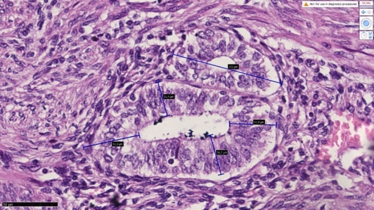 | Figure 1. Sample-5. The patient is 35 years old. Endometriosis. A morphogram with measured height and diameter of the endometrial glands in the myometrial space is presented. NanoZoomer (REFC13140-21.S/N000198/HAMAMATSU PHOTONICS /431-3196 JAPAN) has a scanner. Paint G.E. The size is 40x10 |
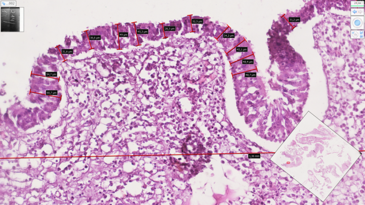 | Figure 2. Sample-8. The patient is 38 years old. Glandular fibrotic polyp of the endometrium. Longitudinal dimensions of polyp surface epithelia are given. NanoZoomer (REF C13140-21.S/N000198/HAMAMATSU PHOTONICS /431-3196 JAPAN) has a scanner. Paint G.E. The size is 40x10 |
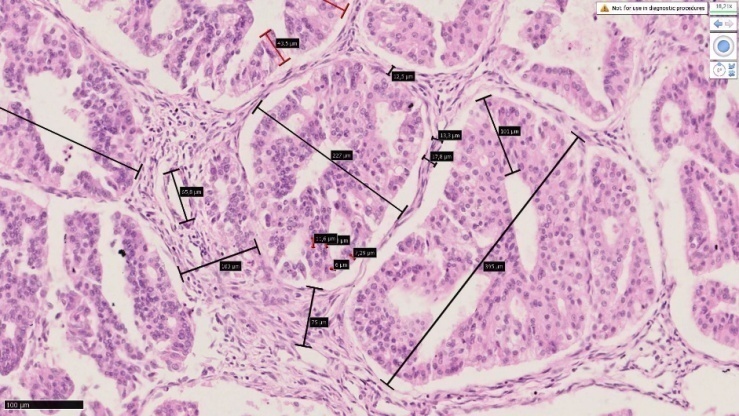 | Figure 3. Sample-10. The patient is 39 years old. Endometrial adenocarcinoma. A morphogram of the endometrial glands, measured in height and diameter, is presented. Polymorphism and metaplasia of epithelial cells were preserved in gland epithelia. NanoZoomer (REF C13140-21.S/N000198/ HAMAMATSU PHOTONICS /431-3196 JAPAN) has a scanner. Paint G.E. The size is 40x10 |
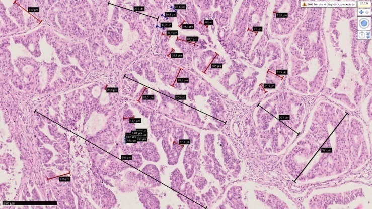 | Figure 4. Sample-13 The patient is 36 years old. Endometrial adenocarcinoma. A morphogram of the endometrial glands, measured in height and diameter, is presented. Polymorphism and metaplasia of epithelial cells were preserved in gland epithelia. NanoZoomer (REF C13140-21.S/N000198/ HAMAMATSU PHOTONICS /431-3196 JAPAN) has a scanner. Paint G.E. The size is 40x10 |
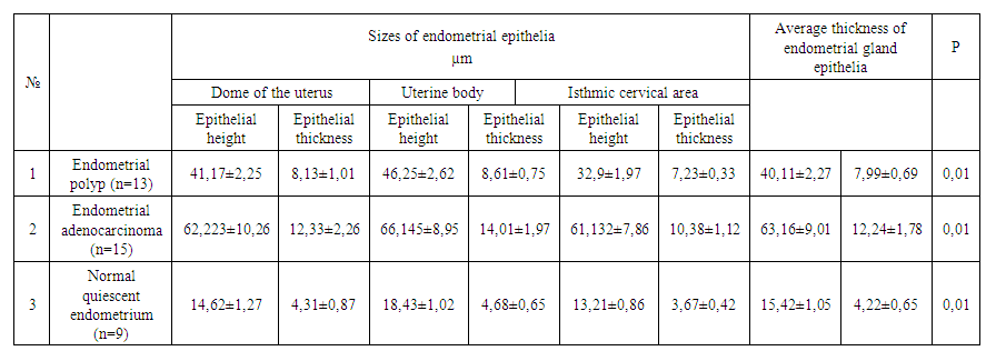 | Table 1. The height and thickness of the epithelial cells of the endometrial gland are measured in μm |
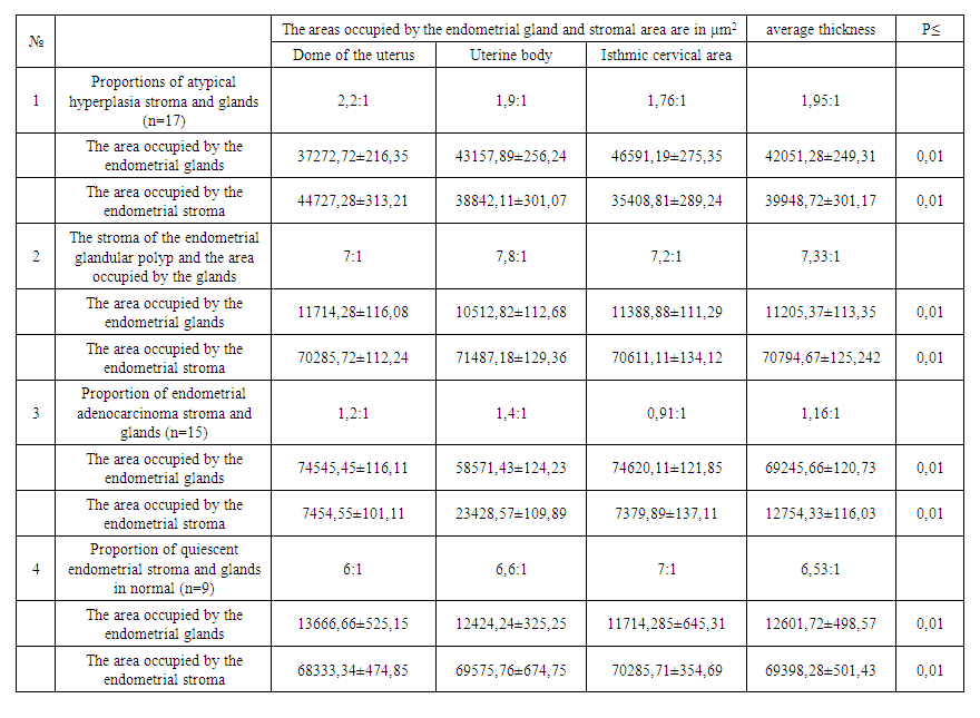 | Table 2. The ratio of endometrial gland and stroma areas per briga is μm2 (82000 μm2) |
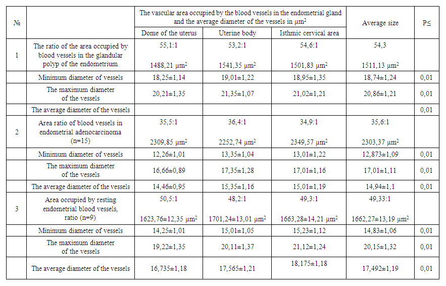 | Table 3. The average size of the area occupied by blood vessels and the diameter of blood vessels in various pathologies of the endometrium is μm2 (82000 µm2) |
4. Summary
- The above compared evidence and its results lead us to the following conclusions:1. In uterine endometrial polyp, the area occupied by blood vessels is 545 of the mucosa, and in about half of the processes related to abnormal bleeding, it is the basis for thinking about the polyp.2. It is characterized by permanent damage to the mucous membrane in various polyps of the endometrium, leading to metaplasia and dysplasia of glandular epithelia.3. In diseases of the female genital organs associated with abnormal bleeding, it is necessary to always consider endometrial polyps and take morphometric indicators into account when determining treatment tactics against it.4. A sharp thickening of the endometrium mainly leads to an increase in gland cells and, in parallel, an increase in vascular networks. This, in turn, confirms the occurrence of processes associated with constant bleeding.