Munisa Yusufova1, Saule Khojanazarova1, Norbek Yusufov2
1Department of Human Anatomy and Operative Surgical Topographic Anatomy, Tashkent Medical Academy, Tashkent, Uzbekistan
2Department of General Medical Sciences, Gulistan State University, Gulistan, Uzbekistan
Correspondence to: Munisa Yusufova, Department of Human Anatomy and Operative Surgical Topographic Anatomy, Tashkent Medical Academy, Tashkent, Uzbekistan.
| Email: |  |
Copyright © 2024 The Author(s). Published by Scientific & Academic Publishing.
This work is licensed under the Creative Commons Attribution International License (CC BY).
http://creativecommons.org/licenses/by/4.0/

Abstract
Morphological aspects of the postnatal ontogeny of the kidneys of offspring born from mother rats fed simultaneously with Fipronil and Jerusalem artichoke powder were studied under experimental conditions. In this group, the histoarchitecture of the kidneys of rats fed Jerusalem artichoke and fipronil at the same time showed almost no signs of developmental delay. Compared to the control group, it was found that almost 85% improved appearance with fully morphofunctionally formed kidneys at 7 days. This shows the effective effect of the substance containing inulin, which has the adsorbent properties of Jerusalem artichoke.
Keywords:
Jerusalem artichoke, Morphology, Mother rat, Pesticides, Developmental ontogeny of kidney, Histology
Cite this paper: Munisa Yusufova, Saule Khojanazarova, Norbek Yusufov, Morphology of Postnatal Ontogenesis of Kidney of Rats Born on a Background of Pesticides Corrected with Topinambur, American Journal of Medicine and Medical Sciences, Vol. 14 No. 7, 2024, pp. 1914-1918. doi: 10.5923/j.ajmms.20241407.39.
1. Relevance of the Topic
Pesticides are any means designed to protect plants. Of course, as science advances, pesticides are becoming less toxic to humans. However, modern means of this variety, unfortunately, cannot be considered completely safe. In addition, pesticides applied to fields 20 or more years ago still remain in the soil and water. Therefore, unfortunately, cases of pesticide poisoning are possible today. Fipronil in the body is mainly found in fat tissue and is broken down into smaller chemicals called metabolites. Fipronil and its metabolites are then removed from the body mainly with feces and urine. Exactly how fipronil passes through the organs of excretion and the mechanism of its effect on the kidneys has not been fully studied. It is precisely in this way that most of the diseases that occur during the postnatal ontogenesis of the kidneys develop secretly and continue with severe complications, the fact that the information about the postnatal ontogenesis of the kidneys during fetal development has not been studied in full dynamics, no specific criteria have been developed, moreover, this research work requires relevance and study.
2. Purpose
Purpose to study the morphological aspects of the formation and growth of the kidneys of the offspring born under the conditions of chronic exposure to Jerusalem artichoke powder and pesticides to the mother’s organism.
3. Material and Methods
As a research object, kidney tissues of 110 generations of rats developed in the background of pesticides were obtained in the experiment. A study of modern multiplex morphometric indicators was carried out in the obtained material.
4. Main Part
Morphological changes of kidneys were studied in different periods of postnatal ontogeny of the kidneys of offspring rats born from mother rats given fipronil and Jerusalem artichoke cake added to their food during pregnancy under experimental conditions. It is due to the pharmacological effect of Jerusalem artichoke used in our research work. In particular, Jerusalem artichoke contains biologically active substances, consisting of “polysaccharide inulin, fructose and some vitamins”, and it is characterized by the ability to bind toxic substances that enter our body with food, since inulin has adsorbent properties. As a result, it is manifested by a sharp reduction in the absorption of pesticides contained in food and a necessary result for the protection of the mother and child.By using the pesticide fipronil with Jerusalem artichoke powder during pregnancy, there was no lethality or fetal death in any of the experimental rats. Offspring born from mother rats that were reduced in the background of pesticides and simultaneously given Jerusalem artichoke cake were decapitated at 3, 7, 14, 21, 30, 60 days, and microsamples prepared from their kidneys were studied. According to the obtained results, the following morphological changes were identified:Kidneys of 3-day-old rats are macroscopically lobulated, the surface is smooth, and the boundaries of the cortex and medulla are clear in cross-section. Signs of acute fullness were not detected. When studied from a microscopic point of view, it was found that the fibrous capsule of the kidneys has the same normal thickness, and the vessels are moderately full. It was found that the proximal and distal tubules make up 4/3 of the visual field of the renal cortex.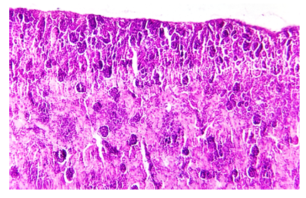 | Figure 1. Kidneys of 3-day-old rat pups born from chronically poisoned mothers given fipranil+topinambur. In the cortical layer, the balls are irregularly tubular and pear-shaped, and appear not yet fully formed. Paint G.E. The size is 40x10 |
The detection of embryonic cells in the balls against the general background is very small, and in the field of 200 field of view, the average number is 5/1 of the parietal cells of the balls. Under the bark, it is determined that the subcapsular balls are small in size, in the form of drops, Shumlyansky Bowman's capsule is composed of tall cellular epithelia with basophilic cytoplasm, and incompletely formed mesangial cells with a fine mesh are composed of cells with round nuclei and eosinophilic nuclei.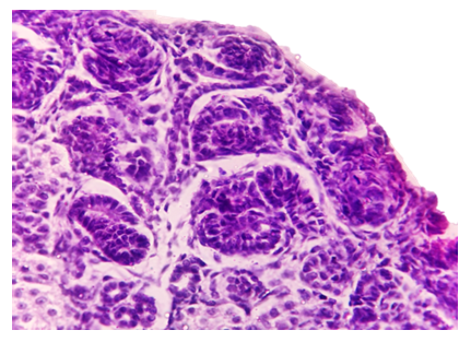 | Figure 2. Kidneys of 3-day-old rat pups born from chronically poisoned mothers given fipranil+topinambur. In the cortical layer, the balls are irregularly tubular and pear-shaped, and appear not yet fully formed. Paint G.E. The size is 40x10 |
Most balls are located in the subcapsular area of the cortex, and most of them are of the same size and location. At this point, we remind you that in the group fed with pesticides, the presence of relatively small droplet and bud-like balls, different levels of development in the postnatal ontogeny were determined.It is determined that in the veins of the wonderful mesh, the same branching and the same filling in the cavity of the ball. The diameter of the proximal tubule is the same, and most of the tubule spaces are narrowed compared to the normal structure, and it is determined that the proximal tubules are formed of tall prismatic epithelia at the beginning. It is determined that epithelial cells are in close contact with each other, their nuclei are large, their borders are clear, and their cytoplasm is darkly eosinophilic.Homogeneous protein structures are not detected in the tubule space. This indicates that the balls are not fully formed from the morphofunctional point of view and the filtration process is not taking place. The basal membrane of the proximal tubules is slightly thicker than usual, darker, its borders and relief are smooth, and the border trajectory is clearly defined.The elbows of the proximal tubules are also identified in the image above, homogeneous pink protein structures are not identified in their cavity. Most of the distal tubules are the same, they have a normal appearance in terms of diameter, the main features of the tubules are narrow, the interior is clean, and most of the epithelial cells are cuboidal, with dark eosinophilic cytoplasm. Nuclei are clear, dark basophilic staining, nuclear boundaries are clearly defined.Pericanalicular and paracanalicular small-caliber blood vessels and capillaries were moderately full, and no drastic changes were detected. At the borders of the medulla and the cortex, hyperglomerular balls were identified, small in size, excellent mesh structures were hypercellular in appearance, spherical in shape, the proximal and distal tubules were evenly distributed around them, and the diameter of most tubules was the same, and the narrowness of the tubules was revealed. The narrowness of these tube spaces is mainly due to the fact that most of the epithelium in the wall of the proximal and distal tubes consists of cylindrical and prismatic cells. The histioarchitectonics of the collecting ducts remained unchanged, and it was found that their main sensory morphological substrates: the presence of subcapsular balls in the form of drops and buds, the majority of the epithelium of the proximal and distal tubules were composed of cylindrical and prismatic epithelium, and the narrowness of the cavity of the tubules was found. Basically, the presence of 1-3 lymphocytes in the peritubular branches of the cortex and medulla, medium fullness of small-caliber blood vessels, a small number of histiocytes, fibroblasts in the distal intertubular spaces are detected in a very small amount in the 200x field of view.In the kidneys of 7-day-old generation rats, macroscopically, the lobular appearance is preserved, the surface is smooth, and the borders of the cortex and medulla are clear in the section. From the microscopic point of view, the fibrous capsule of the kidneys has a normal thickness, and average fullness in its vessels was found.It is determined that drop-shaped balls densely located in the subcapsular area of the kidneys have migrated towards the upper median area of the cortex. It was found that 4/3 of the renal cortex consists of proximal and distal tubules. It is determined that the balls have a semi-oval shape, and the increase in size is clearly developed compared to 3 days.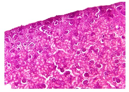 | Figure 3. Kidneys of 7-day-old rat pups born to chronically poisoned mothers given fipranil+topinambur. In the cortical layer, the shape of the balls is tubular and the balls migrated to the middle median layer of the cortex (1), the balls are still in the period of formation. Paint G.E. The size is 40x10 |
In Shumlyansky’s Bowman's capsule, tall cellular epithelia with basophilic cytoplasm are identified, fine mesh networks are relatively widespread, mesangial cells and podocytes are roughly the same size, and the nuclei of mesangial cells are relatively large and slightly elongated, and this appearance is attributed to the changes characteristic of differentiated cells. The changes of 3 days are preserved in the balls located in the subcapsular area, and it is determined that they are of the same size and the same location. It is determined that the subcapsular balls have a perfect network of vessels, the same branching and the same filling in the ball cavity.Ball parietal epithelia are composed of relatively cuboidal epithelia, the cytoplasm of which is stained with dark eosinophils. In the perimeter of the ball, it was found that most of the proximal and intermediate distal tubules are of the same diameter, and most of the tubule epithelia are prismatic and cuboidal in appearance. For the most part, the narrowed appearance of the canal spaces was preserved (recall that even in the rats of the control group, the spaces of all the proximal and distal tubules were narrow in the period of 7-14 days, mainly explained by the fact that in the period of 14-21 days, the tubule epithelia became cuboidal and semi-cuboidal). It is determined that the proximal tubules are formed from tall prismatic epithelia at the beginning. Epithelial cells are relatively cuboidal, tightly packed, the nuclei are kept large, the cytoplasm is dark eosinophilic.A small amount of homogeneous protein structures is detected in the tube cavity. This is considered a morphological marker indicating the beginning of the filtration process of the balls from the morphofunctional point of view. The basal membrane of the proximal tubules is darkly stained, its borders and relief are flat and clearly defined.In the bent branches of the proximal tubules, the same image as above is detected, and homogeneous pink protein structures are not detected in the cavity. Distal tubes have the same appearance in terms of diameter, in the main aspects, the tube cavities are widened, and in their cavity, mesh-like homogeneous structures are determined. Most of the epithelial cells are hemispherical in appearance, and the cytoplasm is dark eosinophilic in appearance. Pericanalicular small-caliber blood vessels and capillaries are moderately full. The boundaries of the core and the cortex are rounded, hypercellularity is sharply reduced in the fine mesh networks, and the spaces of the proximal and distal tubules are clearly visible in the perimeter.In the kidneys of 14-day-old generation rats, the surface is smooth from the macroscopic point of view, and the boundaries of the cortex and medulla are clear in cross-section. Microscopic examination revealed the normal thickness of the fibrous capsule of the kidneys, and medium fullness in the vessels.In the subcapsular area of the kidney tissue, many balls located along the middle median of the cortical layer are identified.It is determined that 4/2 of the cortex of the kidneys is made up of proximal and distal tubules, and 4/2 of it is made up of balls. Most of the balls have dilated Bowman's spaces, which indicates that the filtration process is activated from a morphofunctional point of view. The epithelium of the parietal sheets of the balls is flattened, the shape is round, the fine mesh networks are widely spread and branched, the mesangial cells and podocytes are enlarged in size, blood-shaped elements are detected in the large size fine mesh capillaries, homogeneous protein traces are detected in the Bowman’s cavity of the ball, which indicates that the filtration process is in progress.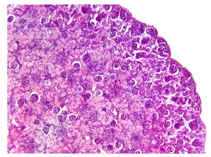 | Figure 4. Kidneys of 14-day-old rat pups born from dams given fipranil + Jerusalem artichoke. In the subcapsular area, it is determined that the formation of balls and the increase in size are well developed compared to those of the previous 3.7 days. The balls formed in the bark layer are increased, the hollows of the tubes are clearly described (1). Paint G.E. The size is 40x10 |
The epithelia of the proximal tubes, which continue after the balls, are cuboidal in shape, the cavity of the tubes is widened, and homogeneous mesh protein structures are detected in the cavity. The basal membranes of the proximal tubules are clear and smooth, along the border of the basal layer, the cytoplasm of the epithelial cells is light eosinophilic, the nuclei are small, dark basophilic, and the perimeter of the nucleus is clearly defined. The sequence of the epithelia of the proximal tubules is flat compared to the period of 3 and 7 days, and is characterized by the tangential arrangement of the nuclei, the decrease in the size of the cells, and the appearance of cuboids and semi-cuboids. This morphological picture indicates the expansion of the space of the proximal tubules and the beginning of the process of reabsorption after filtration.A small amount of homogeneous protein structures is detected in the tube cavity. This is considered a morphological marker indicating the beginning of the filtration process of the balls from the morphofunctional point of view. The basal membrane of the proximal tubules is darkly stained, its borders and relief are flat and clearly defined.In the bent branches of the proximal tubules, the same image as above is detected, and homogeneous pink protein structures are not detected in the cavity. Distal tubes have the same appearance in terms of diameter, in the main aspects, the tube cavities are widened, and in their cavity, mesh-like homogeneous structures are determined. Most of the epithelial cells are hemispherical in appearance, and the cytoplasm is dark eosinophilic in appearance. Pericanalicular small-caliber blood vessels and capillaries are moderately full. The boundaries of the cortex and the cortex are rounded, hypercellularity is sharply reduced in the fine mesh networks, and the spaces of the proximal and distal tubes are clearly described in the perimeter.In the 21-day period, the unformed follicles have shrunk, the projection of peritubular blood vessels is not detected. Hypercellular tubes are detected between the medullary tubes in a vertical view at 100x magnification. Microscopic overview of a 21-day-old rat kidney shows traces of incompletely formed distal and collecting tubules, with no clear lumen. In the area of the cup and cups, the mucous membrane surfaces are mostly covered with a single-layered flat epithelium. In the space between the medulla, weakly formed interstitial swellings are detected. Projection of blood vessels around the hypercellular foci of the distal tubules is poorly defined, and sparse fibrous structures are mainly detected in these areas. The trajectory of arcuate artery and vein branches located along the border of cortex and medullary layers is weakly defined and appears with little blood.
5. Conclusions
It can be concluded that the inulin contained in Jerusalem artichoke absorbs and neutralizes the fipronil pesticide, prevents the accumulation of this poison in the body, and ensures the normal course of the organogenesis process that occurs during the 8-12th week of fetal development. In the experiment presented in our research work, in the postnatal ontogeny of kidney tissue development of the kidney of the offspring born from the mother rat treated with fipronil and topinambur on days 7, 14, 21, compared to the 1st experiment, the cortex and medulla were fully formed on days 7-14, and the balls changed from the drop shape in the period up to 7 days. it was found that it changed to an oval shape. The shape and cellular composition of the morphofunctionalized balls were almost identical to those of the control group, indicating that the effects of fipronil pesticides were significantly reduced under the influence of Jerusalem artichoke. The presence of homogeneous protein structures in the proximal and distal tubules of the kidneys on the 14th and 21st days indicates that the morphofunctional process is taking place in the tubules and that the nephron structure is fully functioning from the 14th day. These indicators strongly reduced the consequences of the organogenesis process under the influence of the fipronil pesticide administered simultaneously with Jerusalem artichoke, and the fact that the development of the kidneys was not delayed, characterized by the absence of signs of premature development from the morphological point of view, confirmed that Jerusalem artichoke sharply reduces the effects of toxic substances by using the natural plant during pregnancy.
References
| [1] | Бобомуродов Н. Л. Возрастные особенности строения желудка крысы и его реактивные изменения при воздействии химикатов // International journal of health systems and medical sciences. – 2022. – Т. 1. – №. 5. – С. 31-44. |
| [2] | Сагатов Т. А. и др. Морфологическое состояние микроциркуляторного русла и тканевых структур матки при хронической интоксикации пестицидом" Вигор" // Проблемы науки. – 2019. – №. 2 (38). – С. 56-60. |
| [3] | Садиков А. У., Юсупова Д. Ю., Курбанова М. Б. Влияние гербицида далзлак на состояние углеводного обмена в крови и печени при лечении топинамбуром // Журнал теоретической и клинической медицины. – 2014. – №. 1. – С. 48-51. |
| [4] | Семеряк Е. В. Патоморфологические признаки токсичности и отдаленные эффекты действия ивермектина на организм животных // Автореферат дисс. канд. вет. наук. Омск. – 2009. – Т. 19. |
| [5] | Ситдыкова М. Э., Аллазов С. А., Саяпова Д. Р. Влияние хлорорганических пестицидов на некоторые урологические заболевания // Казанский медицинский журнал. – 2010. – Т. 91. – №. 3. |
| [6] | Смолякова С. П. и др. Влияние компонентов декоративной косметики, парфюмерии и средств бытовой химии на организм беременной // Успехи современного естествознания. – 2013. – №. 9. – С. 102-103. |
| [7] | Кочевенко А. С. Клинические и патоморфологические изменения в органах и тканях крыс под влиянием карбендазима // Науковий вісник Львівського національного університету ветеринарної медицини та біотехнологій імені СЗ Ижицького. – 2014. – Т. 16. – №. 2-2. – С. 175-179. |
| [8] | Соколова М. О. Морфофункциональные изменения фильтрационного барьера почек крыс при интоксикации параоксоном. Научный доклад // Санкт-Петербург, 2023. -С. |
| [9] | Соколова М. О., Соболев В. Е., Гончаров Н. В. Ультраструктурные изменения почек и биохимические показатели крови и мочи крыс при острой интоксикации о, о-диэтил-о-(4-нитрофенил) фосфатом // Журнал эволюционной биохимии и физиологии. – 2022. – Т. 58. – №. 6. – С. 540–548-540–548. |
| [10] | Кирилова А. С. и др. Обогащения хлебобулочных изделий микронутриентами топинамбура (Helianthus tuberosus L) // Проблемы и перспективы студенческий науки. – 2017. – №. 1. – С. 68-69. |
| [11] | Zafar R. Acute Renal Failure due to Organophosphate Poisoning: A Case Report / R. Zafar, K. Munawar, A. Nasrullah, S. Haq, H. Ghazanfar, A.B. Sheikh, A.Y. Khan // Cureus. – 2017. – Vol. 9. – No. 7. – e1523. |
| [12] | Zhou S. Paraoxon Attenuates Vascular Smooth Muscle Contraction through Inhibiting Ca2+ Influx in the Rabbit Thoracic Aorta / S. Zhou, L. Liu, X. Yang, S. Wu, G. Chen // Journal of Biomedicine and Biotechnology. – 2010. – Vol. 2010. – A: 829190. |

 Abstract
 Reference
 Full-Text PDF
 Full-text HTML