-
Paper Information
- Next Paper
- Paper Submission
-
Journal Information
- About This Journal
- Editorial Board
- Current Issue
- Archive
- Author Guidelines
- Contact Us
American Journal of Medicine and Medical Sciences
p-ISSN: 2165-901X e-ISSN: 2165-9036
2024; 14(7): 1869-1872
doi:10.5923/j.ajmms.20241407.31
Received: Jun. 21, 2024; Accepted: Jul. 15, 2024; Published: Jul. 22, 2024

Immunohistochemical Features of Malignant Tumors of the Thyroid Gland in Aral Region
Rajapov Adilbek Anvarbekovich
Researcher, Urganch Branch of Tashkent Medical Academy, Uzbekistan
Correspondence to: Rajapov Adilbek Anvarbekovich, Researcher, Urganch Branch of Tashkent Medical Academy, Uzbekistan.
Copyright © 2024 The Author(s). Published by Scientific & Academic Publishing.
This work is licensed under the Creative Commons Attribution International License (CC BY).
http://creativecommons.org/licenses/by/4.0/

The results obtained by modern immunohistochemical methods of thyroid gland malignant tumors are important for determining the level of tumor malignancy, target therapy for immunoactive bodies of the cells in its components, or indicators of active development of secondary changes in the tumor. By studying the results obtained in this research work, it will be the basis for pre-planning and development of treatment tactics. At the same time, it consists in studying the dynamic changes of the tumor and the level of expression of immunomarkers through immunohistochemical examination.
Keywords: Immunohistochemical examination, Thyroid tumor, Carcinoma, Follicular tumor, Expression, Proliferation
Cite this paper: Rajapov Adilbek Anvarbekovich, Immunohistochemical Features of Malignant Tumors of the Thyroid Gland in Aral Region, American Journal of Medicine and Medical Sciences, Vol. 14 No. 7, 2024, pp. 1869-1872. doi: 10.5923/j.ajmms.20241407.31.
Article Outline
1. Introduction
- The new edition of the WHO Classification of Endocrine Tumors is based on the decision of MAIR v Lyon, France, 26-28 April 2016, by an international expert working group of 166. The publication of a new edition of the classifications is based on the scientific achievements of recent years and is confirmed by the specificity of immunohistochemical indicators of carcinogenesis of thyroid tumors. Based on this classification, the following forms of malignant thyroid tumors are distinguished: papillary, follicular, medullary (oncocytic) and poorly differentiated anaplastic carcinomas.Purpose: to study specific immunoexpression characteristics in immunohistochemical examinations of malignant tumors of the thyroid gland.
2. Materials and Methods
- The Khorezm Province Bureau of Pathological Anatomy organized immunohistochemical and morphological examinations of 85 malignant thyroid tumor tissues obtained during 3 years.
3. Research Results and Their Discussion
- Ki-67 is a marker of cell proliferation and is expressed at different levels in all phases of cell activation, i.e. G1, S, G2, M. From the initial phase of cell activation, from G1 to M phase, this marker increases and reaches a maximum by the metaphase of mitosis. In the early G1 phase, the Ki-67 marker is located at the centromere of the satellite DNA and at the telomere of the chromosome. In the middle phases of cell activation, the Ki-67 marker appears in the nucleus, and by the G2 phase, it is expressed both in the nucleus and in the karyoplasm. When the cell enters post-mitotic G0, the Ki-67 marker is degraded by proteosomes, undergoes complete catabolism, and is not expressed in interphase cells [1].Therefore, this immunohistochemical marker is of great importance not only in tumor processes, but also in hyperplasia, metaplasia, regeneration, dysplasia, which continues with cell proliferation, as it shows the activity of cell proliferation.Cell proliferation index can be calculated based on the Ki-67 marker. A total of 500 cells were counted in the calculation, and it was determined how many of them had the positive expression of this marker in the nucleus, and what percentage of all cells were positive. 10% - low level,10-20% - medium level, More than 20% is considered high expression [2 p. 67].Taking into account the above discussion, the aim of this study was to determine the level of expression of Ki-67 and the proliferation index in papillary, follicular, medullary and undifferentiated carcinomas of the thyroid gland.The next task is to study the protein R53 as a transcription factor and a protein that controls the cell cycle by immunohistochemical method. R53 is a gene suppressor in tumorigenesis. This protein is mutated in 50% of malignant tumor cells. This gene has several names: r53 cellular tumor antigen, tumor suppressor r53, r53 phosphoprotein, NY-CO-13 antigen. Normally, the R53 tumor suppressor gene is expressed in all cells of the body. If the genetic apparatus of the cell is not damaged, p53 is inactive, and if DNA is damaged, p53 is activated. Thus, p53 is activated when DNA-damaging factors accumulate. Activation of R53 results in cell cycle arrest and apoptosis. R53 is known to be activated in several precancerous diseases, including chronic inflammatory, dysregenerative, and preneoplastic diseases of the bladder.The concentration of p53 protein increases especially in proliferating and multiplying cells, especially when hyperplasia and proliferation of cells with a dysregenerative process increases [3].The significance of the increased concentration of R53 is that it rapidly replicates with DNA and damages the gene machinery, and this state is the preparation of the cell for DNA damage. In intact, well-differentiated cells, the latent r53 protein is located in the cytoplasm of the cell. When the proliferative activity of the cell increases, this p53 protein moves to the nucleus, and in the absence of stress effects on the cells, this protein is degraded in 5-20 minutes. Our next task was to study thyroid transcription protein TTF-1, a protein product expressed in thyroid follicular epithelium and S-cells. This protein is a protein that induces the expression of a group of genes that ensure thyroid follicular epithelium and S-cell differentiation. Therefore, due to the development of malignant tumors from follicular cells and interstitial S-cells in malignant tumors of the thyroid gland, it is of great importance to study the group of genes that ensure their differentiation by immunohistochemical method [4].The results of the investigation showed that the expression of the cell proliferation factor in papillary carcinoma was found to have different indicators in the epithelial cells and interstitial cells of the tumor we studied. It is determined that it is expressed in light brown in most of the tumor epithelial cells, and in some cells in dark brown. It was found that expression of Ki-67 marker in tumor cells was moderate, proliferative index was 21.4%. Expression in both the karyoplasm and nucleolus of epithelial cells confirms that the cells are in the middle phase of activation, i.e., in the G2 phase.Immunohistochemical examination of the Ki-67 marker showed that hyperplastic, irregularly located papillary carcinoma cells were expressed in the nucleus of a significant number of cells in the form of dark brown color. In this case, some nuclei are enlarged and this marker is expressed both in the karyoplasm and the nucleus. It is determined that the nuclei of other cells are relatively small in size and appear in a relatively light brown color. The expression of the Ki-67 marker at different levels indicates that these cells are activated at different levels of mitotic and proliferative activity [5, p. 127].In order to determine the pathomorphological and immunohistochemical changes occurring in the papillary carcinoma epithelium, biopsy material obtained from people without any pathology in the thyroid gland was studied as a control group. Then, the pathomorphological and immunohistochemical changes in the epithelium of different histological variants of papillary carcinoma were compared. It is determined that the epithelium of the thyroid gland follicles of the control group consists of a single-layered prismatic epithelium, and its epithelial cells located in the basal layer are relatively large, hyperchromic, arranged in the basement membrane, and most of the nuclei are elongated and elongated. In the surface layers of the epithelium, it is observed that the cells are relatively sparse, their nuclei are smaller in size, their color is lighter, and their location is flattened.The results of the immunohistochemical examination of r53-protein, a gene suppressor that controls the cycle of cell division and reproduction of the epithelial cell, preventing it from turning into a tumor, showed that in the control group, in the control group, some cells of the cambial level, located only in the basement membrane, are relatively young and have the characteristic of dividing and multiplying in the cytoplasm, and in the nucleus of others, at a very low level. it is observed that this protein is expressed, but not expressed in the cells of other intermediate and surface layers.Microscopic examination showed that in the cells of the basal layer of the thyroid epithelium in the control group, the r53-gene suppressor was located in the cytoplasm of some cells, and in the nucleus of most cells (Figure 1). This situation indicates that some of the cambial cells located in the basal layer are in a proliferative active state and it is confirmed that the r53 gene suppressor is replicated with the DNA protein in their nucleus.A study of the thyroid transcription protein TTF-1, a protein product expressed in the thyroid follicular epithelium, was obtained. In this papillary carcinoma, it was observed that a protein that induces the expression of a group of genes that ensures the differentiation of thyroid follicular epithelial cells is expressed. Here, TTF-1 protein expressed at a low level in follicular epithelial cells located in papillary carcinoma nipples indicates a low level of differentiation of these cells.
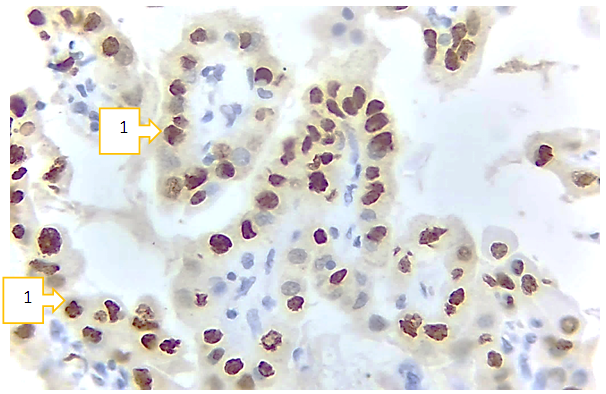 | Figure 1. Papillary carcinoma, positive expression of the r53 marker (1). Paint: Dab chromogenic. Floor: 10x40 |
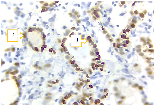 | Figure 2. Follicular carcinoma, r53 marker expression level (1). Paint: Dab chromogenic. Floor: 10x40 |
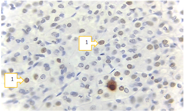 | Figure 3. Medullary carcinoma, Ki-67 marker expression level(1). Paint: Dab chromogenic. Floor: 10x40 |
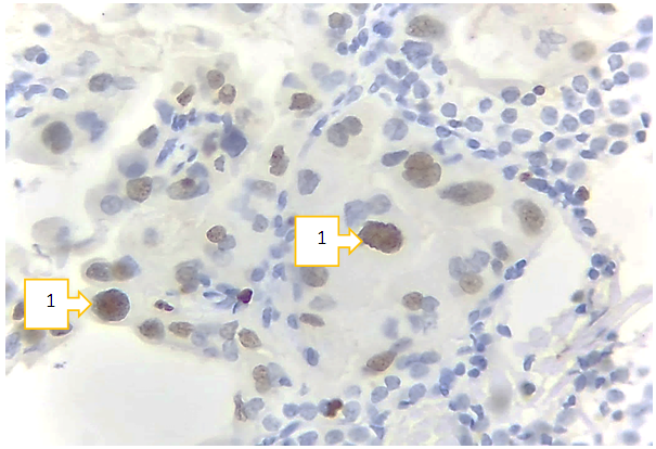 | Figure 4. Undifferentiated carcinoma, positive expression of Ki-67 marker (1). Paint: Dab chromogenic. Floor: 10x40 |
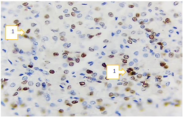 | Figure 5. Low-differentiated carcinoma, TTF-1 marker expression level. Paint: Dab chromogenic. Floor: 10x40 |
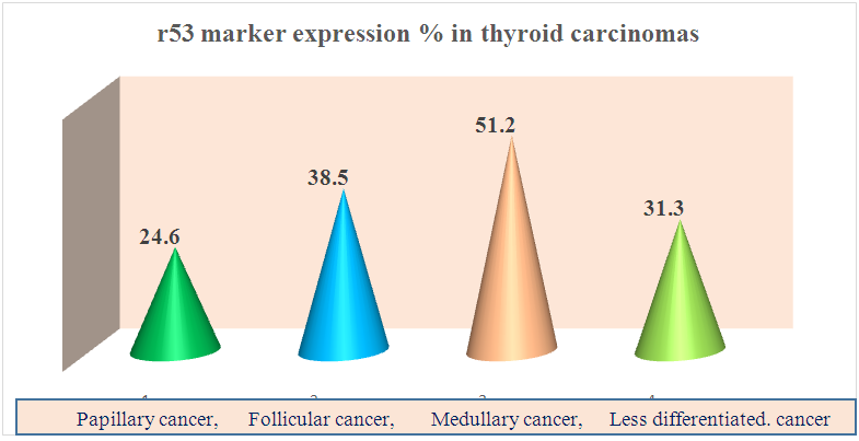 | Figure 6. The expression of a group of genes that ensures the differentiation of tumor cells |
4. Conclusions
- In papillary carcinoma of the thyroid gland, Ki-67, a marker of cell proliferation index, showed an average level, r53 marker gene suppressor was expressed at a high level, thyroid transcription protein TTF-1 was observed to be expressed at a low level in the follicular epithelium.In follicular carcinoma, the cell proliferation index was very high, the gene suppressor r53 marker was moderately expressed, and TTF-1 was proved to be low-expressed.All immunohistochemical markers, including Ki-67, p53, and TTF-1, were found to be underexpressed in medullary carcinoma.In poorly differentiated carcinoma of the thyroid gland, it was confirmed that the cell proliferation index was moderately expressed, the r53 gene suppressor was moderately expressed, and the thyroid transcription protein TTF-1 was moderately expressed.
 Abstract
 Reference
 Full-Text PDF
 Full-text HTML