American Journal of Medicine and Medical Sciences

p-ISSN: 2165-901X    e-ISSN: 2165-9036

2024;  14(7): 1862-1868

doi:10.5923/j.ajmms.20241407.30

Received: Jun. 25, 2024; Accepted: Jul. 19, 2024; Published: Jul. 20, 2024


Clinical, Hormonal and Imaging Features of Inactive Pituitary Adenomas with Recurrent and Stable Course

Kholova Dilorom Sharifovna, Halimova Zamira Yusufovna

Republican Specialized Scientific and Practical Medical Center of Endocrinology named after. Academician Y.Kh. Turakulov, Tashkent, Republic of Uzbekistan

Copyright © 2024 The Author(s). Published by Scientific & Academic Publishing.

This work is licensed under the Creative Commons Attribution International License (CC BY).
http://creativecommons.org/licenses/by/4.0/

Abstract

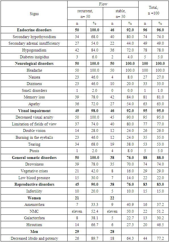
Inactive pituitary adenoma (IPA) or silent pituitary adenoma (Silient Pituitary Adenomas) refers to tumors that secrete one or more hormones or their transcription factors (TFs), detected by immunohistochemical examination (IHC), but this amount of expressed hormones has no clinical significance and is not expressed by symptom complexes [2]. Purpose of the study. To analyze in a comparative aspect the clinical, hormonal and imaging features of inactive pituitary adenomas with a recurrent and stable course. Material and methods. In total, the data of 100 patients with NAH were analyzed: group 1 included 50 patients with a recurrent course, group 2 included 50 patients with a stable course. Results. Both groups were predominantly men under 45 years of age. In the group with a recurrent course, nausea (46% vs. 4.0%; OR 9.80; 95%CI 3.06-31.4; p <0.0001) and dizziness (46.0% vs. 20.0%; OR 3.41; 95% CI 1.40-8.29; p = 0.006) were significantly more common than in the group with a stable course. Along with this, in the group with a recurrent course of the disease, endocrine (100% versus 92.0%) and general somatic (100% versus 76.0%) disorders predominated. A comparative analysis showed that significant factors in the epigenetic modification of the risk of relapse are stress (OR 17.5; 95%CI 6.31-48.4; p<0.0001), brain injury (OR 3.02; 95%CI 1 .27-7.21; p=0.01), obesity (OR 4.89; 95%CI 1.76-13.6; p=0.002) and emotional stress (OR 2.79; 95%CI 1, 11-7.01; p=0.03).

Keywords: Inactive pituitary adenomas, Recurrent course, Stable course

Cite this paper: Kholova Dilorom Sharifovna, Halimova Zamira Yusufovna, Clinical, Hormonal and Imaging Features of Inactive Pituitary Adenomas with Recurrent and Stable Course, American Journal of Medicine and Medical Sciences, Vol. 14 No. 7, 2024, pp. 1862-1868. doi: 10.5923/j.ajmms.20241407.30.

1. Introduction

Inactive “silent” pituitary adenomas account for about 10–20% of all intracranial tumors and 15–30% of all pituitary adenomas [7]. These tumors are observed in different age groups; the manifestation of the disease, as a rule, occurs in the 4th–8th decade of life [1]. According to literature reviews, there is a statistically slight male predominance in the detection of IPA, although not all studies observed this trend of sexual differentiation [4,6,26]. There are studies showing that men, as a rule, live longer, thereby the risk of developing tumors is greater than that of women [4].
Clinical and morphological diagnosis of IPA is difficult, which affects the progression, treatment and prognosis. In most cases, due to the small size and intrasellar location of IPA, incapable of invasive-infiltrative growth, the practical impossibility of their visualization using CT and MRI for tumors up to 1 mm in size, as well as the absence of reliable biochemical tumor markers in the blood, is difficult to reliably assess tactics of therapy, dynamics of the disease, the effectiveness of the treatment [11,18].
IPA are mainly characterized as benign tumors and rarely metastasize to nearby structures, however, a significant proportion of tumors demonstrate invasive growth and a recurrent course [23]. The risk of relapse is higher for tumors where there is residual tumor tissue or residual tissue after transsphenoidal adenomectomy (TAG), in cases where there is invasive adenoma growth in the structures of the cavernous sinus [5,11,24].
Unlike hormonally active adenomas, IPA does not have a clinical syndrome due to unregulated secretion of hormones, but it should also be noted that the cytoplasm of adenoma cells may contain pituitary hormones and, in this case, they are called silent adenomas - “Silent” adenomas " [2,3]. IPA, compared with functional pituitary adenomas, has unique clinical characteristics in various aspects. First, IPA is usually observed in older age groups compared to hormonally active functional adenomas. Secondly, patients mainly have signs and symptoms of compression or mass effect. Third, a large number of patients have hypopituitarism in one or more adenohypophyseal cell lines. On the other hand, many patients with IPA may not have clinical symptoms for many years and are detected by chance, which leads to difficulties in early diagnosis and compilation of epidemiological characteristics of this disease. The frequency of asymptomatic IPA varies according to the literature from both local and foreign sources. In connection with the above data, clinically non-functioning or inactive pituitary adenomas are adenomas that do not lead to excessive secretion of pituitary hormones leading to corresponding dishormonal clinical syndromes [8].

2. Purpose of the Study

To analyze in a comparative aspect the clinical, hormonal and imaging features of inactive pituitary adenomas with a recurrent and stable course.

3. Materials and Methods

In total, the data of 100 patients with NAH were analyzed: group 1 included 50 patients with a recurrent course, group 2 included 50 patients with a stable course.

4. Statistical Analysis

Statistical processing of the results was carried out using Microsoft programs Excel, IBM SPSS Statistica 23 and MedCalc version 18.5. The initial data were assessed for compliance with normal distribution using the Kolmogorov-Smirnov test. Results are presented as median (Me) [interquartile range Q 25; Q 75], as well as M± SD. Differences were considered statistically significant at p <0.05.

5. Results and Discussions

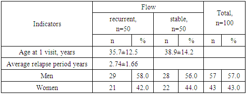
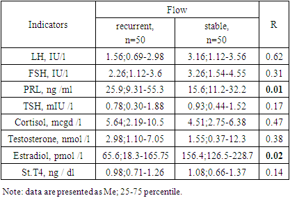
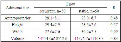
Data from 100 patients with IPA were analyzed: group 1 included 50 patients with a recurrent course, group 2 included 50 patients with a stable course. By age (group 1 – 35.7±12.5 years; group 2 – 38.9±14.2 years; p=0.24) and gender group (group 1 – men – 58.0% and women – 42.0%; 2 – men – 56.0% and women – 44.0%; OR 1.09; 95% 0.49-2.40) were comparable (Table 1)).
Table 1. Clinical characteristics of those examined
     
Both groups were predominantly men under the age of 45 (Fig. 1) and these data coincided with data from literature sources [1,4,6,26]. In the group with a recurrent course, the average period of relapse was 2.74±1.66 years. All patients with NAH, both recurrent and stable, had neurological disorders in the form of headaches (Table 2). In the group with a recurrent course, nausea (46% vs. 4.0%; OR 9.80; 95%CI 3.06-31.4; p <0.0001) and dizziness (46.0% vs. 20.0%; OR 3.41; 95% CI 1.40-8.29; p = 0.006) were significantly more common than in the group with a stable course. Along with this, patients with relapse showed apathy, not significantly but more often (72.0% vs. 54.0%; OR 2.19; 95%CI 0.95-5.03; p=0.06), then as in the case of a stable course of the disease, memory loss had an advantage (84.0% vs. 78.0%; OR 0.68; 95%CI 0.25-1.85; p=0.45).
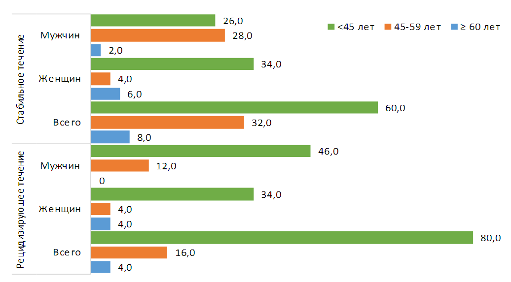
Figure 1. Gender and age composition of those surveyed
Table 2. Frequency of occurrence of various disorders in patients with NAH depending on the course of the disease
     
Along with this, in the group with a recurrent course of the disease, endocrine (100% versus 92.0%) and general somatic (100% versus 76.0%) disorders predominated. In patients with a stable course, it was not significant, but secondary hypothyroidism was more common (80.0% versus 68.0%; OR 0.53; 95%CI 0.21-1.32; p=0.17). While secondary adrenal insufficiency (54.0% vs. 44.0%; OR 1.49; 95%CI 0.68-3.29; p=0.32) and hypogonadism (84.0% vs. 72.0%; OR 2.04; 95%CI 0.77-5.42; p=0.15) were significantly higher in the recurrent course of the disease. When studying general somatic disorders, vegetative crises (42.0% versus 16.0%; OR 3). .80; 95%CI 1.48-9.75; p=0.005) were significantly more often recorded in patients with a recurrent course. Visual (98.0% vs. 92.0%; OR 4.26; 95%CI 0.46 -39.5; p=0.14) and reproductive (90.0% vs. 76.0%; OR 2.84; 95%CI 0.92-8.79; p=0.06) disorders were not significantly more common in the group with a relapsing course. Decreased visual acuity was a characteristic complaint of all (100%) patients with relapse and slightly less (90.0%) with stable disease. Burning sensation in the eyeballs (46.0% versus 24.0%; OR 2.70; 95% CI 1.15-6.34; p=0.02) and lacrimation (68.0% vs. 38.0%; OR 3.47; 95%CI 1.52-7.91; p=0.003) significantly more often were observed in patients with a relapsing course, compared with those with a stable course of the disease.
As for reproductive disorders, infertility was recorded 2 times more often in cases of recurrent disease (20.0% vs. 10.0%; OR 2.25; 95%CI 0.71-7.14; p=0.16) of the disease.
Of the 29 men with a recurrent course, 26 (89.7%) complained of decreased libido and/or potency; similar complaints in the group with a stable course were made by 18 (64.3%; OR 4.82; 95% CI 1.16- 20.0; p=0.02) out of 28 men.
Women with a recurrent course significantly more often complained of excess body hair growth (66.7% vs. 27.3%; OR 5.53; 95%CI 1.45-19.7; p=0.01) compared with those from the group with a stable course of the disease.
In general, menstrual irregularities and galactorrhea were more common in recurrent cases (OR 2.09; 95%CI 0.55-7.91; p=0.27), and amenorrhea - in stable cases (OR 0.7; 95% CI 0.21-2.50; p=0.61).
Next, we analyzed the factors of epigenetic modification of the risk of relapse of IPA in the studied groups (Table 3). Epigenetic mechanisms are factors that change the function of the genome under exogenous influence and at the same time provide a mechanism for the stable spread of a new cell state from one generation to the next. Works in recent years indicate the possible role of nutrition, stress, bad habits, previous brain injuries, genetic diseases in relatives and even night work as factors in the epigenetic modification of the risk of developing pituitary adenomas [6,9,14,16,17,18,19,22,23,27].
Table 3. Frequency of occurrence of various disorders in patients with NAH depending on the course of the disease
     
Among the studied factors in the group with a recurrent course, stress (74.0%), brain injuries (46.0%), obesity (40.0%) and emotional stress (38.0%) predominated. It should be noted that a significant proportion of men in both groups suffered from bad habits (smoking and drinking alcohol), without a significant difference between them.
The vast majority (96.0%) of patients with a relapsing course had one or more factors recorded, compared with those from the group with a stable course (78.0% - OR 6.77; 95% CI 1.42-32.4; p=0.008) (Fig. 2).
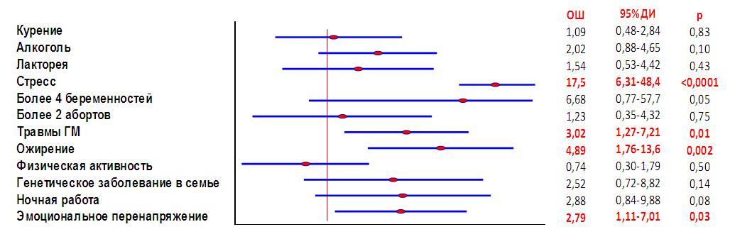
Figure 2. " Forest " factor diagram epigenetic modification of the risk of IPA relapse
A comparative analysis showed that significant factors in the epigenetic modification of the risk of relapse are stress (OR 17.5; 95%CI 6.31-48.4; p<0.0001), brain injury (OR 3.02; 95%CI 1 .27-7.21; p=0.01), obesity (OR 4.89; 95%CI 1.76-13.6; p=0.002) and emotional stress (OR 2.79; 95%CI 1, 11-7.01; p=0.03). Similar data were obtained in studies by foreign authors [21,29,30].
Hormonal studies are highly informative regarding the diagnosis of pituitary adenomas, both hormonally active and inactive, and are markers of the effectiveness of therapy in this category of patients. Based on the above, as well as the assigned tasks, we conducted a comparative analysis to study the level of blood hormones in the study groups (Table 4). Of all the studied hormones in the group with a relapsing course, the concentration of PRL was statistically significantly different in the direction of increase (61.1±108.2 ng /ml versus 21.3±16.9 ng /ml; p=0.01) and estradiol – decrease (107.0±110.7 p m o l / l versus 250.8±235.1 pmol / l p=0.02). Thus, based on the data obtained, we can conclude that in patients with IPA in both groups, the detected hyperprolactinemia is caused by a violation of the integrity of the hypothalamic-pituitary region, as noted in the literature by a violation of dopaminergic regulation in conditions of compression of the portal vessels and nerve fibers by a growing tumor [3], but it should be noted that this symptom is not a specific sign for prognosis of the course of postoperative residual tissue of the pituitary adenoma.
Table 4. Hormonal profile of patients with IPA
     
We studied the visualization parameters of pituitary adenomas in the compared groups. For the clinical presentation and diagnosis of IPA, the size of the tumor is important. To calculate the volume of a pituitary tumor, the formula Di was used Chiro - Nelson [10,15]:
V (volume mm3) = 0.5233 x H (vertical dimension (height)) x W (transverse dimension (width)) x L (sagittal dimension (length))
According to an MRI study of the brain, the majority of IPA had supra -infrapasellar (69.0%) and supra- infralaterosellar (23.0%) tumors.
Moreover, the supra-infralateral sellar formation with growth into the cavernous sinus prevailed in patients with a stable course (32.0% vs. 14.0%; OR 2.89; 95%CI 1.07-7.82; p=0.03). Tumors with supra-infra- pasellar (76.0% vs. 62.0%; OR 1.94; 95%CI 0.82-4.61; p=0.13) and supra-infra-para- antesellar (10.0% versus 6.0%; OR 1.74; 95%CI 0.39-7.71; p=0.46) by the direction of growth, insignificantly, but more often recorded in patients with a recurrent course (Fig. 3).
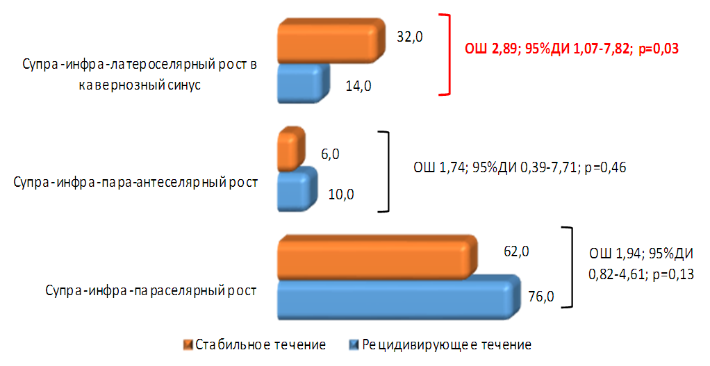
Figure 3. Direction of tumor growth in patients with NAH
The analysis of adenoma sizes did not reveal significant differences in the indicators of the studied groups (Table 5).
Table 5. Visualization characteristics of adenoma sizes in the studied groups
     
It was found that MRI images in the same number of patients had both isointense (46.0%) and hypointense (36.0%) signal on T1-weighted images.
In the group with a recurrent course, a hyperintense signal was recorded significantly more often (82.0% versus 4.0; χ 2 =62.1; p <0.0001) on T2-weighted images, while with a stable course 78.0% had an isointense signal, the frequency of cases with a hypointense signal was the same (18.0%) in both groups.

6. Conclusions

The diagnosis of IPA, as a rule, is established when the tumor reaches a significant size, causing visual impairment, endocrine, neurological, and autonomic disorders associated with compression of the structures of the anterior pituitary gland [3,24]. In this regard, IPA represents one of the difficult problems for neuroendocrinologists, since it is diagnosed late, usually at the stage of symptoms of extrasellar tumor spread [3]. Despite the fact that clinical, hormonal and imaging indicators of inactive pituitary adenomas with recurrent and stable courses had a diverse picture, they did not have comparative features. The study proves that large adenomas exhibit polysymptoms, vivid clinical manifestations of symptoms, and, moreover, have many complications leading to disruption not only of the endocrine system, but also of other organs and systems. The results of these data do not have prognostic significance in relation to indicators of early diagnosis of the course of the disease and in particular the criteria for recurrence and/or stabilization of tumor formation processes, assessing the prediction of treatment outcomes for this group of tumors. And this indicates the importance of using immunohistochemical studies to prevent a recurrent course. The next stage in the development of neuroendocrinology and pituitary surgery should be the introduction of IHC - studies that allow us to determine hormonal, mitotic activity, as well as determine genetic indicators in tumor tissues rather than in the blood, which will allow us to clearly determine the program of measures for patients in the postoperative period and predict their unfavorable outcomes in the form of tumor recurrence, apoplexy, aggressive growth, parasellar neurovascular formations, reducing the incidence of development and often ex juvantibus use of anti-relapse radiation therapy, which contributes to the development of post-radiation encephalopathy, optic nerve atrophy, increased frequency of stroke, panhypopituitarism, chaining these patients for lifelong disability.

References

[1]  Eremkina A.K., Dzeranova L.K., Pigarova E.K., Mokrysheva N.G., Dedov I.I. Morphofunctional features of hormonally inactive pituitary adenomas. Archives of Pathology, 1, 2019 https://doi.org/10.17116/patol20198101171.
[2]  Lipatenkova A.K., Dzeranova L.K., Pigarova E.A., Astafieva L.I., Grigoriev A.Yu., Shishkina L.V., Cherebilo V.Yu., Ektova A.P. “Silent” pituitary adenomas: review of the literature and description of a series of clinical cases. Obesity and metabolism. 2015; 12(2): 40-46.
[3]  Asa Sylvia, Ezzat Shereen. The Pathogenesis of Pituitary Tumors. 2009. 4: 97–126 The Annual Review of Pathology: Mechanisms of Disease is online at pathmechdis.annualreviews.org. This article’s doi: 10.1146/annurev.pathol.4.110807.092259.
[4]  Agustsson TT, Baldvinsdottir T, Jonasson JG, Olafsdottir E, Steinthorsdottir V, Sigurdsson G, et al. The epidemiology of pituitary adenomas in Iceland, 1955-2012: a nationwide population-based study. European journal of endocrinology / European Federation of Endocrine Societies. 2015; 173(5): 655-64.
[5]  Brochier S, Galland F, Kujas M, Parker F, Gaillard S, Raftopoulos C, Young J, Alexopoulou O, Maiter D, Chanson P. Factors predicting relapse of nonfunctioning pituitary macroadenomas after neurosurgery: a study of 142 patients. Eur J Endocrinol. 2010; 163(2): 193-200. https://doi.org/10.1530/EJE-10-0255.
[6]  Daly AF, Tichomirowa MA, Beckers A: The epidemiology andgenetics of pituitary adenomas. Best Pract Res ClinEndocrinolMetab 2009, 23:543-54.
[7]  Das B, Batool S, Khoja A, Islam N. Presentation, management, and outcomes of nonfunctioning pituitary adenomas: An experience from a developing country. Cureus. 2019; 11(9): e5759. DOI: 10.7759/cureus.5759.
[8]  Dashwood Rh, Ho E. Dietary histone deacetylase inhibitors: from cells to mice to man. Semin Cancer Biol. 2007; 17(5): 363–369.
[9]  Di Chiro G, Nelson KB. The volume of the sella turcica. Am J Roentgenol Radium Ther Nucl Med 1962; 87:989-1008.
[10]  Druesne N, Pagniez A, Mayeur C, et al. Diallyl disulfide (DADS) increases histone acetylation and p21(waf1/cip1) expression in human colon tumor cell lines. Carcinogenesis. 2004; 25(7): 1227–1236.
[11]  Greenman Y, Stern N. Non-functioning pituitary adenomas. Best Pract Res Clin Endocrinol Metab. 2009; 23(5): 625-638. https://doi.org/10.1016/j.beem.2009.05.005
[12]  Gonzalez S, Huerta Jm, Alvarez-Uria J, Fernandez S, Patterson Am, Lasheras C. Serum selenium is associated with plasma homocysteine concentrations in elderly humans. J Nutr. 2004; 134(7): 1736–1740.
[13]  Giovannucci E. Epidemiologic studies of folate and colorectal neoplasia: a review. J Nutr. 2002; 132(8 Suppl): 2350S–2355S.
[14]  James M. Provenzale. Approaches to Imaging of the Sella: Notes on “The Volume of the Sella Turcica” American Journal of Roentgenology 2006 186:4, 931-932.
[15]  Jacob Ra, Gretz Dm, Taylor Pc, et al. Moderate folate depletion increases plasma homocysteine and decreases lymphocyte DNA methylation in postmenopausal women. J Nutr. 1998; 128(7): 1204–1212.
[16]  Johnson It, Belshaw Nj. Environment, diet and CpG island methylation: epigenetic signals in gastrointestinal neoplasia. Food Chem Toxicol. 2008; 46(4): 1346–1359.
[17]  Kim Jm, Hong K, Lee Jh, Lee S, Chang N. Effect of folate deficiency on placental DNA methylation in hyperhomocysteinemic rats. J Nutr Biochem. 2009; 20(3): 172–176.
[18]  Mayson SE, Snyder PJ. Silent pituitary adenomas. Endocrinology and metabolism clinics of North America. 2015; 44(1): 79-87.
[19]  Nakahata Y, Grimaldi B, Sahar S, Hirayama J, Sassone-Corsi P. Signaling to the circadian clock: plasticity by chromatin remodeling. Curr Opin Cell Biol. 2007; 19 (2): 230–237.
[20]  Rampersaud Gc, Kauwell Gp, Hutson Ad, Cerda Jj, Bailey Lb. Genomic DNA methylation decreases in response to moderate folate depletion in elderly women. Am J Clin Nutr. 2000; 72(4): 998–1003.
[21]  Ross Sa, Dwyer J, Umar A, et al. Introduction: diet, epigenetic events and cancer prevention. Nutr Rev. 2008; 66(Suppl 1): S1–6.
[22]  Sadik Z.H.A., Voormolen E.H.J., P.R.A.M. Depauw, B. Burhani, W.A. Nieuwlaat, J. Verheul, S. Leenstra, R. Fleischeuer, P.E.J. Hanssens. Treatment of Nonfunctional Pituitary Adenoma Postoperative Remnants: Adjuvant or Delayed Gamma Knife Radiosurgery? World Neurosurg. (2017) 100: 361-368. http://dx.doi.org/10.1016/j.wneu.2017.01.028.
[23]  Saeger W, Lüdecke DK, Buchfelder M, Fahlbusch R, Quabbe HJ, Petersenn S. Pathohistological classification of pituitary tumors: 10 years of experience with the German Pituitary Tumor Registry. Eur J Endocrinol. 2007 Feb; 156(2): 203-16. doi: 10.1530/eje.1.02326. PMID: 17287410].
[24]  Sanmillan JL, Torres-Diaz A, Sanchez-Fernandez JJ, Lau R, Ciller C, Puyalto P, Gabarros A. Radiological predictors for extent of resection in pituitary adenoma surgery. A single-center study. World Neurosurg. 2017; 108: 436-446. https://doi.org/10.1016/j.wneu.2017.09.017.
[25]  Shin W, Yan J, Abratte Cm, Vermeylen F, Caudill Ma. Choline intake exceeding current dietary recommendations preserves markers of cellular methylation in a genetic subgroup of folate-compromised men. J Nutr. 2010; 140(5): 975–980.
[26]  Tjornstrand A, Gunnarsson K, Evert M, Holmberg E, Ragnarsson O, Rosen T, et al. The incidence rate of pituitary adenomas in western Sweden for the period 2001-2011. European journal of endocrinology / European Federation of Endocrine Scieties. 2014; 171(4): 519-26.
[27]  Weaver Ic. Epigenetic programming by maternal behavior and pharmacological intervention. Nature versus nurture: let’s call the whole thing off. Epigenetics. 2007; 2(1): 22–28.
[28]  Weaver Ic, Cervoni N, Champagne Fa, et al. Epigenetic programming by maternal behavior. Nat Neurosci. 2004; 7(8): 847–854.
[29]  Zingg Jm, Jones Pa. Genetic and epigenetic aspects of DNA methylation on genome expression, evolution, mutation and carcinogenesis. Carcinogenesis. 1997; 18(5): 869–882.