-
Paper Information
- Previous Paper
- Paper Submission
-
Journal Information
- About This Journal
- Editorial Board
- Current Issue
- Archive
- Author Guidelines
- Contact Us
American Journal of Medicine and Medical Sciences
p-ISSN: 2165-901X e-ISSN: 2165-9036
2024; 14(7): 1738-1743
doi:10.5923/j.ajmms.20241407.04
Received: Jun. 16, 2024; Accepted: Jul. 2, 2024; Published: Jul. 4, 2024

To the Problem of Diagnosis and Treatment of Acute "Catarrhal" Appendicitis
 Abstract
 Reference
 Full-Text PDF
 Full-text HTML
Sh. A. Tadjibayev1, E. K. Sobirov2, F. Sh. Abdurashidov1
1Andijan State Medical Institute, Andijan, Republic of Uzbekistan
2Medical Hospital ALMOZN, (Hospital Director Dr Ali Alejenef), g. Zintan, Arabic Republic Libya
Correspondence to: Sh. A. Tadjibayev, Andijan State Medical Institute, Andijan, Republic of Uzbekistan.
| Email: |  |
Copyright © 2024 The Author(s). Published by Scientific & Academic Publishing.
This work is licensed under the Creative Commons Attribution International License (CC BY).
http://creativecommons.org/licenses/by/4.0/

To the article by Doctor of Medical Sciences, Associate Professor Tadzhibaev Sh.A., Sobirov E.K., Abdurashidov F.Sh., on the topic: “On the problem of diagnosis and treatment of acute “catarrhal” appendicitis”. Relevance: Despite progress in urgent abdominal surgery, based on the introduction of new advances in medical technology and, as a consequence, the development of non-invasive and minimally invasive diagnostic and treatment technologies, the problem of diagnosis and treatment of acute appendicitis, in particular the “catarrhal” form, remains relevant and is actively discussed in periodical literature. Aim - Based on a clinical analysis of our own material and the results of non-invasive and minimally invasive methods for the diagnosis and treatment of acute appendicitis and its complications, to identify the effectiveness of these methods in the diagnosis of pathomorphological forms of acute appendicitis, especially “catarrhal” appendicitis. Materials and methods: The study is based on an analysis of observations of 128 patients with acute appendicitis who underwent appendectomy using endovideolaparoscopic technique. The age of the patients ranged from 16 to 64 years. Men – 71 (55.5%), women – 57 (44.5%). Additionally, 24 patients with separate abdominal pathology were studied for surgical correction, which is possible using endovideolaparoscopy, to assess the non-inflamed appendix (according to ultrasound and endovideolaparoscopy). Results: Of 128 patients, 22 (17.2%) were diagnosed with a catarrhal form, 89 (69.5%) patients with a phlegmonous form, and 17 (13.3%) with a gangrenous form. Analysis of the clinical material showed that in the group of patients diagnosed with acute “catarrhal” appendicitis, the variability of clinical symptoms made clinical diagnosis difficult. The results of the use of non-invasive and minimally invasive diagnostic methods showed that the sensitivity of ultrasound examination in acute appendicitis was 87.6%; there are certain difficulties in diagnosing the “catarrhal” form of acute appendicitis. Accurate diagnosis of this method is directly proportional to destructive changes in the appendix. In 123 patients, at the stage of diagnostic laparoscopy, the diagnosis was not in doubt, that is, the diagnostic accuracy of endovideolaparoscopy for acute appendicitis was 96%. Conclusions: Endovideolaparoscopy is effective and definitive in diagnosing the “catarrhal” form of acute appendicitis. The use of endovideolaparoscopy in the complex diagnosis of acute appendicitis will allow, when confirming the diagnosis, to transfer diagnostic endovideolaparoscopy to the treatment category, which allows optimizing the diagnosis of acute appendicitis and shortening the preoperative diagnostic period. It is fundamentally important that it is possible to simultaneously solve the problem, both diagnosis and treatment, in particular acute “catarrhal” appendicitis.
Keywords: Acute appendicitis, Diagnostic video laparoscopy, Laparoscopic appendectomy, Acute "catarrhal" appendicitis
Cite this paper: Sh. A. Tadjibayev, E. K. Sobirov, F. Sh. Abdurashidov, To the Problem of Diagnosis and Treatment of Acute "Catarrhal" Appendicitis, American Journal of Medicine and Medical Sciences, Vol. 14 No. 7, 2024, pp. 1738-1743. doi: 10.5923/j.ajmms.20241407.04.
- The study was carried out within the framework of scientific theme No. 012000267 “Development of advanced technologies in the diagnosis, treatment and prevention of acute surgical diseases of the abdominal organs”, Andijan State Medical Institute. The study has no sponsorship.
1. Relevance
- Despite the progress in urgent abdominal surgery based on the introduction of new achievements in medical technology and, as a result, the development of non-invasive and minimally invasive diagnostic and treatment technologies, the problem of diagnosis and treatment of acute appendicitis, in particular the "catarrhal" form, remains relevant and is actively discussed in the periodical literature.This fact is related to the frequency of occurrence, which is 22.8 per 10,000 population [1], and of all urgent patients operated on, appendectomies account for about 40%, and it should be noted that 4-42% of patients have complicated appendicitis [2]. Every year in the world, from 50 to 70 thousand people die from acute appendicitis and its complications. a person [3]. In addition, according to the data of the Chief Surgeon of the Ministry of Health of Russia A. Sh. According to Revishvili, 148,763 patients diagnosed with Acute appendicitis were treated in the Russian Federation in 2022, and surgical activity in 2020 was 98.1%, mortality-0.17% [4]. All these data together emphasize the urgency of the problem.In the diagnosis of acute appendicitis, in most cases, preference is given to non-invasive diagnostic methods such as ultrasound, which has established itself as an effective method in the diagnosis of this pathology. Number errors at stages diagnosis of acute appendicitis without use cases modern non-invasive and minimally invasive methods diagnostics services, it reaches up to 31%, and the frequency of unjustified appendectomies reaches 35-40%, while a high percentage (32.3 -50%) of postoperative complications remains. In the early stages of inflammation of the appendix, there is a low reliability of ultrasound examination up to 50-63%, with destructive forms it reaches 92-96% [5].As for computed tomography, the accuracy of this study in the diagnosis of acute appendicitis reaches 94-100%, and the proportion of "futile" appendectomies is 3-8% [6].It is generally accepted that if there are differential diagnostic difficulties in acute appendicitis, diagnostic laparoscopy should be performed. Laparoscopy has a high accuracy, up to 92.0–95.8%, is often the final diagnostic method, it reduces the number of diagnostic errors and eliminates the catarrhal form of acute appendicitis [7].Since 1983, laparoscopic appendectomy has gained wide popularity, and up to 75% of operations are performed laparoscopically in the world. Moreover, some authors advocate granting laparoscopic appendectomy the status of the "gold standard" for surgical treatment of acute appendicitis [8]. At the same time, although in Russia the role of laparoscopic technologies in the surgical treatment of acute appendicitis tends to increase, the frequency of their use remains low and amounts to 27% [9]. An all-Russian survey of surgeons showed that the widespread use of laparoscopic appendectomy is also hindered by low motivation for the introduction of laparoscopic technologies [10].Problematic issues related to acute appendicitis are unified in the National Clinical Guidelines (NCR) and international recommendations of various surgical societies – the World Community for Emergency Surgery (WSES), European Association of Endoscopic Surgeons (EAES) [11]. Issues related to the definition of tactical approaches to complex clinical situations that arise in acute appendicitis were debatable during the NCR discussions [12]. In this regard, an interesting opinion Timerbulatova Sh. V., in with respect to" simple "or" catarrhal " appendicitis, who believes that pFor the most part, the problem of "wasted" appendectomies is related to the attitude of surgeons and pathologists to the so-called "simple" or "catarrhal" acute appendicitis, especially with minimal, dubious findings during laparoscopy and surgery. So what are the pathohistological signs (criteria) of simple appendicitis that could confirm the validity of the performed appendectomy? What are the surgeon's actions in this situation? National clinical guidelines do not provide answers to these questions [13].At the same time, first of all, acute appendicitis is a clinical diagnosis, and the clinical manifestations of this pathology, together with the results of diagnostic methods, determine the vector of the direction of patient management and treatment.Research objective: Based on a clinical analysis of our own material to identify the effectiveness of these methods in the diagnosis of pathomorphological forms of acute appendicitis, especially "catarrhal" appendicitis, and the results of non-invasive and minimally invasive methods for the diagnosis and treatment of acute appendicitis and its complications.Task: Based on the obtained data from the analysis of clinical material, specify the indications for appendectomy in acute appendicitis of the "catarrhal" form using endovideolaparadjustment.
2. Materials and Methods
- Based on the Department of Surgery of a Medical hospital ALMOZSo, from 2019 to 2024, 182 patients underwent surgery for acute appendicitis using endovideolaparoscopic techniques. This work is based on the analysis of observations of 128 patients with acute appendicitis and its complications who underwent appendectomy using endovideolaparoscopic techniques, 54 (29.7%) patients out of 182, the technique of performing operations had its own characteristics and they are the object of our further research. Additionally, 24 patients with a separate abdominal surgical pathology were examined: surgical correction, which is possible using endovideolaparotomy, to assess the non-inflamed appendix (according to ultrasound and endovideolaparoscopy) as a comparison group with the group of patients diagnosed with acute "catarrhal" appendicitis.The age of the patients ranged from 16 to 64 years. Men – 71 (55.5%), women – 57 (44.5%). All patients underwent a comprehensive examination, while the basic ones were anamnesis collection, clinical and laboratory examination, ultrasound examination of the abdominal cavity, and diagnostic laparoscopy (DL). Ultrasound examination of the abdominal cavity was performed using a scanner SonoScape – P20, manufactured in Germany. Multispiral computed tomography (MSCT) is indicated. This study was performed on a CT scanner manufactured in the USA, General Electric, model 2022. Diagnostic laparoscopy and surgical interventions were performed using the company's endovideolaparoscopic complex COMEG, made in Japan, and a set of Ka toolsrl Storz manufactured in Germany. Histological studies were performed at the Attasami Diagnostic Services Center in Tripoli, Libya. Statistical processing of the material included the calculation of extensive indicators. The diagnosis of acute appendicitis was made on the basis of a set of data obtained.According to Figure 1, out of 128 patients with acute appendicitis, 22 (17.2%) were diagnosed with catarrhal form, 89 patients (69.5%) were diagnosed with phlegmonous form, and 17 (13.3%) with gangrenous form. Patients were operated on within 3-4 hours of admission after a short-term intensive training, taking into account their general condition. Leukocytosis in the operated patients varied from 9.7 to 19.4 thousand / µl. A relatively high percentage of destructive forms of acute appendicitis, namely 106 or 82.8%, is associated with late hospitalization, remote residence of patients and local social conditions.
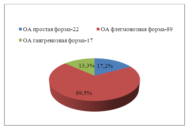 | Figure 1. Distribution of patients by pathomorphological diagnosis |
3. Results and Discussion
- A thorough analysis of the available clinical material revealed a certain sequence of treatment from the moment of admission to discharge of patients. The first stage was a general clinical examination. We drew parallels between the clinical manifestations of acute appendicitis and surgical findings, which allowed us to generalize the clinic depending on the postoperative pathomorphological diagnosis. Thus:1. Simple (catarrhal) appendicitis 22 (17,2%) patients. Statute of limitations diseases up to 6-7 hours. The attack began with sudden pain in the right iliac region or epigastric region, followed by their movement to the right iliac region. Other symptoms of acute appendicitis were intermittent, making clinical diagnosis difficult. Nausea and single vomiting were observed in 19 patients. 11 patients had normal temperature, while the rest had subfebrile temperature. Pulse corresponds to temperature. The stomach is soft, participates in breathing. There is moderate leukocytosis in the blood without shifting the formula to the left or with a slight shift. The variability of clinical symptoms in this group of patients made clinical diagnosis difficult, and what is quite important, reduces the surgeon's alertness to acute appendicitis.2. Acute phlegmonous appendicitis - 89 (69.5%) patients. Statute of limitations diseases up to 1.5-2 days. The main complaint is persistent, gradually increasing pain in the right iliac region. Often a single and much less frequent - repeated vomiting. The tongue is dry and overlaid. The abdomen is of the usual shape, poorly participates in the act of breathing, palpation of moderate muscle tension in the right iliac region and sharp soreness. Schetkin-Blumberg and Rovsing symptoms are positive. In the blood – leukocytosis with a shift of the formula to the left. The classic version of the clinic does not create difficulties in the diagnosis of acute appendicitis.3. Acute gangrenous appendicitis -17 (13.3%) patients. Statute of limitations diseases of 2 days or more. The clinical picture was different. In most cases, the same symptoms were observed as in phlegmonous appendicitis. At the same time, low-intensity abdominal pain and abdominal muscle tension are not very pronounced. Intoxication symptoms prevailed in all patients. Clinic of perforated appendicitis (5 cases), developed as follows: The onset of the disease differs little from the onset of phlegmonous or even simple appendicitis. Only after some time, from 2 to 3 days, a picture of severe general intoxication due to the development of peritonitis developed.The next stage of the examination was the use of ultrasound examination of the abdominal cavity. It should be noted that in the comparison group consisting of 24 patients, 19 or 79.2% managed to visualize the appendix. The general characteristics were as follows: the cross-sectional diameter averaged 5.5 mm, wall layering is visualized, mobility when pressed by the sensor is preserved, rigidity and soreness are absent, the nature of the contents cannot be determined. Classic ultrasound shows no signs of acute inflammation. There are no changes in vascularization according to color Doppler and energy mapping of blood flow.
 | Figure 2. Ultrasound image of the appendix without signs of inflammation |
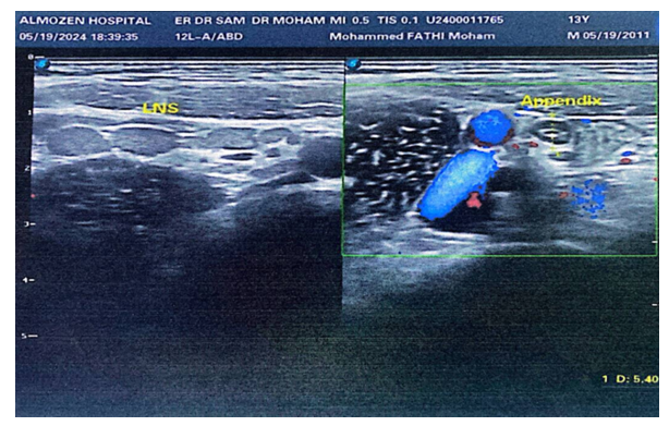
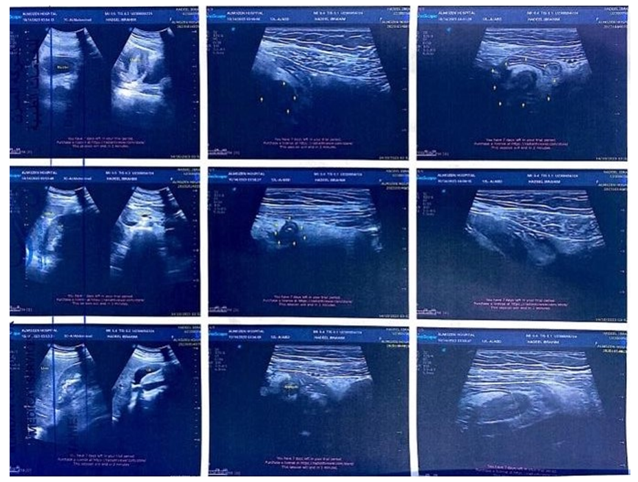 | Figure 3. Ultrasound image of acute appendicitis |
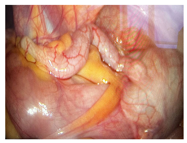 | Figure 4. Unchanged vermiform process |
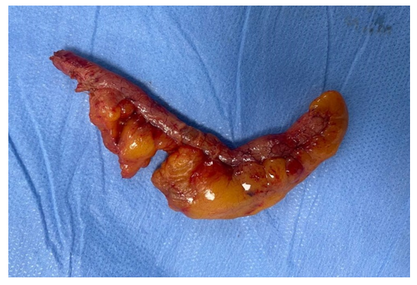 | Figure 5. Acute appendicitis, catarrhal form |
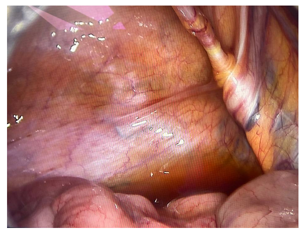 | Figure 6. Turbid abdominal effusion |
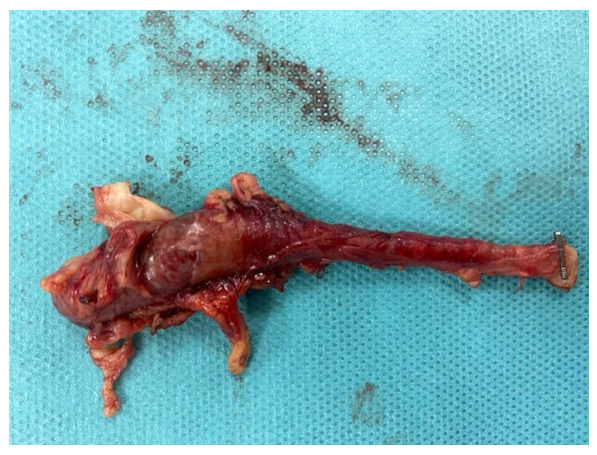 | Figure 7. Acute appendicitis, phlegmonous form. Empyema of the appendix |
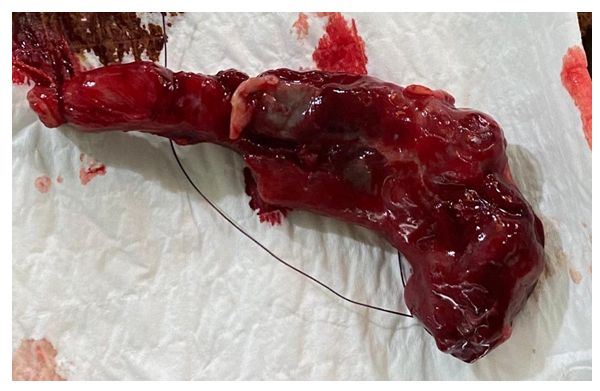 | Figure 8. Acute appendicitis, gangrenous form |
4. Conclusions
- Clinical analysis of the material showed that the variability of clinical symptoms in the group of patients with acute "catarrhal" appendicitis made clinical diagnosis difficult, and what is quite important, reduces the surgeon's alertness to acute appendicitis.The results of non-invasive diagnostic methods showed that the sensitivity of ultrasound examination in acute appendicitis was 87.6%, but there are certain difficulties in diagnosing the "catarrhal" form of acute appendicitis. Accurate diagnosis of this method is directly proportional to the destructive changes in the appendix.In 123 patients out of 128 at the stage of diagnostic laparoscopy, the diagnosis of acute appendicitis was not in doubt, that is, the diagnostic accuracy of endovideolaparoscopy in acute appendicitis was 96%. This diagnostic method is effective and definitive in the diagnosis of the "catarrhal" form of acute appendicitis.The use of endovideolaparoscopy in the complex diagnosis of acute appendicitis will allow, when confirming the diagnosis, to transfer diagnostic endovideolaparoscopy to the therapeutic category, which allows optimizing the diagnosis of acute appendicitis and its pathomorphological forms, and reducing the preoperative diagnostic period. It is fundamentally important that there is a possibility of simultaneous solution of the problem, both diagnosis and treatment.Clinical manifestations of acute appendicitis in conjunction with the results of diagnostic methods and determine the vector direction of management and treatment of the patient.