-
Paper Information
- Next Paper
- Previous Paper
- Paper Submission
-
Journal Information
- About This Journal
- Editorial Board
- Current Issue
- Archive
- Author Guidelines
- Contact Us
American Journal of Medicine and Medical Sciences
p-ISSN: 2165-901X e-ISSN: 2165-9036
2024; 14(6): 1624-1626
doi:10.5923/j.ajmms.20241406.31
Received: May 14, 2024; Accepted: Jun. 6, 2024; Published: Jun. 19, 2024

Specific Characteristics of Pathomorphological Changes in the Myocardial Floor of Dichorial Twins with Premature Birth and Death
Jumanov Ziyadulla Eshmamatovich1, Urunova Mashhura Allamurodovna2, Mamadiyarova Dilfuza Umirzakovna3
1Associate Professor of the Department of Pathological Anatomy of Samarkand State Medical University, Doctor of Medical Sciences (DSc), Uzbekistan
2Independent Researcher and Assistant of the Department of Pathological Anatomy of Samarkand State Medical University, Uzbekistan
3Associate Professor, Department of Pedagogy and Psychology, Samarkand State Medical University, Samarkand, Uzbekistan
Correspondence to: Jumanov Ziyadulla Eshmamatovich, Associate Professor of the Department of Pathological Anatomy of Samarkand State Medical University, Doctor of Medical Sciences (DSc), Uzbekistan.
| Email: |  |
Copyright © 2024 The Author(s). Published by Scientific & Academic Publishing.
This work is licensed under the Creative Commons Attribution International License (CC BY).
http://creativecommons.org/licenses/by/4.0/

Explanation. The purpose of the study is to determine the morphological characteristics of the myocardial structures of twins born prematurely. To achieve this goal, a pathomorphological study of the hearts of 24 deceased twins (12 pairs) was conducted. In the study, tissue fragments of 1x1x0.5 cm were taken from the wall of the left ventricle of the heart of the babies who were born prematurely and lived up to 3 days. The obtained tissue fragments were fixed in 10% neutral formalin, passed through an alcohol battery, and paraffin blocks were prepared. The prepared histological sections were stained with hematoxylin and eosin, according to Van-Gieson. Differences in the myocardial structures of twins with a relatively small heart size and with a large heart size are explained morphologically.
Keywords: Dichorial, Twins, Myocardium, Vascular, Morphology
Cite this paper: Jumanov Ziyadulla Eshmamatovich, Urunova Mashhura Allamurodovna, Mamadiyarova Dilfuza Umirzakovna, Specific Characteristics of Pathomorphological Changes in the Myocardial Floor of Dichorial Twins with Premature Birth and Death, American Journal of Medicine and Medical Sciences, Vol. 14 No. 6, 2024, pp. 1624-1626. doi: 10.5923/j.ajmms.20241406.31.
Article Outline
1. Introduction
- One of the problems facing today's medicine all over the world is multiple pregnancy. The relevance of this problem is due to the high number of pregnancy complications, specificity, severity of surgical delivery, postpartum complications, high perinatal mortality and morbidity in surviving babies [1]. Even with the current level of development of perinatal medicine, neonatal mortality is 5 times higher than in multiple singleton pregnancies [3]. The rate of stillbirth is 2-10 times higher than in patients with single pregnancies. Perinatal morbidity and mortality are primarily associated with preterm birth [4]. In multiple pregnancies, half of births (56%) occur before 37 weeks of gestation, compared to 6% in singletons [3]. The problem of reducing the death of twins is an urgent problem facing today's medicine.
2. The Purpose of the Study
- Study of morphological characteristics of changes in myocardial structures in the death of preterm dichorionic twins.
3. Materials and Methods
- In order to study the morphological characteristics of the myocardial structures of premature twins, a microscopic examination of the hearts of 24 twins who died in 2022-2023 was carried out at the Samarkand Branch of the Scientific Practical Center for Motherhood and Childhood of the Republic of Uzbekistan. For this, clinical data on the birth history of babies and autopsy materials were obtained. Of these, 8 cases are male twins, 4 cases are female twins. In the study, tissue fragments of 1x1x0.5 cm were taken from the wall of the left ventricle of the heart of the babies who were born prematurely and lived up to 3 days. The obtained tissue fragments were fixed in 10% neutral formalin, passed through an alcohol battery, and paraffin blocks were prepared. The prepared histological sections were stained with hematoxylin and eosin, according to Van-Gieson. Microphotographic techniques were conducted.Histological preparations were studied and photographed using a LeicaGME microscope (Leica, India) coupled with a LeicaEC3 digital camera (Leica, Singapore) and a Pentium IV computer. Photo processing was carried out using Windows Professional applications.
4. Results and Discussion
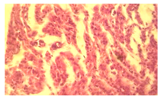
- Macroscopic studies of dichorionic twins who died prematurely show a significant difference in the size of the hearts of the twins. When microscopic examination is carried out, karyopyknotic conditions are noted in most of the cardiomyocytes in the myocardial layer of twins with a relatively small heart size, and karyorrhexis and karyolysis are visible in a small number of cells. Weak intermuscular swelling is prominent (Figure 1).
 | Figure 1. Dichorionic twins with a relatively small heart size have severe intermuscular swelling in the myocardial layer. Stained in hematoxylin-eosin. Ob.40, ok.10 |
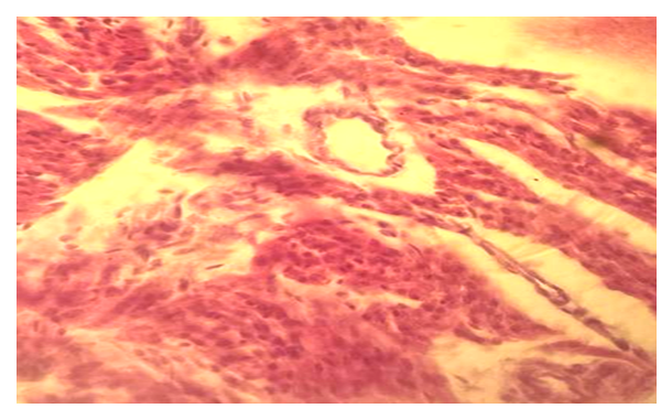
 | Figure 2. Intramyocardial blood vessels in the myocardial layer of twins with relatively small heart size. Stained in hematoxylin-eosin. Ob.40, ok.10 |
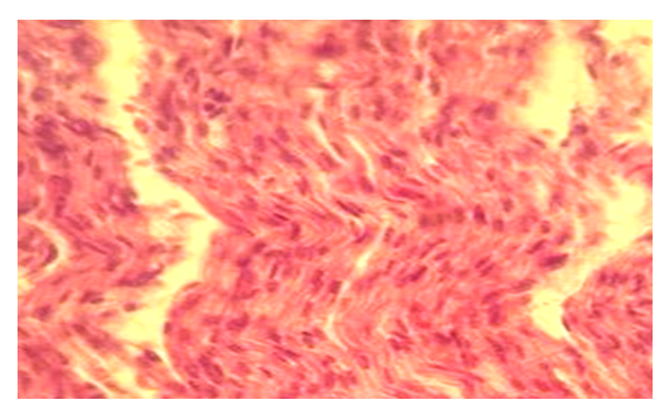
 | Figure 3. Intermuscular swelling in the myocardial layer of dichorionic twins with a relatively large heart size. Stained in hematoxylin-eosin. Ob.40, ok.10 |
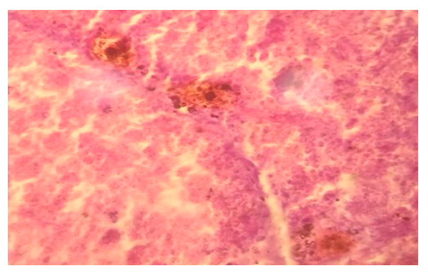
 | Figure 4. Intramyocardial blood vessels in the myocardial layer of twins with relatively large heart size. Stained in hematoxylin-eosin. Ob.40, ok.10 |
5. Conclusions
- Thus, most twins with a separate placenta have a small heart size, and in twins with a large heart size, pathomorphological changes in myocardial structures are weakly manifested. This should be taken into account in the practice of neontology. In particular, it is necessary to carry out scientific research on the study of the structures of the myocardium of babies who were born and died prematurely.
 Abstract
 Reference
 Full-Text PDF
 Full-text HTML