American Journal of Medicine and Medical Sciences

p-ISSN: 2165-901X    e-ISSN: 2165-9036

2024;  14(5): 1451-1466

doi:10.5923/j.ajmms.20241405.68

Received: Apr. 19, 2024; Accepted: May 17, 2024; Published: May 30, 2024


Assessing the Effectiveness of Topical Treatments for Recurrent Aphthous Stomatitis: A Network Meta-Analysis

Yakubova Farida Khaldarovna, Shukurova Gulnora Raxmanovna

Associate Professor at Tashkent State Pediatric Institute, Uzbekistan

Correspondence to: Yakubova Farida Khaldarovna, Associate Professor at Tashkent State Pediatric Institute, Uzbekistan.

Email:

Copyright © 2024 The Author(s). Published by Scientific & Academic Publishing.

This work is licensed under the Creative Commons Attribution International License (CC BY).
http://creativecommons.org/licenses/by/4.0/

Abstract

Background and Objectives: The aim was to compare the effectiveness and safety of various topical interventions for recurrent aphthous stomatitis. Methods: Following the PRISMA guidelines, this network meta-analysis examined randomized controlled trials retrieved from PubMed, Web of Science (WOS), Cochrane Central Register of Controlled Trials, and Embase. Quality assessment was conducted using Cochrane Handbook criteria. Data on healing, size reduction, symptom alleviation, recurrence, and safety were independently extracted by two authors. Network meta-analysis was performed using ADDIS and RevMan. Results: 72 trials (5272 subjects) involving 29 topical treatments were analyzed. Honey, insulin liposome gel, laser, amlexanox, glycyrrhiza, and triamcinolone showed superior efficacy. Probiotics and chlorhexidine extended ulcer intervals and reduced recurrence. Doxycycline and penicillin carried high adverse event risks. Hematologic evaluation showed no bias. Laser was ranked high for short-term size and symptom reduction, while probiotics showed long-term benefits. Conclusion: Laser is recommended for short-term intervention during exacerbation, and probiotics for long-term management during both exacerbation and remission phases of recurrent aphthous stomatitis.

Keywords: Recurrent aphthous stomatitis, Network meta-analysis, Topical intervention

Cite this paper: Yakubova Farida Khaldarovna, Shukurova Gulnora Raxmanovna, Assessing the Effectiveness of Topical Treatments for Recurrent Aphthous Stomatitis: A Network Meta-Analysis, American Journal of Medicine and Medical Sciences, Vol. 14 No. 5, 2024, pp. 1451-1466. doi: 10.5923/j.ajmms.20241405.68.

1. Introduction

Recurrent aphthous ulcer (RAU), also known as recurrent aphthous stomatitis (RAS), is a prevalent condition affecting the oral mucosa. Its prevalence varies widely, ranging from 1.4% to 21.4% [1,2,3,4,5] as indicated by retrospective population-based studies across different countries and regions. Extensive research has been conducted on the etiology and pathogenesis of RAS. There is speculation that the oral microbiota [6], including organisms like Streptococcus [7], Helicobacter pylori [8], cytomegalovirus [9], and various other microorganisms [10], may play significant roles in ulcer formation. Additionally, systemic diseases can manifest with ulcers as a prominent phenotype [11], further complicating etiological investigations.
Despite extensive research, the precise etiology and pathogenesis of RAS remain incompletely understood. Consequently, specific treatments for RAU have yet to be identified in clinical and basic trials [12]. Presently, treatment for RAS primarily focuses on symptom management, aiming to alleviate pain, facilitate lesion healing, and prolong the interval between episodes. Topical treatments, including medication, laser therapy, cryotherapy, and cautery, are considered effective for managing minor recurrent aphthous ulcers (MiRAU) and as adjuncts for major recurrent aphthous ulcers (MaRAU) [13]. Topical glucocorticoids, such as dexamethasone and triamcinolone, are commonly prescribed as first-line agents due to their anti-inflammatory and immunosuppressive properties [14]. Tetracyclines and derivatives, particularly doxycycline, are believed to inhibit ulcer formation and tissue destruction by targeting matrix metalloproteinases in the inflammatory pathway [15]. Amlexanox, recognized for its anti-inflammatory and anti-allergic effects, has gained attention in recent studies [16].
Emerging therapeutic modalities, including biological and laser treatments, hold promise for managing mucosal diseases like RAS [17]. Systematic analyses have underscored the role of oral flora alterations in RAS progression [18], paving the way for topical probiotic interventions. Laser therapy has shown efficacy in accelerating tissue repair and pain relief, while traditional treatments like freezing [19] and cautery [20] remain prevalent for their beneficial effects on cell metabolism and tissue regeneration. Low serum zinc levels have been identified as a risk factor for RAS [21], prompting the recommendation for topical zinc supplementation. Moreover, natural extracts such as curcumin [22], glycyrrhiza [23], honey [24], quercetin [25], chitosan [26], aloe [27], berberine gelatin [28], diosmectite [29], allicin [30], and others have shown promise as potential topical interventions for ulcers.
RAS presents as a self-limiting condition characterized by varying durations and manifestations among individuals. While most cases of recurrent aphthous stomatitis resolve within a few days, they can significantly impact daily life due to localized mucosal lesions causing discomfort, pain from chemical-mechanical irritation, and recurrent episodes [31]. Traditional meta-analyses by various authors have primarily focused on comparing the efficacy of local interventions for RAS against ineffective placebos [32,33,34]. However, these approaches limit the comparison to pairwise estimates of effect, preventing simultaneous assessment of the relative efficacy of multiple interventions.
The question of which local intervention is most effective for RAS remains contentious, with a dearth of robust research evidence. Despite extensive literature searches, we have not identified a comprehensive systematic evaluation and ranking of multiple local interventions for RAS treatment. Hence, we conducted a systematic review, incorporating numerous randomized controlled trials (RCTs), to assess the efficacy and safety of up to 29 local interventions for RAS treatment. Our objective was to furnish a dependable reference for clinicians in selecting more effective topical treatment options for RAS patients.

2. Materials and Methods

Assertion
Network meta-analysis (NMA) represents a novel, high-quality analytical approach founded on the principles of homogeneity, transferability, and consistency. This method facilitates simultaneous comparisons among multiple interventions and offers a potential ranking of their effectiveness [35]. Consequently, NMA has become a cornerstone in numerous studies, enabling the provision of more robust evidence and aiding in the selection of optimal solutions. Our study adhered rigorously to the criteria and protocols governing NMA [36]. We followed relevant guidelines and standards throughout the study, ensuring compliance with software requirements, including Addis, Revman, Endnote, and others. Furthermore, our study was registered in the PROSPERO International Prospective Register of Systematic Reviews before implementation, in accordance with established guidelines (registration number: CRD42021251154).
Data Sources and Search Strategy
We conducted a comprehensive search across four databases—PubMed, Web of Science (WOS), Cochrane Central Register of Controlled Trials, and Embase—during the study period. The search encompassed literature published from the inception of each database up to October 1, 2021. Medical terms such as "Stomatitis, Aphthous," "Oral Ulcer," and "Clobetasol" were employed as primary search terms. Additionally, synonyms and abbreviations such as "Canker sore," "Corticosteroid," and "LLLT*" were included as keywords to broaden the search scope. The search process was independently executed by two individuals, and the Endnote literature management software facilitated the organization of search results. Further details regarding the search strategy are outlined in Tables S1–S5.
Selection Criteria
The randomized controlled trials (RCTs) included in this study adhered to the following criteria: (1) Confirmation of recurrent aphthous ulcers through clinical or histopathological examination, with visible ulcer-like lesions observed on any area of the oral mucosa. (2) Inclusion of simple ulcerative lesions of undetermined origin, excluding oral manifestations of systemic diseases like leukoaraiosis or diabetes mellitus, as well as specific ulcerative lesions resulting from trauma or radiotherapy. (3) Participants received only local interventions or placebos during the trial, without undergoing any other treatments that could potentially affect recurrent aphthous stomatitis (RAS) prior to or during the trial, such as systemic steroids or immunosuppressants. (4) For studies involving patients with multiple oral mucosal diseases, data related specifically to RAS were extracted. If extraction was not feasible, the study was excluded from analysis.
Outcomes
In this study, clinical efficacy and safety were chosen as primary outcome measures. Clinical efficacy was evaluated based on healing efficacy, size reduction effect, and symptom reduction effect. Healing efficacy was assessed by the time taken for complete healing, measured from the enrollment in the local intervention to the total resolution of ulcer-like lesions. The size reduction effect was determined using the efficacy index (EI), calculated as the ratio of ulcer reduction area to baseline ulcer area. The cumulative reduction in ulcer size across various examination days during the trial was recorded. Symptom reduction effect was also assessed using the efficacy index (EI), calculated as the ratio of reduction in pain score to baseline ulcer pain score. Individual subjects' pain levels were measured using visual analog scale (VAS) or decile scale scores on different examination days, and cumulative pain relief was calculated accordingly (VAS score: 10 cm horizontal line, marked 0 = no pain to 10 = worst pain; Decile scale: 0 for no pain, 10 for most pain). Safety evaluation included monitoring adverse events and assessing blood levels of the intervention drug. Additionally, the study extracted data related to the impact on the recurrence of RAS.
Data Collection and Risk of Bias Assessment
Two researchers independently conducted literature screening against predefined inclusion and exclusion criteria, assessed literature quality according to established criteria, and extracted relevant information. This process was carried out independently by the researchers and subsequently cross-verified, with any discrepancies resolved through consultation with a third party or discussion among the research team. Extracted information included details such as the first author of the included studies, publication time, country of origin, sample size, demographics (gender and age), interventions, and outcome indicators.
The quality of the included studies was evaluated using RevMan 5.3 software provided by the Cochrane Collaboration. This evaluation encompassed criteria such as random sequence generation, allocation concealment, blinding of participants and personnel, blinding of outcome assessment, completeness of outcome data, selective reporting, and other potential biases.
Furthermore, potential publication bias within the included studies was examined utilizing a funnel plotting approach.
Data Synthesis and Statistical Analysis
Traditional meta-analysis was conducted using RevMan 5.3 and ADDIS 1.16. Risk difference (RD) served as the efficacy indicator for dichotomous variables, while mean difference (MD) was employed for continuous variables. Each effect size estimate was accompanied by a 95% confidence interval (95% CI). Heterogeneity of test results was quantified using the I2 statistic. Heterogeneity was deemed small if p ≥ 0.1 and I2 ≤ 50%, warranting the use of a fixed-effects model for combining. Conversely, if p < 0.1 and I2 > 50%, indicating significant heterogeneity, a random-effects model was applied for combining, with sensitivity analysis conducted through subgroup analysis or individual literature exclusion.
A random-effects network within a Bayesian framework model was established using ADDIS 1.16 [37]. Networks were tailored to different outcome indicators to accommodate a wide array of local interventions. Direct and indirect comparisons between interventions were made to ensure comprehensive and complete results. Statistical significance was considered at p < 0.05. ADDIS 1.16 also estimated ranking probabilities for interventions. MD for each local intervention was compared to an arbitrary control, with the convergence of the model evaluated through the number of Markov chain iterations. Variance calculations and node splitting analyses were performed to assess inconsistency in the network meta-analysis. Results were deemed inconsistent if the random effects variance significantly deviated from the inconsistency or if the discrepancy between direct and indirect evidence was deemed significant (p < 0.05).

3. Results

Study Selection The initial database search yielded 11,962 records. Following the removal of 2388 duplicate articles, the titles and abstracts of 9574 articles were screened. Among these, 9314 articles were excluded as they did not meet the inclusion criteria. A full-text review was conducted for 260 articles, of which 186 were subsequently excluded. Ultimately, 72 eligible studies were included for qualitative and quantitative analysis (see Figure 1).
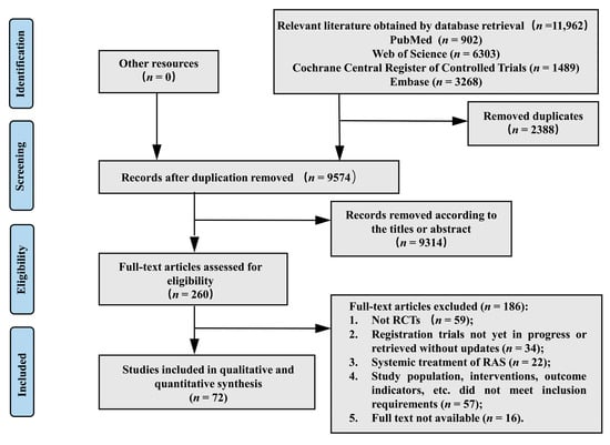
Figure 1. Flow chart of the study selection process
Characteristics and Quality of Studies
The 72 studies included in this analysis are outlined in Table S6. Among them, three were three-arm studies, two were four-arm studies, and one study sub-grouped participants into adult and child groups, which we merged for comparison purposes. The majority of participants had definite recurrent aphthous stomatitis (RAS), mostly classified as definite minor RAS, with a few studies providing information on ulcer size, meeting our inclusion criteria. All participants were histologically and clinically confirmed to have RAS and presented with definite symptoms at the onset of the study. The minimum mean age among participants was 6.82 years, while the oldest participant was 71 years old. Female participants outnumbered male participants in most studies. Treatment durations ranged from a few days to several months, with intervention forms including mucoadhesive matrices, pastes, liquids, etc. Specific interventions and treatment regimens are detailed in Table S6, while study outcomes are summarized in Tables S7 and S8.
Overall, 69 studies contributed data for the assessment of four evaluation criteria: (a) healing effect, with data from 26 randomized controlled trials (RCTs) involving 1306 participants; (b) size-reducing effect, with data from 37 RCTs involving 3587 participants; (c) symptom-reducing effect, with data from 46 RCTs involving 4020 participants; and (d) adverse effects, with data from 36 RCTs involving 2787 participants. The network structure is depicted in Figure 2. Three studies provided incomplete data and were evaluated descriptively for recurrence outcomes. Additionally, four of the 72 studies reported hematologic values, which were also evaluated descriptively. Risk of bias assessments are illustrated in Figures S1 and S2. The majority of studies demonstrated a low risk of bias in terms of "incomplete outcome data," "selective reporting," and "other bias." However, due to insufficient detail regarding allocation, randomization, and measurement processes in some articles, the risk estimates for "random sequence generation," "allocation concealment," and "blinding of outcome assessment" were deemed unclear. Some studies were single-blinded due to the nature of interventions such as laser therapy, cauterization, and freezing, which did not fully conceal participants and personnel, resulting in a high risk for "blinding of participants and personnel." Nevertheless, this does not necessarily imply low quality in the included studies. Utilizing RevMan 5.3 software, funnel plots were generated to aid in the assessment of publication bias (Tables S3–S6).
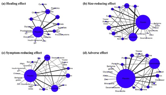
Figure 2. Network plots for the main outcomes considered in the review. (a) Healing effect; (b) size-reducing effect; (c) symptom-reducing effect; (d) adverse effect. Nodes and edges are weighted according to volume of studies, including that treatment or comparison
Pairwise Meta-Analysis
Data from 14 randomized controlled trials (RCTs) involving 820 participants were pooled for assessing the healing effect (Chapter S1). Triamcinolone, laser therapy, silver nitrate, and honey demonstrated significant efficacy compared to placebo. However, comparisons between curcumin vs. triamcinolone and triamcinolone vs. placebo did not reveal a clear preference.
For the size-reducing effect, data from 23 RCTs (n = 1807) were analyzed, resulting in eight pairwise comparisons (Chapter S2). Triamcinolone, probiotics, glycyrrhiza, and amlexanox were found to be significantly more effective than placebo. Conversely, comparisons between aloe vs. placebo, curcumin vs. triamcinolone, laser vs. placebo, and probiotics vs. triamcinolone did not yield statistically significant differences.
Similarly, data from 32 RCTs (n = 2940) with nine pairwise comparisons were analyzed for the symptom-reducing effect (Chapter S3). Triamcinolone, laser therapy, glycyrrhiza, amlexanox, and aloe were deemed superior to placebo. Laser therapy was preferred over triamcinolone. However, comparisons between curcumin vs. triamcinolone, doxycycline vs. placebo, and probiotics vs. placebo showed no distinguishable differences.
Regarding adverse effects, data from 22 RCTs (n = 1748) with nine pairwise comparisons were pooled (Chapter S4). No statistically significant differences were observed among the interventions.
Furthermore, it is important to note an additional point regarding the size-reducing effect and symptom-reducing effect. Given that the reporting times of the various studies were relatively close and did not meet the criteria for dividing into long-term and short-term comparisons, pairwise comparisons were only conducted at the treatment endpoint. However, more detailed and precise comparisons of daily changes were also performed, as outlined in Chapters S2 and S3.
Network Meta-Analysis
Healing Effect
Data from 26 randomized controlled trials (RCTs) [20,24,26,38,39,40,41,42,43,44,45,46,47,48,49,50,51,52,53, 54,55,56,57,58,59,60] encompassing 20 pairwise comparisons among 18 interventions were synthesized. Honey, insulin liposome gel, and laser therapy demonstrated shorter healing times and superior healing effects compared to placebo (refer to Table 1 for estimated values). However, no significant differences were noted in the remaining comparisons (further elaboration provided in Chapter S1). Our analysis suggests that these 18 local interventions may exhibit consistent or similar performance regarding healing effect. Regarding rank probability (with only the top five listed in Table 2, more detailed information available in Chapter S1), the most effective intervention is insulin-liposomal gel (p-core, 0.24), followed by honey (p-core, 0.15), laser therapy (p-core, 0.11), penicillin (p-core, 0.09), and aloe (p-core, 0.06), when compared to placebo.
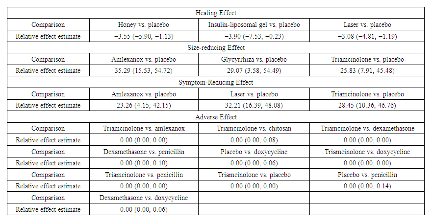
Table 1. Significantly different estimates for healing effect
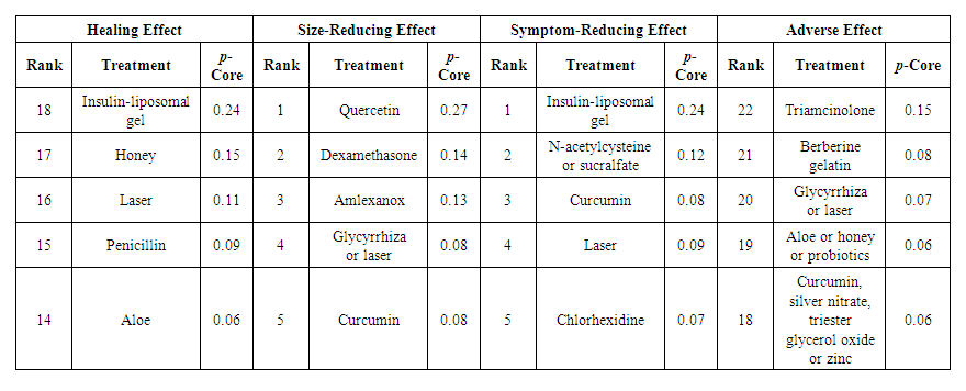
Table 2. Outcome p-cores for the best five interventions
Size-Reducing Effect
Data from 37 randomized controlled trials (RCTs) [19,22,25,27,28,29,30,40,42,48,50,55,61,62,63,64,65,66, 67,68,69,70,71,72,73,74,75,76,77,78, 79,80,81,82,83,84] comprising 30 pairwise comparisons among 19 interventions were analyzed. Network meta-analysis revealed that amlexanox, glycyrrhiza, and triamcinolone exhibited greater effectiveness than placebo (see Table 1). However, no statistically significant differences in size-reduction effect were observed for the other interventions (further details provided in Chapter S2). According to the rank probability (refer to Table 2, Chapter S2), quercetin emerged as the optimal solution (p-core, 0.27). The remaining potentially efficient interventions, listed in order of priority, include dexamethasone (p-core, 0.14), amlexanox (p-core, 0.13), glycyrrhiza or laser therapy (p-core, 0.08), and curcumin (p-core, 0.08). Many studies not only evaluated efficacy at the end of the intervention but also conducted multiple measurements throughout the treatment period. To leverage these data fully and compare the change in ulcer size on each day of the treatment period, a more detailed ranking was conducted (refer to Chapter S2). This ranking utilized days as the time unit rather than the entire treatment duration. We extracted the probability of each intervention being ranked first (p-core) and represented it as a "Time-Rank 1 probability" line chart to provide a more precise reflection of the intervention's impact on ulcer size (see Chart 1). Laser therapy, glycyrrhiza, and zinc were deemed the most promising interventions for significantly reducing ulcer size in the short term.
Symptom-Reducing Effect
Data from 46 randomized controlled trials (RCTs) [20,25,28,29,30,38,43,44,46,48,50,51,52,53,55,61,62,63, 64,65,66,67,69,70,71,72,73,74,75,76,77, 78,79,80,81,82,83,84,85,86,87,88,89,90,91] comprising 32 pairwise comparisons among 23 interventions were analyzed. Amlexanox, laser therapy, and triamcinolone exhibited a significant advantage over placebo (see Table 1). However, no statistically significant differences were observed among the other interventions (refer to Chapter S3 for further details). According to the rank probability (refer to Table 2, Chapter S3), insulin-liposomal gel emerged as the preferred option (p-core, 0.24), followed by N-acetylcysteine or sucralfate (p-core, 0.12), curcumin (p-core, 0.08), laser therapy (p-core, 0.09), and chlorhexidine (p-core, 0.07).
Furthermore, we generated a "Time-Rank 1 probability" line chart to illustrate the daily improvement in pain during treatment for the symptom-reducing effect (see Chart 2). Laser therapy, insulin-liposomal gel, and sucralfate were identified as potential optimal solutions for short-term relief of pain caused by ulcers.
Adverse Effect
Data from 36 randomized controlled trials (RCTs) [20,22,24,26,28-30,40,41,42,47,48,50,53,54,63,64, 65,70,71,75,76,77,80,82,83,85,86,87,88,89,92-96] comprising 32 pairwise comparisons among 22 interventions were analyzed. Triamcinolone exhibited superior performance in terms of possible adverse events, with none reported in 259 subjects, compared to amlexanox, chitosan, dexamethasone, doxycycline, penicillin, and placebo. Additionally, dexamethasone showed significant improvement compared to penicillin and doxycycline, with 4 adverse events reported in 120 subjects, while penicillin and doxycycline exhibited weaknesses compared to placebo (refer to Table 1 for details). No significant differences were observed among the other interventions (further information available in Chapter S4).
Regarding the rank probability (refer to Table 2, Chapter S4), triamcinolone emerged as the optimal choice (p-core, 0.15). Other interventions with notable performance in rank possibility included berberine gelatin (p-core, 0.08), glycyrrhiza or laser therapy (p-core, 0.07), aloe, honey, or probiotics (p-core, 0.06), and curcumin, silver nitrate, triester glycerol oxide, or zinc (p-core, 0.06). Detailed information on possible adverse events is compiled and presented in Chapter S4.
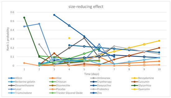
Chart 1. “Time-Rank 1 probability” folding line chart of side-reducing effect
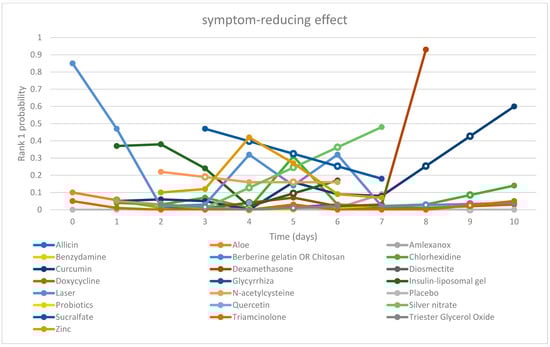
Chart 2. “Time-Rank 1 probability” folding line chart of symptom-reducing effect
Other Outcome Indicators
Hematologic Values
Four randomized controlled trials (RCTs) [30,77,80,82] included in the study provided data on the blood levels of the intervention drug or blood laboratory findings (refer to Table 3). However, no significant results were discerned. Hematologic safety hazards were not associated with dexamethasone, aloe, allicin, or amlexanox.
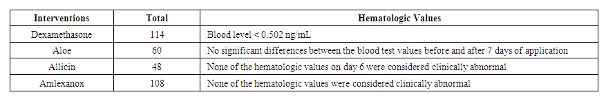
Table 3. Hematologic values
Relapse
Considerations regarding recurrent aphthous stomatitis (RAS) recurrence were outlined in four randomized controlled trials (RCTs) [58,61,97,98] (refer to Table 4). These trials evaluated four interventions: probiotics, chlorhexidine, benzydamine, and triamcinolone, with outcome indicators including outbreak frequency, number of new ulcers, and interval between ulcers. The findings from the investigations were as follows: (a) probiotics demonstrated efficacy in reducing the frequency of ulcer outbreaks in children but exhibited differing performance in adults; (b) chlorhexidine extended the interval between ulcers but did not significantly decrease the number of new ulcers during the trial period; (c) benzydamine did not exhibit efficacy in reducing the number of new ulcers; (d) regardless of whether administered in media-based form or in liquid form, the reduction in the number of new ulcers by triamcinolone was not statistically significant.
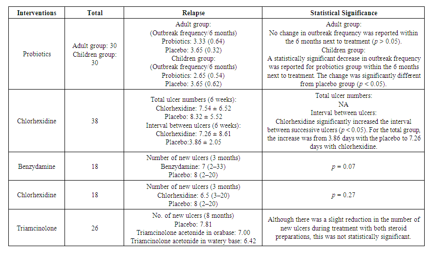
Table 4. Recurrence and statistical significance
Consistency and Sensitivity
Analysis ADDIS 1.16 software was employed to evaluate the consistency and sensitivity of the network meta-analysis comprehensively. We employed the consistency model, inconsistency model, and node split model to ensure thorough examination. The results indicated the absence of statistical heterogeneity within the study, with no observed inconsistency across the four comparisons: healing effect, size-reducing effect, symptom-reducing effect, and adverse effect. The inconsistency between direct and indirect comparisons was minimized to the lowest possible level.
Regarding sensitivity analysis, we utilized both the method of systematically excluding literature for sensitivity comparison and the subgroup analysis based on the timing of outcome measurement. These approaches aimed to optimize the analysis to the greatest extent possible.
Our Viewpoint
The efficacy and safety of an intervention are pivotal factors determining its suitability for use. In this extensive analysis encompassing multiple studies and interventions, we prioritized efficacy and safety as key comparison criteria. Upon evaluation, we found that in terms of safety alone, all interventions included in the study demonstrated substantial compliance, with no instances of severe adverse events or hematologic effects. Therefore, our primary focus shifted to assessing efficacy when selecting interventions. Taking into account healing promotion, pain reduction, and relapse prevention as evaluation metrics, alongside the crucial consideration of timing, we recommend laser therapy as a short-term "shock therapy" intervention and probiotics as a long-term "maintenance" intervention.

4. Discussion

Recurrent aphthous stomatitis (RAS) exhibits inherent self-healing properties [99]. However, due to its clinical features of frequent recurrence, local tissue damage, and pain [31], interventions are commonly employed. Given the unclear etiology and significant individual variability, the treatment of RAS primarily revolves around symptomatic management, with the overarching goals of promoting healing, alleviating pain, and reducing recurrence [13]. Local interventions are frequently utilized in patients with minor RAS and major RAS unaccompanied by systemic symptoms [100]. A plethora of local interventions are available, many of which have shown promising outcomes in clinical trials compared to placebo. Yet, the optimal intervention remains elusive, necessitating a thorough assessment and comparison of available topical treatments.
This study represents the first systematic evaluation and network meta-analysis of topical interventions for RAS, encompassing a broad array of potential options. Both pairwise analyses and network comparisons were employed, supplemented by descriptive analyses for outcome indicators with limited sample sizes and varied evaluation metrics. A total of 29 topical interventions, including placebo, were scrutinized, ranging from allicin to zinc. Notably, clobetasol and minocycline were solely evaluated for adverse events and were not part of the primary outcome analysis. To maximize data acquisition, we conducted comprehensive searches across four major databases, complemented by gray literature sources. We established stringent inclusion and exclusion criteria, particularly excluding traumatic ulcers, radiological ulcers, and ulcers associated with systemic diseases. Seventy-two studies involving 5272 subjects were ultimately included in the analysis. Our assessment focused on four efficacy outcomes and one safety outcome. Healing effect, size-reducing effect, and symptom-reducing effect served as the primary efficacy evaluation metrics, with relapse analysis limited to descriptive examination. Safety evaluation primarily centered on adverse effects, with hematologic values serving as supplementary indicators. Additionally, rank possibility, based on p-score, served as a screening tool to aid in intervention selection.
As delineated in the findings, amalgamating the four efficacy assessments along with an additional safety appraisal, we advocate our recommendation to clinicians: employ laser therapy as a short-term "shock therapy" during episodes of ulcer exacerbation, and utilize probiotics as a long-term "modifier" throughout the entire ulcer cycle.
Laser therapy stands out as a pivotal milestone in clinical practice for treating oral mucosal ailments. Its efficacy extends beyond common oral mucosal conditions to encompass precancerous lesions like oral mucositis resulting from radiotherapy and chemotherapy, oral submucosal fibrosis, oral lichen planus, oral leukoplakia, and burning mouth syndrome. The therapeutic effect of laser on the oral mucosa is attributed to its stimulating biological impact. Controlled laser light within specific wavelength and power parameters engages in local metabolic processes through various physicochemical mechanisms, thereby exerting analgesic, anti-inflammatory, and pro-repair effects without causing thermal damage. Undoubtedly, its role in treating RAS is gaining recognition. Current laser treatment modalities include carbon dioxide laser, crystal laser, diode laser, and low-level laser therapy (LLLT). Our evaluation encompassed 14 randomized controlled trials (RCTs) involving laser interventions. Among these, five studies utilized diode lasers, six employed CO2 lasers, one utilized Er, Cr: YSGG lasers, one used Nd: YAG lasers, and one involved LLLT. Variations in laser modality, wavelength, and power output may impact efficacy, yet this aspect remains underexplored due to limited studies and a lack of treatment standards. Consequently, we grouped different laser modalities under the umbrella term "laser" in our study. Across efficacy evaluations, lasers emerged as superior in promoting healing, reducing ulcer size, and alleviating symptoms throughout the treatment duration. Notably, in daily assessments, laser therapy demonstrated unparalleled short-term efficacy. Furthermore, laser therapy boasts impeccable safety credentials. A recent retrospective evaluation by Valerie G. A. Suter and colleagues echoed similar expectations for laser interventions in RAS while highlighting concerns regarding laser standardization. The advantages of laser therapy over other topical interventions include: (a) short treatment duration, often yielding significant outcomes with minimal interventions; (b) superior efficacy in accelerating wound healing and pain reduction; and (c) proven safety profile, with several retrospective studies reporting no significant adverse events or hematologic abnormalities. Hence, laser therapy presents a compelling option for routine cases, steroid-intolerant individuals, and patients with severe ulcers necessitating systemic treatment. We advocate for laser therapy's use as a short-term intervention during ulcer exacerbations to expedite healing and alleviate pain.
The oral cavity functions as a natural habitat for various microorganisms, with probiotics playing a crucial role. These beneficial microorganisms contribute to the construction of the oral microbial community, which is vital for maintaining oral microecological balance and counteracting pathogenic bacteria. Maintaining this delicate microbial balance is essential for oral health, as imbalance may predispose individuals to conditions such as dental caries, periodontal disease, and fungal infections. Numerous studies support the use of probiotics, either alone or in combination with other agents, to modulate the composition and structure of oral flora in diseased states, thereby intervening in conditions like dental caries, periodontal disease, and halitosis. This also extends to recurrent aphthous stomatitis (RAS). Despite the unclear etiology of RAS, evidence from microbiological and immunological studies suggests the involvement of microbial factors in its pathogenesis [10,127,128]. Consequently, probiotic therapy has been explored and implemented in RAS management [129].
In our study, probiotics were administered topically for RAS management, with four randomized controlled trials (RCTs) involving Lactobacillus and one with Bacillus Clausii probiotics. While one trial utilized a mouthwash containing lactic acid, the others utilized solid tablets, with trial durations ranging from 7 to 90 days. Initially, probiotics did not demonstrate significant advantages in terms of healing, size reduction, or symptom alleviation. However, by day 7, they began to exhibit efficacy in promoting healing. Regarding recurrence, one study by Lotfy Aggour and colleagues [61] investigated the use of L. acidophilus lozenges in adult and pediatric subjects compared to placebo. They observed a significantly lower frequency of outbreaks in children using probiotics compared to controls, although this effect was not observed in adults. Notably, further studies on recurrence are warranted. Safety evaluations yielded satisfactory results.
Potential mechanisms underlying the involvement of probiotics in RAS management include: (a) competition mechanism, wherein probiotics outcompete pathogenic microorganisms, leading to the establishment of harmless or beneficial biofilms [130,131,132]; (b) pro-repair effects, involving the reduction of pro-inflammatory cytokines and promotion of local tissue repair [133,134]; and (c) regulation of the microenvironment, achieved through the secretion of metabolites and active molecules that modulate the local physicochemical and immune environments, thereby promoting tissue resistance and repair [131,133,135]. These effects collectively contribute to the regulation of the microenvironment conducive to RAS, thereby reducing recurrence. However, the limited short-term healing promotion may be attributed to insufficient metabolites and active substances.
Based on these considerations, we recommend the use of probiotics as a long-term intervention during both the exacerbation and remission phases of ulcers to prolong the inter-episode interval and reduce recurrence.
Nevertheless, our study has several limitations. Firstly, the scarcity of RCTs on certain interventions compromises the study's credibility. Secondly, grouping different types of lasers with varying wavelengths into a single intervention category precluded exploring their differences. Additionally, more studies investigating RAS recurrence are warranted. The evidence supporting conclusions about probiotics is weakened by non-direct evidence articles and a limited number of clinical trials. Thus, future high-quality studies with larger sample sizes and standardized outcome evaluation criteria are imperative.
In summary, this extensive network meta-analysis, which takes into account various considerations, indicates that the majority of local interventions did not demonstrate significant disparities in efficacy or safety. From the evidence available, we suggest employing laser therapy for short-term intervention to enhance healing and alleviate pain during the active phase of RAS, while recommending probiotics for long-term use to extend the interval between episodes and mitigate recurrence throughout both active and remission phases of RAS. We advocate for further large-scale RCTs adhering to rigorous standards to enhance the credibility of future research in this domain.

Medicina-1747928-Supplementary

References

[1]  Kaur R., Behl A.B., Punia R.S., Nirav K., Singh K.B., Kaur S. Assessment of Prevalence of Recurrent Aphthous Stomatitis in the North Indian Population: A Cross-Sectional Study. J. Pharm. Bioallied Sci. 2021; 13: S363–S366. doi: 10.4103/jpbs.JPBS_581_20. [PMC free article] [PubMed] [CrossRef] [Google Scholar]
[2]  Darjani A., Joukar F., Naghipour M., Asgharnezhad M., Mansour-Ghanaei F. Lifetime prevalence of recurrent aphthous stomatitis and its related factors in Northern Iranian population: The PERSIAN Guilan Cohort Study. Clin. Oral. Investig. 2021; 25: 711–718. doi: 10.1007/s00784-020-03611-y. [PubMed] [CrossRef] [Google Scholar]
[3]  Xu K., Zhou C., Huang F., Duan N., Wang Y., Zheng L., Wang X., Wang W. Relationship between dietary factors and recurrent aphthous stomatitis in China: A cross-sectional study. J. Int. Med. Res. 2021; 49: 675888644. doi: 10.1177/03000605211017724. [PMC free article] [PubMed] [CrossRef] [Google Scholar]
[4]  Hariyani N., Bramantoro T., Nair R., Singh A., Sengupta K. Depression symptoms and recurrent aphthous stomatitis—Evidence from a population-based study in Indonesia. Oral Dis. 2020; 26: 948–954. doi: 10.1111/odi.13303. [PubMed] [CrossRef] [Google Scholar]
[5]  Queiroz S., Silva M., Medeiros A., Oliveira P.T., Gurgel B., Silveira É. Recurrent aphthous ulceration: An epidemiological study of etiological factors, treatment and differential diagnosis. An. Bras. Dermatol. 2018; 93: 341–346. doi: 10.1590/abd1806-4841.20186228. [PMC free article] [PubMed] [CrossRef] [Google Scholar]
[6]  Gasmi B.A., Noor S., Menzel A., Gasmi A. Oral Aphthous: Pathophysiology, Clinical Aspects and Medical Treatment. Arch. Razi Inst. 2021; 76: 1155–1163. [PMC free article] [PubMed] [Google Scholar]
[7]  Bankvall M., Sjöberg F., Gale G., Wold A., Jontell M., Östman S. The oral microbiota of patients with recurrent aphthous stomatitis. J. Oral Microbiol. 2014; 6: 25739. doi: 10.3402/jom.v6.25739. [PMC free article] [PubMed] [CrossRef] [Google Scholar]
[8]  Kazanowska-Dygdała M., Duś I., Radwan-Oczko M. The presence of Helicobacter pylori in oral cavities of patients with leukoplakia and oral lichen planus. J. Appl. Oral Sci. 2016; 24: 18–23. doi: 10.1590/1678-775720150203. [PMC free article] [PubMed] [CrossRef] [Google Scholar]
[9]  Irani S. New Insights into Oral Cancer-Risk Factors and Prevention: A Review of Literature. Int. J. Prev. Med. 2020; 11: 202. doi: 10.4103/ijpvm.IJPVM_403_18. [PMC free article] [PubMed] [CrossRef] [Google Scholar]
[10]  Slebioda Z., Szponar E., Kowalska A. Etiopathogenesis of recurrent aphthous stomatitis and the role of immunologic aspects: Literature review. Arch. Immunol. Ther. Exp. 2014; 62: 205–215. doi: 10.1007/s00005-013-0261-y. [PMC free article] [PubMed] [CrossRef] [Google Scholar]
[11]  Ludovichetti F.S., Signoriello A.G., Girotto L., Del Dot L., Piovan S., Mazzoleni S. Oro-dental lesions in paediatric patients with coeliac disease: An observational retrospective clinical study. Rev. Esp. Enferm. Dig. 2022. Online ahead of print. [PubMed] [CrossRef]
[12]  Scully C., Porter S. Oral mucosal disease: Recurrent aphthous stomatitis. Br. J. Oral Maxillofac. Surg. 2008; 46: 198–206. doi: 10.1016/j.bjoms.2007.07.201. [PubMed] [CrossRef] [Google Scholar]
[13]  Saikaly S.K., Saikaly T.S., Saikaly L.E. Recurrent aphthous ulceration: A review of potential causes and novel treatments. J. Dermatolog. Treat. 2018; 29: 542–552. doi: 10.1080/09546634.2017.1422079. [PubMed] [CrossRef] [Google Scholar]
[14]  Ahluwalia A. Topical glucocorticoids and the skin—Mechanisms of action: An update. Mediat. Inflamm. 1998; 7: 183–193. doi: 10.1080/09629359891126. [PMC free article] [PubMed] [CrossRef] [Google Scholar]
[15]  Golub L.M., Ramamurthy N.S., Mcnamara T.F., Greenwald R.A., Rifkin B.R. Tetracyclines inhibit connective tissue breakdown: New therapeutic implications for an old family of drugs. Crit. Rev. Oral Biol. Med. 1991; 2: 297–321. doi: 10.1177/10454411910020030201. [PubMed] [CrossRef] [Google Scholar]
[16]  Baccaglini L., Lalla R.V., Bruce A.J., Sartori-Valinotti J.C., Latortue M.C., Carrozzo M., Rogers R.R. Urban legends: Recurrent aphthous stomatitis. Oral Dis. 2011; 17: 755–770. doi: 10.1111/j.1601-0825.2011.01840.x. [PMC free article] [PubMed] [CrossRef] [Google Scholar]
[17]  Yuan H., Qiu J., Zhang T., Wu X., Zhou J., Park S. Quantitative changes of Veillonella, Streptococcus, and Neisseria in the oral cavity of patients with recurrent aphthous stomatitis: A systematic review and meta-analysis. Arch. Oral Biol. 2021; 129: 105198. doi: 10.1016/j.archoralbio.2021.105198. [PubMed] [CrossRef] [Google Scholar]
[18]  Da Silva J.P., Da Silva M.A., Almeida A.P., Lombardi J.I., Matos A.P. Laser therapy in the tissue repair process: A literature review. Photomed. Laser Surg. 2010; 28: 17–21. doi: 10.1089/pho.2008.2372. [PubMed] [CrossRef] [Google Scholar]
[19]  Arikan O.K., Birol A., Tuncez F., Erkek E., Koc C. A prospective randomized controlled trial to determine if cryotherapy can reduce the pain of patients with minor form of recurrent aphthous stomatitis. Oral Surg. Oral Med. Oral Pathol. Oral Radiol. Endod. 2006; 101: E1–E5. doi: 10.1016/j.tripleo.2005.07.035. [PubMed] [CrossRef] [Google Scholar]
[20]  Soylu Özler G. Silver nitrate cauterization: A treatment option for aphthous stomatitis. J. Cranio-Maxillofac. Surg. 2014; 42: e281–e283. doi: 10.1016/j.jcms.2013.10.006. [PubMed] [CrossRef] [Google Scholar]
[21]  Al-Maweri S.A., Halboub E., Al-Sharani H.M., Shamala A., Al-Kamel A., Al-Wesabi M., Albashari A., Al-Sharani A., Abdulrab S. Association between serum zinc levels and recurrent aphthous stomatitis: A meta-analysis with trial sequential analysis. Clin. Oral Investig. 2021; 25: 407–415. doi: 10.1007/s00784-020-03704-8. [PubMed] [CrossRef] [Google Scholar]
[22]  Manifar S., Obwaller A., Gharehgozloo A., Boorboor Shirazi Kordi H.R., Akhondzadeh S. Curcumin gel in the treatment of minor aphthous ulcer: A randomized, placebo-controlled trial. J. Med. Plants. 2012; 11: 40–45. [Google Scholar]
[23]  Martin M.D., Sherman J., Van Der Ven P., Burgess J. A controlled trial of a dissolving oral patch concerning glycyrrhiza (licorice) herbal extract for the treatment of aphthous ulcers. Gen. Dent. 2008; 56: 206–210. [PubMed] [Google Scholar]
[24]  El-Haddad S.A., Asiri F.Y., Al-Qahtani H.H., Al-Ghmlas A.S. Efficacy of honey in comparison to topical corticosteroid for treatment of recurrent minor aphthous ulceration: A randomized, blind, controlled, parallel, double-center clinical trial. Quintessence Int. 2014; 45: 691–701. [PubMed] [Google Scholar]
[25]  Pandya M., Kalappanavar A.N., Annigeri R.G., Rao D.S. Relative Efficacy of Quercetin Compared with Benzydamine Hydrochloride in Minor Aphthae: A Prospective, Parallel, Double Blind, Active Control, Preliminary Study. Int. J. Dent. 2017; 2017: 7034390. doi: 10.1155/2017/7034390. [PMC free article] [PubMed] [CrossRef] [Google Scholar]
[26]  Rahmani F., Moghadamnia A.A., Kazemi S., Shirzad A., Motallebnejad M. Effect of 0.5% Chitosan mouthwash on recurrent aphthous stomatitis: A randomized double-blind crossover clinical trial. Electron. Physician. 2018; 10: 6912–6919. doi: 10.19082/6912. [PMC free article] [PubMed] [CrossRef] [Google Scholar]
[27]  Babaee N., Zabihi E., Mohseni S., Moghadamnia A.A. Evaluation of the therapeutic effects of Aloe vera gel on minor recurrent aphthous stomatitis. Dent. Res. J. 2012; 9: 381–385. [PMC free article] [PubMed] [Google Scholar]
[28]  Jiang X.W., Zhang Y., Zhu Y.L., Zhang H., Lu K., Li F.F., Peng H.Y. Effects of berberine gelatin on recurrent aphthous stomatitis: A randomized, placebo-controlled, double-blind trial in a Chinese cohort. Oral Surg. Oral Med. Oral Pathol. Oral Radiol. 2013; 115: 212–217. doi: 10.1016/j.oooo.2012.09.009. [PubMed] [CrossRef] [Google Scholar]
[29]  Jiang X.W., Zhang Y., Zhang H., Lu K., Yang S.K., Sun G.L. Double-blind, randomized, controlled clinical trial of the effects of diosmectite and basic fibroblast growth factor paste on the treatment of minor recurrent aphthous stomatitis. Oral Surg. Oral Med. Oral Pathol. Oral Radiol. 2013; 116: 570–575. doi: 10.1016/j.oooo.2013.07.003. [PubMed] [CrossRef] [Google Scholar]
[30]  Jiang X.W., Zhang Y., Song G.D., Li F.F., Peng H.Y., Yang S.K., Sun G.L. Clinical evaluation of allicin oral adhesive tablets in the treatment of recurrent aphthous ulceration. Oral Surg. Oral Med. Oral Pathol. Oral Radiol. 2012; 113: 500–504. doi: 10.1016/j.oooo.2011.09.007. [PubMed] [CrossRef] [Google Scholar]
[31]  Al-Omiri M.K., Karasneh J., Alhijawi M.M., Zwiri A.M., Scully C., Lynch E. Recurrent aphthous stomatitis (RAS): A preliminary within-subject study of quality of life, oral health impacts and personality profiles. J. Oral Pathol. Med. 2015; 44: 278–283. doi: 10.1111/jop.12232. [PubMed] [CrossRef] [Google Scholar]
[32]  Cheng B., Zeng X., Liu S., Zou J., Wang Y. The efficacy of probiotics in management of recurrent aphthous stomatitis: A systematic review and meta-analysis. Sci. Rep. 2020; 10: 21181. doi: 10.1038/s41598-020-78281-7. [PMC free article] [PubMed] [CrossRef] [Google Scholar]
[33]  Al-Maweri S.A., Halboub E., Ashraf S., Alqutaibi A.Y., Qaid N.M., Yahya K., Alhajj M.N. Single application of topical doxycycline in management of recurrent aphthous stomatitis: A systematic review and meta-analysis of the available evidence. BMC Oral Health. 2020; 20: 231. doi: 10.1186/s12903-020-01220-5. [PMC free article] [PubMed] [CrossRef] [Google Scholar]
[34]  Cheng L.L. Limited Evidence Suggests That Patients with Recurrent Aphthous Stomatitis May Benefit from Using Sodium Lauryl Sulfate-free Dentifrices. J. Evid. Based Dent. Pract. 2019; 19: 101349. doi: 10.1016/j.jebdp.2019.101349. [PubMed] [CrossRef] [Google Scholar]
[35]  Watt J., Tricco A.C., Straus S., Veroniki A.A., Naglie G., Drucker A.M. Research Techniques Made Simple: Network Meta-Analysis. J. Invest. Dermatol. 2019; 139: 4–12. doi: 10.1016/j.jid.2018.10.028. [PubMed] [CrossRef] [Google Scholar]
[36]  Hutton B., Salanti G., Caldwell D.M., Chaimani A., Schmid C.H., Cameron C., Ioannidis J.P., Straus S., Thorlund K., Jansen J.P., et al. The PRISMA extension statement for reporting of systematic reviews incorporating network meta-analyses of health care interventions: Checklist and explanations. Ann. Intern. Med. 2015; 162: 777–784. doi: 10.7326/M14-2385. [PubMed] [CrossRef] [Google Scholar]
[37]  van Valkenhoef G., Tervonen T., Zwinkels T., de Brock B., Hillege H. ADDIS: A decision support system for evidence-based medicine. Decis. Support Syst. 2013; 55: 459–475. doi: 10.1016/j.dss.2012.10.005. [CrossRef] [Google Scholar]
[38]  Huo X., Han N., Liu L. Effect of different treatments on recurrent aphthous stomatitis: Laser versus medication. Lasers Med. Sci. 2021; 36: 1095–1100. doi: 10.1007/s10103-020-03166-0. [PubMed] [CrossRef] [Google Scholar]
[39]  Shi Y., Wei K., Lu J., Wei J., Hu X., Chen T. A Clinic Trial Evaluating the Effects of Aloe Vera Fermentation Gel on Recurrent Aphthous Stomatitis. Can. J. Infect. Dis. Med. Microbiol. 2020; 2020: 8867548. doi: 10.1155/2020/8867548. [PMC free article] [PubMed] [CrossRef] [Google Scholar]
[40]  Ibrahim S.A., Elkot R.A., Soliman H.E. Lactic acid 5% mouth wash vs Kenalog in Orabase 0.1% for treatment and prophylaxis of recurrent aphthous ulcer. J. Cosmet. Dermatol. 2020; 19: 964–969. doi: 10.1111/jocd.13110. [PubMed] [CrossRef] [Google Scholar]
[41]  Owlia M.B., Mirzadeh M., Mehrpoor G. Penicillin in oral aphthosis, new insight for an old drug: A randomized, double-blind, controlled clinical trial. J. Res. Med. Sci. 2020; 25: 95. [PMC free article] [PubMed] [Google Scholar]
[42]  Raman P., Pitty R., Krithika C.L., Anand S.P.N., Subramani G.P. Topical Curcumin and Triamcinolone Acetonide in Recurrent Minor Aphthous Ulcers: A Pilot Trial. J. Contemp. Dent. Pract. 2020; 21: 884–890. [PubMed] [Google Scholar]
[43]  Halboub E., Alkadasi B., Alakhali M., Alkhairat A., Mdabesh H., Alkahsah S., Abdulrab S. N-acetylcysteine versus chlorhexidine in treatment of aphthous ulcers: A preliminary clinical trial. J. Dermatol. Treat. 2019; 32: 649–653. doi: 10.1080/09546634.2019.1688231. [PubMed] [CrossRef] [Google Scholar]
[44]  El-Wakeel N.M., Dawoud M.H.S. Topical insulin-liposomal formulation in management of recurrent aphthous ulcers: A randomized placebo-controlled trial. J. Investig. Clin. Dent. 2019; 10: e12437. doi: 10.1111/jicd.12437. [PubMed] [CrossRef] [Google Scholar]
[45]  Tavangar A., Aslani A., Nikbakht N. Comparative Study of Punica granatum Gel and Triadent Oral Paste Effect on Recurrent Aphthous Stomatitis, a Double Blind Clinical Trial. J. Dent. 2019; 20: 184–189. [PMC free article] [PubMed] [Google Scholar]
[46]  Zeini J.N., Ghapanchi J., Pourshahidi S., Zahed M., Ebrahimi H. Clinical Evaluation of High and Low-Level Laser Treatment (CO2vsInGaAlP Diode Laser) for Recurrent Aphthous Stomatitis. J. Dent. 2017; 18: 17–23. [PMC free article] [PubMed] [Google Scholar]
[47]  Rodríguez-Archilla A., Raissouni T. Randomized clinical trial of the effectiveness of complementary therapies for recurrent aphthous stomatitis. Med. Clin. 2017; 149: 55–60. doi: 10.1016/j.medcli.2016.12.031. [PubMed] [CrossRef] [Google Scholar]
[48]  Raeesi V., Arbabi-Kalati F., Akbari N., Hamishekar H. Comparison effectiveness of the bioadhesive paste containing licorice 5% with bioadhesive paste without drug in the management of recurrent aphthous stomatitis. Acta Med. Mediterr. 2015; 31: 1331–1335. [Google Scholar]
[49]  Aggarwal H., Pal Singh M., Nahar P., Mathur H., Sowmya G.V. Efficacy of low-level laser therapy in treatment of recurrent aphthous ulcers—A sham controlled, split mouth follow up study. J. Clin. Diagn. Res. 2014; 8: 218–221. doi: 10.7860/JCDR/2014/7639.4064. [PMC free article] [PubMed] [CrossRef] [Google Scholar]
[50]  Deshmukh R.A., Bagewadi A.S. Comparison of effectiveness of curcumin with triamcinolone acetonide in the gel form in treatment of minor recurrent aphthous stomatitis: A randomized clinical trial. Int. J. Pharm. Investig. 2014; 4: 138–141. doi: 10.4103/2230-973X.138346. [PMC free article] [PubMed] [CrossRef] [Google Scholar]
[51]  Soylu Ö.G., Okuyucu Ş., Akoğlu E. The Efficacy of Sucralfate and Chlorhexidine as an Oral Rinse in Patients with Recurrent Aphthous Stomatitis. Adv. Med. 2014; 2014: 986203. [PMC free article] [PubMed] [Google Scholar]
[52]  Prasad R.S., Pai A. Assessment of immediate pain relief with laser treatment in recurrent aphthous stomatitis. Oral Surg. Oral Med. Oral Pathol. Oral Radiol. 2013; 116: 189–193. doi: 10.1016/j.oooo.2013.02.011. [PubMed] [CrossRef] [Google Scholar]
[53]  Vijayabala G.S., Kalappanavar A.N., Annigeri R.G., Sudarshan R., Shettar S.S. Single application of topical doxycycline hyclate in the management of recurrent aphthous stomatitis. Oral Surg. Oral Med. Oral Pathol. Oral Radiol. 2013; 116: 440–446. doi: 10.1016/j.oooo.2013.06.015. [PubMed] [CrossRef] [Google Scholar]
[54]  Zand N., Fateh M., Ataie-Fashtami L., Djavid G.E., Fatemi S.M., Shirkavand A. Promoting wound healing in minor recurrent aphthous stomatitis by non-thermal, non-ablative CO2 laser therapy: A pilot study. Photomed. Laser Surg. 2012; 30: 719–723. doi: 10.1089/pho.2012.3301. [PubMed] [CrossRef] [Google Scholar]
[55]  Moghadamnia A.A., Motallebnejad M., Khanian M. The efficacy of the bioadhesive patches containing licorice extract in the management of recurrent aphthous stomatitis. Phytother. Res. 2009; 23: 246–250. doi: 10.1002/ptr.2601. [PubMed] [CrossRef] [Google Scholar]
[56]  Garnick J.J., Singh B., Winkley G. Effectiveness of a medicament containing silicon dioxide, aloe, and allantoin on aphthous stomatitis. Oral Surg. Oral Med. Oral Pathol. Oral Radiol. Endod. 1998; 86: 550–556. doi: 10.1016/S1079-2104(98)90344-4. [PubMed] [CrossRef] [Google Scholar]
[57]  Taylor L.J., Walker D.M., Bagg J. A clinical trial of prostaglandin E2 in recurrent aphthous ulceration. Brit. Dent. J. 1993; 175: 125–129. doi: 10.1038/sj.bdj.4808250. [PubMed] [CrossRef] [Google Scholar]
[58]  Hunter L., Addy M. Chlorhexidine gluconate mouthwash in the management of minor aphthous ulceration. A double-blind, placebo-controlled cross-over trial. Brit. Dent. J. 1987; 162: 106–110. doi: 10.1038/sj.bdj.4806042. [PubMed] [CrossRef] [Google Scholar]
[59]  Addy M., Carpenter R., Roberts W.R. Management of recurrent aphthous ulceration. A trial of chlorhexidine gluconate gel. Br. Dent. J. 1976; 141: 118–120. doi: 10.1038/sj.bdj.4803798. [PubMed] [CrossRef] [Google Scholar]
[60]  Addy M., Tapper-Jones L., Seal M. Trial of astringent and antibacterial mouthwashes in the management of recurrent aphthous ulceration. Br. Dent. J. 1974; 136: 452–455. doi: 10.1038/sj.bdj.4803212. [PubMed] [CrossRef] [Google Scholar]
[61]  Aggour R.L., Mahmoud S.H., Abdelwhab A. Evaluation of the effect of probiotic lozenges in the treatment of recurrent aphthous stomatitis: A randomized, controlled clinical trial. Clin. Oral Invest. 2021; 25: 2151–2158. doi: 10.1007/s00784-020-03527-7. [PubMed] [CrossRef] [Google Scholar]
[62]  Kavita K., Singh R., Singh R., Gonuguntla S., Luke A.M., Jois H.S. Assessment of efficacy of 5% topical amlexanox and 0.1% topical triamcinolone acetonide in management of recurrent aphthous stomatitis. J. Pharm. Bioallied Sci. 2020; 12: S444–S447. [PMC free article] [PubMed] [Google Scholar]
[63]  Shao Y., Zhou H. Clinical evaluation of an oral mucoadhesive film containing chitosan for the treatment of recurrent aphthous stomatitis: A randomized, double-blind study. J. Dermatol. Treat. 2020; 31: 739–743. doi: 10.1080/09546634.2019.1610548. [PubMed] [CrossRef] [Google Scholar]
[64]  Ghorbani A., Akbari J., Boorboor M., Nekoukar Z., Eslami G. Evaluation of zinc sulfate mucoadhesive formulation on recurrent aphthous stomatitis: A randomized double-blind, placebo-controlled clinical trial. BMC Oral Health. 2020; 20: 212. doi: 10.1186/s12903-020-01194-4. [PMC free article] [PubMed] [CrossRef] [Google Scholar]
[65]  Kia S.J., Mansourian A., Basirat M., Akhavan M., Mohtasham-Amiri Z., Moosavi M.S. New concentration of curcumin orabase in recurrent aphthous stomatitis: A randomized, controlled clinical trial. J. Herb. Med. 2020; 22: 100336. doi: 10.1016/j.hermed.2020.100336. [CrossRef] [Google Scholar]
[66]  Bardellini E., Veneri F., Amadori F., Conti G., Majorana A. Photobiomodulation therapy for the management of recurrent aphthous sto-matitis in children: Clinical effectiveness and parental satisfaction. Med. Oral Patol. Oral. 2020; 25: e549–e553. doi: 10.4317/medoral.23573. [PMC free article] [PubMed] [CrossRef] [Google Scholar]
[67]  Seyyedi S.A., Olyaee P., Fekrazad R., Partovi S., Baghizadeh F.M. The Effect of Carbon Dioxide Laser on Aphthous stomatitis Treatment: A Double-Blind Randomized Clinical Trial. J. Lasers Med. Sci. 2020; 11: S67–S72. doi: 10.34172/jlms.2020.S11. [PMC free article] [PubMed] [CrossRef] [Google Scholar]
[68]  Nirmala M., Smitha S.G., Kamath G.J. A Study to Assess the Efficacy of Local Application of Oral Probiotic in Treating Recurrent Aphthous Ulcer and Oral Candidiasis. Indian J. Otolaryngol. Head Neck Surg. 2019; 71: 113–117. doi: 10.1007/s12070-017-1139-9. [PMC free article] [PubMed] [CrossRef] [Google Scholar]
[69]  Soliman H.A., Mostafaa D. Clinical evaluation of 660 nm diode laser therapy on the pain, size and functional disorders of recurrent aphthous stomatitis. Open Access Maced. J. Med. Sci. 2019; 7: 1516–1522. doi: 10.3889/oamjms.2019.268. [PMC free article] [PubMed] [CrossRef] [Google Scholar]
[70]  Sharma R., Pallagatti S., Aggarwal A., Sheikh S., Singh R., Gupta D. A Randomized, Double-Blind, Placebo-Controlled Trial on Clinical Efficacy of Topical Agents in Reducing Pain and Frequency of Recurrent Aphthous Ulcers. Open Dent. J. 2018; 12: 700–713. doi: 10.2174/1745017901814010700. [PMC free article] [PubMed] [CrossRef] [Google Scholar]
[71]  Ofluoglu D., Ergun S., Warnakulasuriya S., Namdar-Pekiner F., Tanyeri H. An evaluation of the efficacy of a topical gel with Triester Glycerol Oxide (TGO) in the treatment of minor recurrent aphthous stomatitis in a turkish cohort: A randomized, double-blind, placebo-controlled clinical trial. Med. Oral Patol. Oral Cir. Bucal. 2017; 22: e159–e166. doi: 10.4317/medoral.21469. [PMC free article] [PubMed] [CrossRef] [Google Scholar]
[72]  Nasry S.A., El Shenawy H.M., Mostafa D., Ammar N.M. Different modalities for treatment of recurrent aphthous stomatitis. A randomized clinical trial. J. Clin. Exp. Dent. 2016; 8: e517–e522. doi: 10.4317/jced.52877. [PMC free article] [PubMed] [CrossRef] [Google Scholar]
[73]  Abbasi F., Raoof M., Khatami R., Shadman N., Borjian-Boroojeni F., Nazari F. Effectiveness of Amlexanox and Adcortyl for the treatment of recurrent aphthous ulcers. J. Clin. Exp. Dent. 2016; 8: e368–e372. doi: 10.4317/jced.52540. [PMC free article] [PubMed] [CrossRef] [Google Scholar]
[74]  Andishe Tadbir A., Pourshahidi S., Ebrahimi H., Hajipour Z., Memarzade M.R., Shirazian S. The effect of Matricaria chamomilla (chamomile) extract in Orabase on minor aphthous stomatitis, a randomized clinical trial. J. Herb. Med. 2015; 5: 71–76. doi: 10.1016/j.hermed.2015.05.001. [CrossRef] [Google Scholar]
[75]  Mansour G., Ouda S., Shaker A., Abdallah H.M. Clinical efficacy of new aloe vera- and myrrh-based oral mucoadhesive gels in the management of minor recurrent aphthous stomatitis: A randomized, double-blind, vehicle-controlled study. J. Oral Pathol. Med. 2014; 43: 405–409. doi: 10.1111/jop.12130. [PubMed] [CrossRef] [Google Scholar]
[76]  Bhat S., Sujatha D. A clinical evaluation of 5% amlexanox oral paste in the treatment of minor recurrent aphthous ulcers and comparison with the placebo paste: A randomized, vehicle controlled, parallel, single center clinical trial. Indian J. Dent. Res. Off. Publ. Indian Soc. Dent. Res. 2013; 24: 593–598. doi: 10.4103/0970-9290.123382. [PubMed] [CrossRef] [Google Scholar]
[77]  Bhalang K., Thunyakitpisal P., Rungsirisatean N. Acemannan, a polysaccharide extracted from aloe vera, is effective in the treatment of oral aphthous ulceration. J. Altern. Complem. Med. 2013; 19: 429–434. doi: 10.1089/acm.2012.0164. [PubMed] [CrossRef] [Google Scholar]
[78]  Halim D.S., Khalik N.I.B.A., Taib H., Pohchi A., Hassan A., Alam M.K. Novel material in the treatment of minor oral recurrent aphthous stomatitis. Int. Med. J. 2013; 20: 392–394. [Google Scholar]
[79]  Sattayut S., Trivibulwanich J., Pipithirunkarn N., Danvirutai N. A clinical efficacy of using CO2 laser irradiating to transparent gel on aphthous stomatitis patients. Laser Ther. 2013; 22: 283–289. doi: 10.5978/islsm.13-OR-24. [PMC free article] [PubMed] [CrossRef] [Google Scholar]
[80]  Liu C., Zhou Z., Liu G., Wang Q., Chen J., Wang L., Zhou Y., Dong G., Xu X., Wang Y., et al. Efficacy and safety of dexamethasone ointment on recurrent aphthous ulceration. Am. J. Med. 2012; 125: 292–301. doi: 10.1016/j.amjmed.2011.09.011. [PubMed] [CrossRef] [Google Scholar]
[81]  Galal M., Nasry S.A., Mostafa D.M., Ammar N.M. Therapeutic Efficacy of Herbal Formulations for Recurrent Aphthous Ulcer. Correlation with Salivary Epidermal Growth Factor. Life Sci. J. 2012; 9: 2398–2406. [Google Scholar]
[82]  Meng W., Dong Y., Liu J., Wang Z., Zhong X., Chen R., Zhou H., Lin M., Jiang L., Gao F., et al. A clinical evaluation of amlexanox oral adhesive pellicles in the treatment of recurrent aphthous stomatitis and comparison with amlexanox oral tablets: A randomized, placebo controlled, blinded, multicenter clinical trial. Trials. 2009; 10: 30. doi: 10.1186/1745-6215-10-30. [PMC free article] [PubMed] [CrossRef] [Google Scholar]
[83]  Liu J., Zeng X., Chen Q., Cai Y., Chen F., Wang Y., Zhou H., Lin M., Shi J., Wang Z., et al. An evaluation on the efficacy and safety of amlexanox oral adhesive tablets in the treatment of recurrent minor aphthous ulceration in a Chinese cohort: A randomized, double-blind, vehicle-controlled, unparallel multicenter clinical trial. Oral Surg. Oral Med. Oral Pathol. Oral Radiol. Endod. 2006; 102: 475–481. doi: 10.1016/j.tripleo.2005.12.014. [PubMed] [CrossRef] [Google Scholar]
[84]  Khandwala A., Van Inwegen R.G., Alfano M.C. 5% amlexanox oral paste, a new treatment for recurrent minor aphthous ulcers: I. Clinical demonstration of acceleration of healing and resolution of pain. Oral Surg. Oral Med. Oral Pathol. Oral Radiol. Endod. 1997; 83: 222–230. doi: 10.1016/S1079-2104(97)90009-3. [PubMed] [CrossRef] [Google Scholar]
[85]  Pedersen A.M.L., Bukkehave K.H., Bennett E.P., Twetman S. Effect of Lozenges Containing Lactobacillus reuteri on the Severity of Recurrent Aphthous Ulcers: A Pilot Study. Probiotics Antimicrob. Proteins. 2020; 12: 819–823. doi: 10.1007/s12602-019-09586-x. [PubMed] [CrossRef] [Google Scholar]
[86]  Yilmaz H.G., Albaba M.R., Caygur A., Cengiz E., Boke-Karacaoglu F., Tumer H. Treatment of recurrent aphthous stomatitis with Er,Cr:YSGG laser irradiation: A randomized controlled split mouth clinical study. J. Photochem. Photobiol. B Biol. 2017; 170: 1–5. doi: 10.1016/j.jphotobiol.2017.03.011. [PubMed] [CrossRef] [Google Scholar]
[87]  Tezel A., Kara C., Balkaya V., Orbak R. An evaluation of different treatments for recurrent aphthous stomatitis and patient perceptions: Nd:YAG laser versus medication. Photomed. Laser Surg. 2009; 27: 101–106. doi: 10.1089/pho.2008.2274. [PubMed] [CrossRef] [Google Scholar]
[88]  Skulason S., Holbrook W.P., Kristmundsdottir T. Clinical assessment of the effect of a matrix metalloproteinase inhibitor on aphthous ulcers. Acta Odontol. Scand. 2009; 67: 25–29. doi: 10.1080/00016350802526559. [PubMed] [CrossRef] [Google Scholar]
[89]  Zand N., Ataie-Fashtami L., Djavid G.E., Fateh M., Alinaghizadeh M.R., Fatemi S.M., Arbabi-Kalati F. Relieving pain in minor aphthous stomatitis by a single session of non-thermal carbon dioxide laser irradiation. Laser. Med. Sci. 2009; 24: 515–520. doi: 10.1007/s10103-008-0555-1. [PubMed] [CrossRef] [Google Scholar]
[90]  Ylikontiola L., Sorsa T., Häyrinen-Immonen R., Salo T. Doxymycine-cyanoacrylate treatment of recurrent aphthous ulcers. Oral Surg. Oral Med. Oral Pathol. Oral Radiol. Endod. 1997; 83: 329–333. doi: 10.1016/S1079-2104(97)90238-9. [PubMed] [CrossRef] [Google Scholar]
[91]  Miles D.A., Bricker S.L., Razmus T.F., Potter R.H. Triamcinolone acetonide versus chlorhexidine for treatment of recurrent stomatitis. Oral Surg. Oral Med. Oral Pathol. 1993; 75: 397–402. doi: 10.1016/0030-4220(93)90158-Z. [PubMed] [CrossRef] [Google Scholar]
[92]  Albrektson M. Recurrent aphthous stomatitis and pain management with low-level laser therapy: A randomized controlled trial. Oral Surg. Oral Med. Oral Pathol. Oral Radiol. 2014; 117: 590–594. doi: 10.1016/j.oooo.2014.01.228. [PubMed] [CrossRef] [Google Scholar]
[93]  Trinchieri V., Di Carlo S., Bossu’ M., Polimeni A. Use of lozenges containing Lactobacillus brevis CD2 in recurrent aphthous stomatitis: A double-blind placebo-controlled trial. Ulcers. 2011; 2011: 439425. doi: 10.1155/2011/439425. [CrossRef] [Google Scholar]
[94]  Gorsky M., Epstein J., Raviv A., Yaniv R., Truelove E. Topical minocycline for managing symptoms of recurrent aphthous stomatitis. Spec. Care Dent. 2008; 28: 27–31. doi: 10.1111/j.1754-4505.2008.00006.x. [PubMed] [CrossRef] [Google Scholar]
[95]  Rodríguez M., Rubio J.A., Sanchez R. Effectiveness of two oral pastes for the treatment of recurrent aphthous stomatitis. Oral Dis. 2007; 13: 490–494. doi: 10.1111/j.1601-0825.2006.01327.x. [PubMed] [CrossRef] [Google Scholar]
[96]  Greer R.O., Jr., Lindenmuth J.E., Juarez T., Khandwala A., Kaugars G.E. A double-blind study of topically applied 5% amlexanox in the treatment of aphthous ulcers. J. Oral Maxillofac. Surg. 1993; 51: 243–249. doi: 10.1016/S0278-2391(10)80164-8. [PubMed] [CrossRef] [Google Scholar]
[97]  Matthews R.W., Scully C.M., Levers B.G.H., Hislop W.S. Clinical evaluation of benzydamine, chlorhexidine, and placebo mouthwashes in the management of recurrent aphthous stomatitis. Oral Surg. Oral Med. Oral Pathol. 1987; 63: 189–191. doi: 10.1016/0030-4220(87)90310-0. [PubMed] [CrossRef] [Google Scholar]
[98]  Browne R.M., Fox E.C., Anderson R.J. Topical triamcinolone acetonide in recurrent aphthous stomatitis. A clinical trial. Lancet. 1968; 1: 565–567. doi: 10.1016/S0140-6736(68)92833-X. [PubMed] [CrossRef] [Google Scholar]
[99]  Diegelmann R.F., Evans M.C. Wound healing: An overview of acute, fibrotic and delayed healing. Front. Biosci. 2004; 9: 283–289. doi: 10.2741/1184. [PubMed] [CrossRef] [Google Scholar]
[100]  Manfredini M., Guida S., Giovani M., Lippolis N., Spinas E., Farnetani F., Dattola A., Di Matteo E., Pellacani G., Giannetti L. Recurrent Aphthous Stomatitis: Treatment and Management. Dermatol. Pract. Concept. 2021; 11: e2021099. doi: 10.5826/dpc.1104a99. [PMC free article] [PubMed] [CrossRef] [Google Scholar]
[101]  Daugėlaitė G., Užkuraitytė K., Jagelavičienė E., Filipauskas A. Prevention and Treatment of Chemotherapy and Radiotherapy Induced Oral Mucositis. Medicina. 2019; 55: 25. doi: 10.3390/medicina55020025. [PMC free article] [PubMed] [CrossRef] [Google Scholar]
[102]  Gondivkar D., Gadbail D., Sarode D., Gondivkar D., Patil S., Gaikwad D., Dinh-Toi C., Yuwanati D.M. Treatment outcomes of laser therapy in oral submucous fibrosis—A systematic review. J. Oral Biol. Craniofac. Res. 2020; 10: 253–258. doi: 10.1016/j.jobcr.2020.05.004. [PMC free article] [PubMed] [CrossRef] [Google Scholar]
[103]  García-Pola M.J., González-Álvarez L., Garcia-Martin J.M. Treatment of oral lichen planus. Systematic review and therapeutic guide. Med. Clin. 2017; 149: 351–362. doi: 10.1016/j.medcli.2017.06.024. [PubMed] [CrossRef] [Google Scholar]
[104]  Lodi G., Franchini R., Warnakulasuriya S., Varoni E.M., Sardella A., Kerr A.R., Carrassi A., Macdonald L.C., Worthington H.V. Interventions for treating oral leukoplakia to prevent oral cancer. Cochrane Database Syst. Rev. 2016; 7: D1829. doi: 10.1002/14651858.CD001829.pub4. [PMC free article] [PubMed] [CrossRef] [Google Scholar]
[105]  Matos A.L., Silva P.U., Paranhos L.R., Santana I.T., Matos F.R. Efficacy of the laser at low intensity on primary burning oral syndrome: A systematic review. Med. Oral Patol. Oral Cir. Bucal. 2021; 26: e216–e225. doi: 10.4317/medoral.24144. [PMC free article] [PubMed] [CrossRef] [Google Scholar]
[106]  Figueiredo A.L., Lins L., Cattony A.C., Falcão A.F. Laser therapy in the control of oral mucositis: A meta-analysis. Rev. Assoc. Med. Bras. 2013; 59: 467–474. doi: 10.1016/j.ramb.2013.08.003. [PubMed] [CrossRef] [Google Scholar]
[107]  Lino M.D., Carvalho F.B., Oliveira L.R., Magalhães E.B., Pinheiro A.L., Ramalho L.M. Laser phototherapy as a treatment for radiotherapy-induced oral mucositis. Braz. Dent. J. 2011; 22: 162–165. doi: 10.1590/S0103-64402011000200013. [PubMed] [CrossRef] [Google Scholar]
[108]  Silveira P.C., Streck E.L., Pinho R.A. Evaluation of mitochondrial respiratory chain activity in wound healing by low-level laser therapy. J. Photochem. Photobiol. B. 2007; 86: 279–282. doi: 10.1016/j.jphotobiol.2006.10.002. [PubMed] [CrossRef] [Google Scholar]
[109]  Karu T.I., Kolyakov S.F. Exact action spectra for cellular responses relevant to phototherapy. Photomed. Laser Surg. 2005; 23: 355–361. doi: 10.1089/pho.2005.23.355. [PubMed] [CrossRef] [Google Scholar]
[110]  Medrado A.R., Pugliese L.S., Reis S.R., Andrade Z.A. Influence of low level laser therapy on wound healing and its biological action upon myofibroblasts. Lasers Surg. Med. 2003; 32: 239–244. doi: 10.1002/lsm.10126. [PubMed] [CrossRef] [Google Scholar]
[111]  Suter V., Sjölund S., Bornstein M.M. Effect of laser on pain relief and wound healing of recurrent aphthous stomatitis: A systematic review. Lasers Med. Sci. 2017; 32: 953–963. doi: 10.1007/s10103-017-2184-z. [PubMed] [CrossRef] [Google Scholar]
[112]  Han M., Fang H., Li Q.L., Cao Y., Xia R., Zhang Z.H. Effectiveness of Laser Therapy in the Management of Recurrent Aphthous Stomatitis: A Systematic Review. Scientifica. 2016; 2016: 9062430. doi: 10.1155/2016/9062430. [PMC free article] [PubMed] [CrossRef] [Google Scholar]
[113]  Najeeb S., Khurshid Z., Zohaib S., Najeeb B., Qasim S.B., Zafar M.S. Management of recurrent aphthous ulcers using low-level lasers: A systematic review. Medicina. 2016; 52: 263–268. doi: 10.1016/j.medici.2016.07.006. [PubMed] [CrossRef] [Google Scholar]
[114]  Pavlić V., Vujić-Aleksić V., Aoki A., Nežić L. Treatment of recurrent aphthous stomatitis by laser therapy: A systematic review of the literature. Vojnosanit. Pregl. 2015; 72: 722–728. doi: 10.2298/VSP140410028P. [PubMed] [CrossRef] [Google Scholar]
[115]  Bizzini B., Pizzo G., Scapagnini G., Nuzzo D., Vasto S. Probiotics and oral health. Curr. Pharm. Des. 2012; 18: 5522–5531. doi: 10.2174/138161212803307473. [PubMed] [CrossRef] [Google Scholar]
[116]  Bandara H., Panduwawala C.P., Samaranayake L.P. Biodiversity of the human oral mycobiome in health and disease. Oral Dis. 2019; 25: 363–371. doi: 10.1111/odi.12899. [PubMed] [CrossRef] [Google Scholar]
[117]  Nyvad B., Crielaard W., Mira A., Takahashi N., Beighton D. Dental caries from a molecular microbiological perspective. Caries Res. 2013; 47: 89–102. doi: 10.1159/000345367. [PubMed] [CrossRef] [Google Scholar]
[118]  Dye B.A. Global periodontal disease epidemiology. Periodontol. 2000. 2012; 58: 10–25. doi: 10.1111/j.1600-0757.2011.00413.x. [PubMed] [CrossRef] [Google Scholar]
[119]  Telles D.R., Karki N., Marshall M.W. Oral Fungal Infections: Diagnosis and Management. Dent. Clin. North Am. 2017; 61: 319–349. doi: 10.1016/j.cden.2016.12.004. [PubMed] [CrossRef] [Google Scholar]
[120]  Gruner D., Paris S., Schwendicke F. Probiotics for managing caries and periodontitis: Systematic review and meta-analysis. J. Dent. 2016; 48: 16–25. doi: 10.1016/j.jdent.2016.03.002. [PubMed] [CrossRef] [Google Scholar]
[121]  Laleman I., Teughels W. Probiotics in the dental practice: A review. Quintessence Int. 2015; 46: 255–264. [PubMed] [Google Scholar]
[122]  Laleman I., Detailleur V., Slot D.E., Slomka V., Quirynen M., Teughels W. Probiotics reduce mutans streptococci counts in humans: A systematic review and meta-analysis. Clin. Oral Investig. 2014; 18: 1539–1552. doi: 10.1007/s00784-014-1228-z. [PubMed] [CrossRef] [Google Scholar]
[123]  Laleman I., Yilmaz E., Ozcelik O., Haytac C., Pauwels M., Herrero E.R., Slomka V., Quirynen M., Alkaya B., Teughels W. The effect of a streptococci containing probiotic in periodontal therapy: A randomized controlled trial. J. Clin. Periodontol. 2015; 42: 1032–1041. doi: 10.1111/jcpe.12464. [PubMed] [CrossRef] [Google Scholar]
[124]  Shimauchi H., Mayanagi G., Nakaya S., Minamibuchi M., Ito Y., Yamaki K., Hirata H. Improvement of periodontal condition by probiotics with Lactobacillus salivarius WB21: A randomized, double-blind, placebo-controlled study. J. Clin. Periodontol. 2008; 35: 897–905. doi: 10.1111/j.1600-051X.2008.01306.x. [PubMed] [CrossRef] [Google Scholar]
[125]  Wylleman A., Vuylsteke F., Dekeyser C., Teughels W., Quirynen M., Laleman I. Alternative therapies in controlling oral malodour: A systematic review. J. Breath Res. 2021; 15: 026009. doi: 10.1088/1752-7163/abcd2b. [PubMed] [CrossRef] [Google Scholar]
[126]  Georgiou A.C., Laine M.L., Deng D.M., Brandt B.W., van Loveren C., Dereka X. Efficacy of probiotics: Clinical and microbial parameters of halitosis. J. Breath Res. 2018; 12: 46010. doi: 10.1088/1752-7163/aacf49. [PubMed] [CrossRef] [Google Scholar]
[127]  Hernández-Olivos R., Muñoz M., Núñez E., Camargo-Ayala P.A., Garcia-Huidobro J., Pereira A., Nachtigall F.M., Santos L.S., Rivera C. Salivary proteome of aphthous stomatitis reveals the participation of vitamin metabolism, nutrients, and bacteria. Sci. Rep. 2021; 11: 15646. doi: 10.1038/s41598-021-95228-8. [PMC free article] [PubMed] [CrossRef] [Google Scholar]
[128]  Koybasi S., Parlak A.H., Serin E., Yilmaz F., Serin D. Recurrent aphthous stomatitis: Investigation of possible etiologic factors. Am. J. Otolaryngol. 2006; 27: 229–232. doi: 10.1016/j.amjoto.2005.09.022. [PubMed] [CrossRef] [Google Scholar]
[129]  Cappello F., Rappa F., Canepa F., Carini F., Mazzola M., Tomasello G., Bonaventura G., Giuliana G., Leone A., Saguto D., et al. Probiotics Can Cure Oral Aphthous-Like Ulcers in Inflammatory Bowel Disease Patients: A Review of the Literature and a Working Hypothesis. Int. J. Mol. Sci. 2019; 20: 5026. doi: 10.3390/ijms20205026. [PMC free article] [PubMed] [CrossRef] [Google Scholar]
[130]  Piwat S., Sophatha B., Teanpaisan R. An assessment of adhesion, aggregation and surface charges of Lactobacillus strains derived from the human oral cavity. Lett. Appl. Microbiol. 2015; 61: 98–105. doi: 10.1111/lam.12434. [PubMed] [CrossRef] [Google Scholar]
[131]  Takahashi N. Oral Microbiome Metabolism: From “Who Are They?” to “What Are They Doing?” J. Dent. Res. 2015; 94: 1628–1637. doi: 10.1177/0022034515606045. [PubMed] [CrossRef] [Google Scholar]
[132]  Twetman S. Are we ready for caries prevention through bacteriotherapy? Braz. Oral Res. 2012; 26 ((Suppl. S1)): 64–70. doi: 10.1590/S1806-83242012000700010. [PubMed] [CrossRef] [Google Scholar]
[133]  Maldonado G.C., Cazorla S.I., Lemme D.J., Vélez E., Perdigón G. Beneficial Effects of Probiotic Consumption on the Immune System. Ann. Nutr. Metab. 2019; 74: 115–124. doi: 10.1159/000496426. [PubMed] [CrossRef] [Google Scholar]
[134]  Haukioja A. Probiotics and oral health. Eur. J. Dent. 2010; 4: 348–355. doi: 10.1055/s-0039-1697851. [PMC free article] [PubMed] [CrossRef] [Google Scholar]
[135]  Stamatova I., Meurman J.H. Probiotics: Health benefits in the mouth. Am. J. Dent. 2009; 22: 329–338. [PubMed] [Google Scholar]