American Journal of Medicine and Medical Sciences

p-ISSN: 2165-901X    e-ISSN: 2165-9036

2024;  14(5): 1379-1384

doi:10.5923/j.ajmms.20241405.50

Received: Apr. 11, 2024; Accepted: May 13, 2024; Published: May 22, 2024


Comparative Evaluation of Different Treatment Approaches “Thin” Endometrium in the Preconception Period in the IVF Program

Dilnoza Saidzhalilova, Kholida Eshtemirova

Tashkent Medical Academy, Uzbekistan

Copyright © 2024 The Author(s). Published by Scientific & Academic Publishing.

This work is licensed under the Creative Commons Attribution International License (CC BY).
http://creativecommons.org/licenses/by/4.0/

Abstract

The share of the female factor in the structure of the causes of infertility reaches 42-48%, while the share of the “uterine factor” accounts for up to 60%. The frequency of detection of pathological changes in the endometrium during infertility exceeds 40%. These data support the view that the endometrium plays a key role in the processes of implantation and placentation. We examined 122 women with infertility and dysfunction (“thin”) endometrium. Depending on the method of therapy, the women were divided into 2 groups: 67 women who received estradiol valeriat and natural progesterone; and 55 women who received the heparinoid sulodexide in combination. With “thin” endometrium, there is a high frequency (56.7%) of negative results in the IVF program. Hormonal therapy helps to increase positive results in the IVF program (43.3%). Complex therapy with heparinoids ensures growth and perfusion of the endometrium and increases the effectiveness of the IVF program by 1.6 times (69.1%).

Keywords: Pathological changes, Endometrium, Hormonal therapy, Endometrium, Female factor, Uterine factor

Cite this paper: Dilnoza Saidzhalilova, Kholida Eshtemirova, Comparative Evaluation of Different Treatment Approaches “Thin” Endometrium in the Preconception Period in the IVF Program, American Journal of Medicine and Medical Sciences, Vol. 14 No. 5, 2024, pp. 1379-1384. doi: 10.5923/j.ajmms.20241405.50.

1. Introduction

Pathological changes in the endometrium are one of the leading causes of miscarriage, not only in naturally occurring pregnancies, but also in IVF programs [1,4,6,11,17]. The share of the female factor in the structure of the causes of infertility reaches 42-48%, while the share of the “uterine factor” accounts for up to 60%. The frequency of detection of pathological changes in the endometrium during infertility exceeds 40% [2,3,13,21]. These data support the opinion about the key role of the endometrium in the implantation process [5,7,14,25].
The frequency of detection of pathological changes in the endometrium during infertility exceeds 40% [8,26]. However, there is still no consensus on the endometrial indicators necessary to predict implantation in ART programs. In a number of scientific works, researchers have defined criteria for a “thin” endometrium and most opinions agree that for pregnancy to occur, the thickness of the endometrium should be at least 7 mm [4,8,12,15]. It has been established that if the thickness of the endometrium is less than 7 mm, then the probability of embryo implantation is about 15% [10,16,20], but even if pregnancy occurs, the risk of miscarriage remains high and is about 38-45% [3,6,22]. “Thin” endometrium is one of the most common reasons for implantation failure in IVF cycles. According to most researchers, the endometrium is considered “thin” if its thickness is less than 8 mm on the 19-24th day of the menstrual cycle [21,23]. Altered morphological characteristics of the endometrium negatively affect the prognosis of implantation [14,24]. With a “thin” endometrium, the likelihood of IVF success is extremely low, and the procedure is usually not performed [27].
The polyetiology of endometrial dysfunction is noted [18,19]. The condition of the endometrium is determined by factors such as age, lifestyle, the presence of gynecological diseases and endocrine disorders, among which the leading place is occupied by the hormonal factor and circulatory failure in the endometrium. If a decrease in endometrial thickness is caused by endocrine reasons, it is necessary to prescribe hormonal drugs to stimulate endometrial growth. In cryoprotocols, when the endometrium is prepared for the transfer of thawed embryos, estrogen preparations (Proginova, Estrofem, Divigel, Estrogel) are usually used, followed by the administration of progesterone (Utrozhestan, Fetalston, Crinon) [24].
But sometimes there are situations when, even with hormonal therapy, the endometrium does not reach the required thickness. Inflammatory changes in gynecological diseases and iatrogenic interventions are accompanied by the development of regenerative-plastic and functional disorders: decreased perfusion and impaired receptivity. These pathological processes lead to the formation of a “thin” endometrium, which prevents normal implantation and explains the high frequency of reproductive losses in this group of patients [9].
It must be remembered that the endometrium contains a huge number of blood vessels, actively supplying and nourishing important endometrial structures and cells necessary for implantation and physiological functioning of the trophoblast.
A number of patients with miscarriage or infertility, even with normal hormone levels, experience insufficient growth and functioning of the endometrium [9,10]. Thus, the work of some researchers has shown that an increase in peripheral vascular resistance in the uterine vasculature serves as a trigger for a decrease in the rate of proliferation and production of vascular endothelial growth factor in the endometrium and explains the decrease in the growth rate of the glandular layer and vasculogenesis in the presence of a “thin” endometrium [9,10]. The need for sufficient thickness of the endometrium, but also for its full functioning in women with infertility to increase the pregnancy rate in IVF programs is beyond doubt, which determines the continuation of research to study the possibility of influencing these aspects of endometrial dysfunction.
One of the methods of treating “thin” endometrium with an endocrine cause is growth stimulation with the help of hormonal drugs. Estrogens are necessary for endometrial proliferation and the effects of progesterone (P). At the moment, there are many methods for stimulating endometrial growth, which include hysteroscopy adhesiolysis, administration of estrogens, vasoactive agents, intrauterine administration of granulocyte colony-stimulating factor, platelet-rich autologous plasma, stimulation of regeneration by physical methods and with the help of placental preparations, etc. [5,10,20].
Of all the listed methods, only the administration of estrogens and hysteroscopy adhesiolysis are included in the clinical recommendations for the preparation of the endometrium in IVF programs in the world, and the remaining methods continue to be the subject of scientific research. In preparation protocols for IVF, stimulation of endometrial growth is achieved using estrogen-containing drugs followed by the administration of progesterone. It is not uncommon that even with hormonal therapy, the thickness of the endometrium does not reach the required thickness for normal functioning. Apparently, in addition to hormonal causes of endometrial dysfunction, there is also a cause of insufficient angiogenesis and blood circulation in this functional layer.
In this regard, the treatment of endometrial dysfunction in the preconception period remains controversial.
Purpose of the study: in a comparative aspect, to evaluate different approaches to the treatment of endometrial dysfunction in the preconception period in the IVF program.

2. Material and Methods of Research

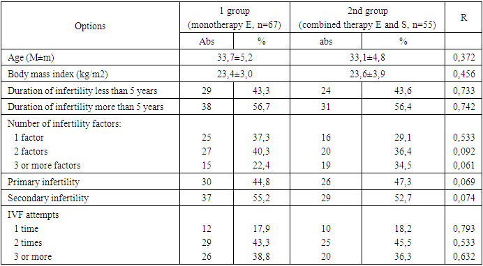
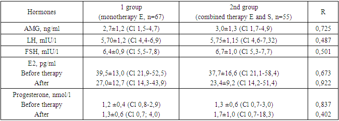
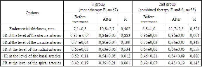
A comparative prospective study was carried out on 122 women with infertility in an IVF program with a diagnosis of dysfunction of the (“thin”) endometrium established during the study (n=122). Depending on the method of therapy, the women were divided into groups: Group 1 consisted of 67 women who were prescribed a drug containing estradiol valeriat at a dose of 2 mg for 3 months (Proginova, France) and a natural progesterone drug (Fetalston) as therapy. , India) at a dose of 100 mg 2 times a day from 16 to 26 days of the cycle; The 2nd group consisted of 55 women who, in combination with hormonal therapy, received the heparinoid sulodexide (Wessel-due F, Italy) at a dose of 1000 units/day (2 tablets 2 times a day) for 3 months.
Inclusion criteria: endometrial thickness less than 7-8 mm in the 2nd phase of the cycle, woman’s age less than 40 years, absence of other uterine pathologies (uterine endometriosis of degree II or more, uterine fibroids larger than 20 mm), severe somatic diseases, absence of contraindications to prescription of estrogens, low risk of thrombotic complications, in the future transfer of no more than 1 embryo of good quality.
We assessed the state of the endometrium after the use of various therapies (endometrial thickness before ET, increase in endometrial thickness (%), the presence of perfusion), the presence of side effects from taking drugs and the outcomes of IVF programs (pregnancy rate, “take baby home” rate and the rate of reproductive losses).
When included in the study at the 2nd-3rd DMC, all women underwent transvaginal ultrasound examination (TVUS) according to the standard method [15,17,18]. To determine the functional activity of the endometrium, its thickness, structure and volume, the nature of the distribution of the color Doppler mapping (CDM) signal in the endometrium and sub endometrial zone were assessed. Monitoring of endometrial growth during the menstrual cycle was carried out at the 8th, 12-14th, 19-24th DMC. On the 3rd day of the menstrual cycle (DMC), the hormonal profile of the patients was assessed. Serum concentrations of anti-Müllerian hormone (AMH), follicle-stimulating hormone (FSH), luteinizing hormone (LH) and estradiol (E2) were determined. At the stage of preparing the endometrium for embryo transfer, patients of group 1 received estradiol vale rate 2 mg (Proginova, France) in tablet form, orally, according to the schedule. Estrogen preparations were used for 3-6 months until a clinical effect was achieved in relation to the thickness and structure of the endometrium, after which an embryo transfer was performed in the subsequent menstrual cycle. In all patients, 14 days after PE, the level of β-hCG in the peripheral blood was determined, and if the value was more than 50 IU/l, the test for pregnancy was considered positive.
Mathematical processing of the results was carried out using descriptive statistics methods using Excel 2010. Descriptive statistics of quantitative variables are presented as medians (Me) and quartiles (Q1; Q3). The confidence interval (CI) is indicated as M (SD), where M is the arithmetic mean and SD is the standard deviation. To determine the statistical significance of differences in quantitative variables in independent samples, nonparametric tests (Mann-Whitney test) were used. Comparative analysis of categorical variables was performed using Student's t test. Differences were considered statistically significant at r.

3. Research Results

A study of the age characteristics of women with infertility who applied for IVF showed that the average age was 33.7±5.2 and 33.1±4.8, respectively (Table 1). This is apparently due to the duration of infertility treatment with other methods. Body mass index indicators in women in the study groups were within unreliably significant limits and amounted to 23.4±3.0 and 23.6±3.9. Determining the duration of infertility in groups revealed that more than half of the women (56% and 55.4%, respectively) who turned to IVF suffered from infertility for more than 5 years.
Table 1. Clinical characteristics of patients
     
Table 2. Hormone levels in the women studied
     
Thus, 62.7% and 71.3% of women had several reasons that led to infertility. Only 1/3 of women had one cause of infertility identified. The most common causes of primary infertility were endocrine factors, and those of secondary infertility were tube-peritoneal factors.
During the history collection, it was found that women had previously attempted IVF: 1 attempt - in 18.2% and 17.4% of women; 2 attempts – in 42.7% and 45.1% of women and 3 or more – in 21.8% and 34.4% of women. The presence of 3 or more attempts indicates a high frequency of unsuccessful attempts, which determines the relevance of research and development of preconception preparation of women for the IVF program.
The hormonal parameters of the women studied after therapy had differences, depending on the correction method, only in relation to estrogens and progesterone. While the study of the level of AMH, LH and FSH was within unreliably significant limits between groups. Thus, the level of AMH was 2.7±1.2 and 3.0±1.3 ng/ml, LH - 5.70±1.2 and 5.75±1.15 mIU/l, FSH - 6.4± 0.9 and 6.7±1.0 mIU/l, which was within unreliably significant limits between groups. The level of estradiol before therapy in women in the study groups averaged 39.5±13.0 and 37.7±16.6 pg/ml, which was within unreliably significant limits between the groups. After monotherapy with estradiol, estrogen levels decreased by 31.6% and amounted to 27.0±12.7 pg.//ml. Whereas, after complex therapy with estradiol and sulodexide, the level of estrogen decreased by 37.9% and amounted to 23.4 pg/ml. Apparently, the combined use of estradiol valerian with sulodexide did not have a negative effect on estrogen levels and did not differ within significantly significant limits from the indicators of women who used estrogen monotherapy.
A study of the effect of various methods of treating “thin” endometrium on progesterone levels showed differences. The values of progesterone level with estrogen monotherapy increased by 8.33% from the initial level, while with complex therapy, the progesterone level increased by 30.8%, which was higher within significantly significant limits of the dynamics in the group with estrogen monotherapy by 22.4%. Consequently, the complex use of estrogen and sulodexide preparations promotes not only the growth of the endometrium, but also improves its functional characteristics by enhancing the processes of angiogenesis in the functionally active layer of the endometrium, which is important for subsequent implantation and placentation.
To confirm our conclusions, at the end of various types of therapy for the “thin” endometrium, we carried out color circulation in the sub endometrial layer and endometrium (Table 3). With estrogen monotherapy and complex therapy with the inclusion of sulodexide, endometrial growth in the groups by the 19th-24th day of the menstrual cycle (DMC) was 10.8±2.7 mm and 11.7±2.5 mm, respectively, and turned out to be comparable (p=0.524). But analysis of endometrial thickness before and after therapy showed a significant increase. Thus, the thickness of the endometrium after complex therapy increased by 1.7 times, and with monotherapy, the endometrium increased by 1.5 times relative to the thickness before treatment.
Table 3. Assessment of the state of the endometrium in the dynamics of various types of therapy
     
Of particular clinical interest were the data we obtained on the relationship between endometrial thickness during the “implantation window” and the nature and distribution of the CDK signal in the sub endometrial layer and endometrium at the end of therapy (Table 3).
Adequate endometrial perfusion was noted in 53 (96.4%) of 55 women receiving combined therapy compared with 44 (65.7%) of 67 women receiving estrogen therapy alone (p < 0.001). Adequate distribution of the CDC signal in the endometrium (at the level of the spiral arteries (SA)) was achieved statistically significantly more often in women who received complex therapy including sulodexide (47 (85.5%) out of 55 compared to 41 (61.2%) out of 67; p=0.046). The peripheral resistance index (RI) at the SA level was statistically significantly lower in patients receiving estrogen monotherapy: 0.39±0.21 compared to 0.43±0.19 (p=0.001), which indicates achievement more adequate perfusion of the endometrium during the “implantation window”.
It is noteworthy that when the thickness of the endometrium was less than 6 mm, it was not possible to register the CDC signal in the subendometrial layer and in the endometrium. With an endometrial thickness of 6.0-8.0 mm, the CDC signal at the level of the basal arteries (BA) and SA was determined in the vast majority of women (81.1%). Whereas, with an endometrial thickness of 8.0-12.0 mm, these blood flow characteristics were determined in 43.6 and 41.8% of patients, respectively. It was noteworthy that with an endometrial thickness of 12.0 mm or more, the opposite trend was observed - a decrease in the number of cases of registration of the color flow signal in the microvasculature from 40.6% to 17.6 and 18.8%, respectively. When analyzing the duration of therapy to restore a functionally adequate endometrium, we identified statistically significant differences between groups. Thus, the average duration of therapy for “thin” endometrium until the moment of PE was statistically significantly shorter in patients receiving complex therapy including sulodexide and amounted to 14.0 ± 4.1 days compared to 16.8 ± 4.7 days in the estrogen monotherapy group (p=0.043). Undesirable moderate side effects of estrogen therapy were noted in 5.5% (n=3) of women in the group receiving complex therapy and in 7.5% (n=5) of women in the group receiving estrogen monotherapy, which was within a non-significantly significant range (p =0.453). Thus, weakness was noted in 2 (2.9%) of 67 women of the 1st group and in 2 (3.6%) of 55 patients of the 2nd group. 3 (4.5%) of 67 women in group 1 and 2 (3.6%) of 55 women in group 2 complained of nausea (p=0.453). Women during therapy also complained of headaches, which in the group with estrogen monotherapy amounted to 6.0% (n=4), in the group with complex therapy - 3.6% (n=2), which was 1.7 times less.
A study of the incidence of clinical pregnancy after 3 months of therapy in groups showed different data. Thus, in the group of women receiving estrogen monotherapy, pregnancy occurred in 43.3% (n=29), while in the group of women receiving therapy including sulodexide, pregnancy occurred in 69.1% (n=38), which is 1. 6 times more often (p<0.05). The onset of clinical pregnancy is an intermediate stage of the IVF program, while the “take baby home” indicator is the final result, which differed in the study groups and amounted to 34.3% (n=23) in the estrogen monotherapy group and 63.6% (n=35) in the group receiving complex therapy (p<0.001). The odds ratio (OR) for pregnancy with endometrial thickness less than 8 mm was 2.0 (95% CI 1.07; 3.81; p=0.031); in the presence of adequate endometrial perfusion - 2.84 (95% CI 1.32; 6.14; p=0.008) and 2.99 (95% CI 0.82; 10.95; p=0.097), respectively.
Thus, the results of our study showed that in case of endometrial dysfunction, the administration of only estrogen preparations solves one problem - it promotes the growth of the endometrium necessary for implantation. Whereas, complex therapy of estrogens with the inclusion of drugs that enhance angiogenesis and the development of spiral arteries, which helps improve blood circulation in the endometrium and sub endometrial layer, which ultimately, in addition to the growth of the endometrium, increases its functional activity.

4. Conclusions

1. When a “thin” endometrium is formed, the implantation process is disrupted, which explains the high frequency of negative results in assisted reproductive technology programs (56.7%). Hormonal therapy increases the thickness of the endometrium, which contributes to an increase in positive results in the IVF program (43.3%).
2. For “thin” endometrium, it is advisable to prescribe estrogen preparations in combination with heparinoids (sulodexide), which ensures adequate growth and perfusion of the endometrium, which increases the effectiveness of the IVF program by 1.6 times (69.1%).

References

[1]  Vartanyan E.V., Devyatova E.A., Tsaturova K.A., Aglyamova D.R. The role of systemic therapy in infertility and failure of reproductive function. // Obstetrics, gynecology and reproduction. 2018; 12(1): 5-16.
[2]  Volkova E.Yu., Korneeva I.E., Silantyeva E.S. The role of uterine hemodynamics in assessing endometrial receptivity. // Problems of reproduction. 2012; 18 (2): 57–62.
[3]  Davydov A.I., Goryachkina V.L., Kuznetsova I.V., Mukhamedova S.G. Molecular biological basis of endometrial decidualization from the standpoint of prevention and treatment of pregnancy loss. // Issues of gynecology, obstetrics and perinatology. 2018; 17(3): 53-8. DOI: 10.20953/1726-1678-2018-3-53-58.
[4]  Krasnopolskaya K.V., Ershova I.Yu., Fedorov A.A. Thin endometrium. Treatment of infertility in women with endometrial hypoplasia. M.: GEOTAR-Media, 2020.
[5]  Kuznetsova I.V., Zemlina N.S., Musina R.A. and others. The use of autologous mesenchymal stem cells to restore the endometrium in patients with reduced fertility. // Issues of gynecology, obstetrics and perinatology. 2019; 18(6): 34-40.
[6]  Motovilova T.M., Kachalina T.S., Zinovieva O.S., Bogatova M.E., Chikalova K.I., Kozlova N.A. The place of endometriopathy in the problem of reproductive failures and options for its correction. // Medical almanac. 2017; 6(51): 80-3.
[7]  Rabadanova A.K., Shalina R.I., Gugushvili N.A., Gavrilina E.N., Denisenko M.V. Pregnancy loss after IVF and its prevention in the first trimester in patients with reduced ovarian reserve. // Issues of gynecology, obstetrics and perinatology. 2017; 16(6): 36-43. DOI: 10.20953/1726-1678-2017-6-36-43.
[8]  Shneiderman M.G. The problem of thin endometrium: possibilities of combined non-hormonal treatment in preparation for the in vitro fertilization procedure. // Gynecology. 2021; 3: 67–71.
[9]  Yashchuk A.G., Maslennikov A.V., Fatkullina I.B., Rakhmatullina I.R., Berg E.A., Berg P.A. The use of sulodexide to improve intrauterine hemodynamics in patients with miscarriage. // Obstetrics and gynecology. 2019; 10: 172-9.
[10]  Yashuk A.G., Maslennikov A.V., Dautova L.A., Fatkullina I.B., Popova E.M., Gazizova G.Kh. and others. Differentiated approach to the management of patients with “thin” endometrial syndrome. // Russian Bulletin of Obstetrician-Gynecologist. 2019; 19(2): 52-6. DOI:0.17116/rosakush20191902152.
[11]  Alfer J., Happel L., Dittrich R. et al. Insufficient Angiogenesis: Cause of Abnormally Thin Endometrium in Subfertile Patients? // Geburtshilfe Frauenheilkd. 2017; 77(7): 756-764. https://doi.org/10.1055/s-0043-111899.
[12]  Azumaguchi A, Henmi H, Ohnishi H et al. Role of dilatation and curettage performed for spontaneous or induced abortion in the etiology of endometrial thinning. // J. Obstet. Gynaecol. Res. 2017; 43(3): 523–9.
[13]  Bashiri A., Halper K.I., Orvietto R. Recurrent Implantation Failure update overview on etiology, diagnosis, treatment and future directions. // Reproductive Biology and Endocrinology: 2018; 16(1): 121. https://doi.org/10.1186/s12958-018-0414-2.
[14]  Bu Z., Wang K., Dai W. et al. Endometrial thickness significantly affects clinical pregnancy and live birth rates in frozen-thawed embryo transfer cycles. // Gynecol Endocrinol 2016; 32 (7): 524–8. 14. Bu Z., Wang K., Dai W. et al. Endometrial thickness significantly affects clinical pregnancy and live birth rates in frozen-thawed embryo transfer cycles. // Gynecol Endocrinol 2016; 32(7): 524–8.
[15]  Check J. The importance of sonographic endometrial parameters in influencing success following embryo transfer in the modern era and therapeutic options – part 1: the importance of late proliferative phase endometrial thickness. // Clin. Exp. Obstet. Gynecol. 2018; 38(3): 197–200.
[16]  Chen S., Wu F., Luo C. et al. Combined analysis of endometrial thickness and pattern in predicting outcome of in vitro fertilization and embryo transfer: a retrospective cohort study. // Reprod. Biol. Endocrinol. 2019; 24: 30.
[17]  Corbacioğlu A., Baysal B. Effects of endometrial thickness and echogenic pattern on assisted reproductive treatment outcome. // Clin. Exp. Obstet. Gynecol. 2019; 36 (3): 145–7.
[18]  Elleuch N., Zidi H., Bellamine Z., Hamdane A., Guerchi M., Jellazi N., CVD study investigators. Sulodexide in Patients with Chronic Venous Disease of the Lower Limbs // Clinical Efficacy and Impact on Quality of Life. Adv. Ther. 2016; 33: 1536-49. DOI: 10.1007/s12325-016-0359-9.
[19]  Fang R., Cai L., Xiong F. et al. The effect of endometrial thickness on the day of hCG administration on pregnancy outcome in the first fresh IVF/ICSI cycle. // Gynecol. Endocrinol. 2016; 32 (6): 473–6.
[20]  Kasius A., Smit J., Torrance H. et al. Endometrial thickness and pregnancy rates after IVF: a systematic review and meta-analysis. // Hum. Reprod. Update 2019; 20 (4): 530–41.
[21]  Khan M., Shaikh A., Ratnani R. Ultrasonography and Doppler Study to Predict Uterine Receptivity in Infertile Patients Undergoing. // Embryo Transfer. J. Obstet. Gynaecol. India 2016; 66 (Suppl. 1): 377–82.
[22]  Kuijsters N.P.M., Methorst W.G., Kortenhorst M.S.Q., Rabotti C., Mischi M., Schoot B.C. Uterine peristalsis and fertility: current knowledge and future perspectives: a review and meta-analysis. // Reproductive Biomedicine Online. 2017; 35(1): 50-71. https://doi.org/10.1016/j.rbmo.2017.03.019.
[23]  Lebovitz O., Orvieto R. Treating patients with "thin" endometrium – an ongoing challenge. // Gynecol. Endocrinol. 2021; 30 (6): 409–14.
[24]  Liu S.M., Zhou Y.Z., Wang H.B., Sun Z.Y., Zhen J.R., Shen K., Deng C.Y., Lang J.H. Factors Associated with Effectiveness of Treatment and Reproductive Outcomes in Patients with Thin Endometrium Undergoing Estrogen Treatment. // Chinese Medical Journal. 2015; 128(23): 3173-3177. https://doi.org/10.4103/0366-6999.170258.
[25]  Ma N., Chen L., Dai W. et al. Influence of endometrial thickness on treatment outcomes following in vitro fertilization/intracytoplasmic sperm injection. // Reprod. Biol. Endocrinol. 2017; 15 (1).
[26]  Park H.J., Kim Y.S., Toon T.K., Lee W.S. Chronic endometritis and infertility. // Clinical and Experimental Reproductive Medicine. 2016; 43(4): 185-192. https://doi.org/10.5653/cerm.2016.43.4.185.
[27]  Yuan X., Saravelos S., Wang Q. et al., Endometrial thickness as a predictor of pregnancy outcomes in 10787 fresh IVF-ICSI cycles. // Reprod. Biomed Online 2016; 33 (2): 197–205.