-
Paper Information
- Paper Submission
-
Journal Information
- About This Journal
- Editorial Board
- Current Issue
- Archive
- Author Guidelines
- Contact Us
American Journal of Medicine and Medical Sciences
p-ISSN: 2165-901X e-ISSN: 2165-9036
2024; 14(5): 1337-1339
doi:10.5923/j.ajmms.20241405.40
Received: Apr. 15, 2024; Accepted: May 10, 2024; Published: May 21, 2024

Morphological Aspects of Adenotonsillar System Changes
 Abstract
 Reference
 Full-Text PDF
 Full-text HTML
Abdusamatova Iroda Ilkhamovna , Tastanova Gulchekhra Eshtayevna , Shamsiev Djakhangir Fazlitdinovich
Tashkent State Dental Institute, Tashkent, Uzbekistan
Copyright © 2024 The Author(s). Published by Scientific & Academic Publishing.
This work is licensed under the Creative Commons Attribution International License (CC BY).
http://creativecommons.org/licenses/by/4.0/

The incidence rate of adenotonsillar system pathology is increasing steadily over the past decade and accounts for 61.2% of all common and primary diseases. This pathology is taking a leading place among otorhinolaryngological diseases in school-age and younger children, constituting 16-50% of upper respiratory tract diseases. It should be emphasized that the incidence rate of adenotonsillar system diseases is increasing annually. These indicators pose significant challenges for ENT specialists.
Keywords: Adenotonsillar system, Nasal breathing impairment, Children, Morphology, Age
Cite this paper: Abdusamatova Iroda Ilkhamovna , Tastanova Gulchekhra Eshtayevna , Shamsiev Djakhangir Fazlitdinovich , Morphological Aspects of Adenotonsillar System Changes, American Journal of Medicine and Medical Sciences, Vol. 14 No. 5, 2024, pp. 1337-1339. doi: 10.5923/j.ajmms.20241405.40.
1. Introduction
- Operations in the adenotonsillar system constitute the main volume of surgical interventions in pediatric otolaryngology. Operations on the palatine tonsils due to adenoid vegetations causing nasal breathing obstruction occupy a leading position in this regard.Although the recurrence of adenoids is monitored in many cases, the need for adenoidectomy surgery, which leads to various complications, is increasing. [2,3] However, it is essential to emphasize the importance of monitoring adenotonsillar tissues as vital lymphoepithelial organs and the high incidence of complications both during and after surgery. The presence of endocrinocytes resembling those located in the anterior part of the pituitary gland in the parenchyma of the palatine tonsils, entering the Pirogov-Valdeyer ring, indicates their difference from other lymphoid organs.Objective: To study the morphological changes of adenotonsillar glandular tissues and the significance of some factors in the development of adenoid vegetations against the background of impaired nasal breathing.
2. Materials and Methods
- A total of 180 children aged 3 to 12 years were divided into two clinical groups. The first clinical group included 85 patients diagnosed with adenoid vegetations of II-III degree along with nasal septum deviations. [4,6] The second clinical group consisted of 96 children with chronic rhinosinusitis (nasal breathing impairment) at various stages of adenoid vegetation.To conduct the planned research, patients were distributed among the main groups based on the "addition/removal" zones.Twenty healthy children aged 3 to 7 years, who were trained in educational institutions in Tashkent city, were selected for the control group. The selected children had no respiratory or allergic conditions in their medical history, and their perinatal period was uneventful. [5,7]All patients underwent comprehensive clinical and anamnestic examination, including history taking, physical and instrumental examination, examination by otolaryngologists and other specialists as necessary, general laboratory, and histological examination methods. Endoscopy, MSCT, 3D X-ray, and histological examination of adenoid biopsy were performed to diagnose and identify concomitant pathologies in patients. Histological examination of adenoid tissues obtained by surgical removal was carried out using standard methods at the PUB center in Tashkent.
3. Results and Discussions
- A total of 180 children aged 3 to 12 years were divided into two clinical groups. The first clinical group included 85 patients diagnosed with adenoid vegetations of 2-3 degrees along with nasal septum deviations. Of the total number of patients, 45 (57.2%) were boys, and 40 (45.1%) were girls; the average age was 4.8±1.25 years. [7,8] According to age indicators, in the first group, on average, 4 years – 30 (34.4%) children, among them girls – 19 (21.7%), were observed; boys (11 children) accounted for 12.6%. The second clinical group consisted of 96 children with chronic rhinosinusitis (nasal breathing impairment) at various stages of adenoid vegetations. Of them, 60 (72%) were boys and 36 (42.3%) were girls; the average age was 4.6±1.17 years. According to age indicators, in the second group, on average, 4 years – 35 (41.6%) children, among them boys – 22 (26.2%), were observed; girls (10 children) accounted for 11.4%.
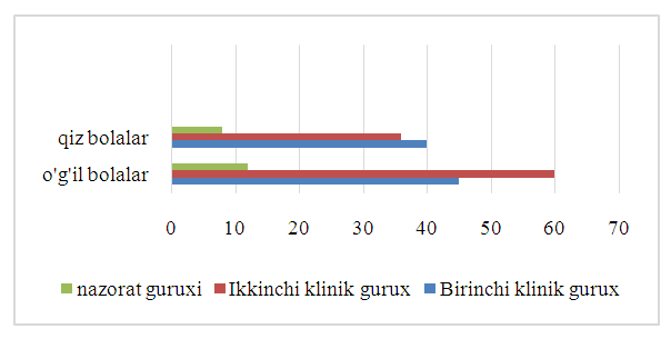 | Figure 1. Grouping of Children by Gender |
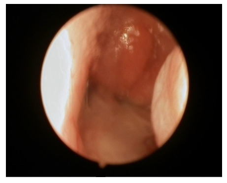 | Figure 2. Nasopharynx before endoscopy. Patient aged 3, disease history № 14714/705 |
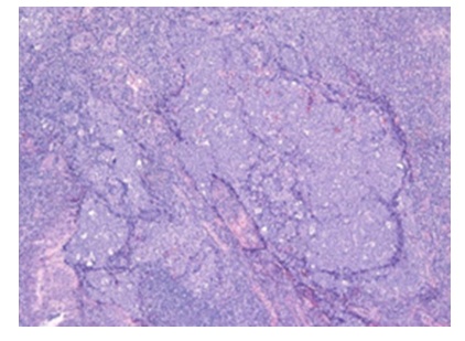 | Figure 3. Bifollicular aberration of the tonsil in a 3-year-old boy. Stained with hematoxylin and eosin, magnification 1×10 |
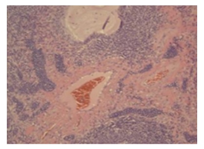 | Figure 4. The patient, 7 years old, a male child, with lymphatic vessels expanded with lymphocytic stasis and proliferation of delicate collagen bundles in the stroma |
4. Conclusions
- Various factors influencing the hypertrophy of adenotonsillar system lymphoid tissues in the context of nasal breathing disorders were identified through the conducted research. Based on the results of questionnaires administered to parents of children with adenoid vegetation and adenoiditis, prognostic aspects of prenatal factors in the development of the disease were investigated, revealing hereditary predispositions of children to the harmful habits of their parents (smoking, alcohol consumption), as well as a hereditary predisposition of parents to rhinosinusitis and chronic tonsillitis.It was found that 80% of parents of healthy control group children did not report allergic conditions. A significant correlation was identified between allergic conditions and pet ownership among parents in both the first and second clinical groups (r<0.001, Z=-8.250), which was not observed in healthy control group parents (r=0.041, Z=-2.041).The results of the questionnaires administered to parents provided insights into the transition of parental harmful behavior and the peculiarities of the early postnatal period in children. In the healthy control group, 80.6% of parents reported no harmful behaviors during the perinatal period. Meanwhile, among the children with nasal breathing disorders, this indicator constituted 42.3% (r<0.001). The impact of harmful behaviors on the perinatal period, maternal toxemias, the risk of premature birth, and maternal susceptibility to infectious diseases during the perinatal period contributed to the prognostic significance (r<0.001).