-
Paper Information
- Next Paper
- Previous Paper
- Paper Submission
-
Journal Information
- About This Journal
- Editorial Board
- Current Issue
- Archive
- Author Guidelines
- Contact Us
American Journal of Medicine and Medical Sciences
p-ISSN: 2165-901X e-ISSN: 2165-9036
2024; 14(5): 1281-1284
doi:10.5923/j.ajmms.20241405.27
Received: Apr. 20, 2024; Accepted: May 2, 2024; Published: May 14, 2024

Possibilities of Modern Radiation Methods in the Diagnosis of Liver Echinococcosis and Its Complications
 Abstract
 Reference
 Full-Text PDF
 Full-text HTML
Mamanov Muxammad Chorievich, Arziev Ismoil Alievich, Arzieva Gulnora Borievna
Samarkand State Medical University, Samarkand, Uzbekistan
Copyright © 2024 The Author(s). Published by Scientific & Academic Publishing.
This work is licensed under the Creative Commons Attribution International License (CC BY).
http://creativecommons.org/licenses/by/4.0/

All 118 patients suffering from liver echinococcosis and its complications underwent comprehensive diagnostics. In each of the 118 cases, the presence of liver echinococcosis and its complications was confirmed using combined ultrasound and computed tomography (CT) methods. The diagnostic algorithms that have been developed make it possible to choose different surgical tactics, taking into account the location, size and connection of echinococcal cysts with the vascular and secretory structures of the liver, the stage of the parasite’s life cycle and the type of complications that have developed. Differentiated surgical tactics depending on the type of echinococcal cysts, their location, number and presence of complications allows the priority use of low-traumatic methods as the final method of treatment or as a stage of preliminary sanitation of a purulent focus in case of endogenous intoxication due to suppuration of cysts and breakthrough into the bile ducts.
Keywords: Liver echinococcosis, Consequences, Diagnosis, Minimally invasive methods, Radiation methods
Cite this paper: Mamanov Muxammad Chorievich, Arziev Ismoil Alievich, Arzieva Gulnora Borievna, Possibilities of Modern Radiation Methods in the Diagnosis of Liver Echinococcosis and Its Complications, American Journal of Medicine and Medical Sciences, Vol. 14 No. 5, 2024, pp. 1281-1284. doi: 10.5923/j.ajmms.20241405.27.
1. Relevance
- Radiation methods such as ultrasound, computed tomography (CT) and magnetic resonance imaging (MRI) are the main diagnostic methods for liver echinococcosis (EP) [4]. Their use makes it possible to more accurately visualize the cyst, determine its interaction with vascular structures and bile ducts, as well as determine the size, which helps to choose the most appropriate access for surgery [1,3,9]. These methods have the advantage that they allow the disease to be detected at an early stage, when clinical manifestations are minimal [2,5], which allows operations to be performed with less risk to the patient. Some researchers believe that intraoperative ultrasound is necessary to identify hidden cysts and prevent the development of residual echinococcosis [6,7].Compared to ultrasound (ultrasound), computed tomography (CT) offers a more sensitive image and a panoramic image. Thanks to this method, it is possible to carry out detailed differential and topical diagnostics, determine the location, volume and number of the cyst, as well as evaluate its interaction with blood vessels and bile ducts. Thanks to these data, the doctor can choose the most appropriate method, access and volume of surgery, as well as anticipate possible risks of intraoperative complications.Magnetic resonance imaging (MRI) studies in echinococcosis are considered comparable in their diagnostic effectiveness to computed tomography (CT), while MRI does not use ionizing radiation [10]. Radiation diagnostic methods such as ultrasound, computed tomography (CT) and magnetic resonance imaging (MRI) can determine the location, size, number and stage of development of the parasite, as well as its interaction with surrounding structures. This contributes to the correct choice of access and method of surgical intervention [8].It is important to note that in addition to diagnosing echinococcosis itself, it is important to identify its complications, such as infection and suppuration of the cyst, its breakouts, the presence of cystobiliary fistulas, as well as determining the type and degree of liver damage. The purpose of the study. Modern methods of radiation examination improve the diagnosis of liver echinococcosis and its complications.
2. Material and Methods
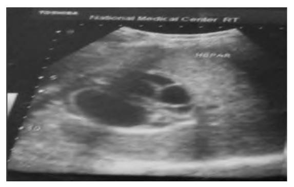
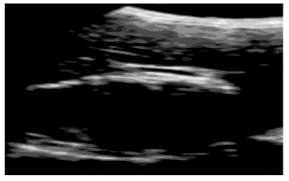
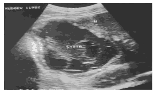
- The study was conducted at the Faculty of Surgical Pathology No. 1 of the University of Samarkand. Analytical processing of diagnostic and surgical results was performed on 118 patients with liver echinococcosis and its complications.The study period covers the years 2018 - 2023. All 118 patients admitted to the hospital underwent ultrasound examination, which was an effective method of diagnosing liver echinococcosis and its complications. Comprehensive ultrasound diagnostics (ultrasound) made it possible to determine the size, location and nature of the contents of cysts, the thickness of the walls of cysts, the structure of the liver, echogenicity and their connection with large intrahepatic vessels and ducts. The data obtained helped to decide on the best access and type of surgery.Ultrasound signs of echinococcal cysts appear as rounded formations containing liquid, with obvious negative echogenicity. The brushes have clear borders, a thickened wall, a double contour and calcification of the fibrous capsule. In addition, daughter blisters are often found inside the cyst; this usually helps to distinguish them from cysts of a different nature.An echinococcal cyst of the liver has a compacted and thick wall around its contour. As a result of the presence of fibrous and chitinous capsule shells, two-circuit is usually observed.According to the BO3 classification, CL type echinococcal bones are characterized by low echogenicity and a clear, even (rarely moderately uneven) contour. (Fig. 1). The echinococcal cyst does not have an echogenic suspension or other inclusions, and it does not have a noticeable hyperechoic contour. The symptom of "fish scales" was detected in 61% of cases and is one of the characteristic signs of EP and its complications. The formation of a thickened fibrous capsule and the presence of several daughter cysts are signs of long-term echinococcal cysts. On ultrasound, daughter cysts appear as heterogeneous rounded inclusions that are in close contact with each other, and the maternal cyst often has destroyed contents. (Fig. 2).
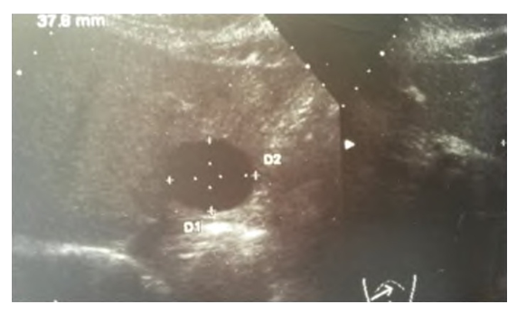 | Figure 1. Sonogram. Echinococcal cyst of the liver (4 cm) type CL |
 | Figure 2. Sonogram. Multiple echinococcal liver cysts type CE2 |
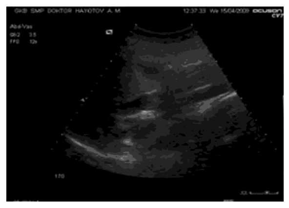
 | Figure 3. Sonogram. Echinococcal cyst type CE3 |
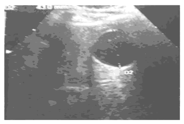
 | Figure 4. Sonogram. Echinococcal cyst type CE4 |
 | Figure 5. Sonogram. Echonegative cysts type CE5 |
 | Figure 6. Sonogram. Suppurated echinococcal cyst of the liver with a thinned capsule and daughter vesicles type CE2. |
 | Figure 7. Sonogram. The breakthrough of an echinococcal cyst into the bile ducts |
 | Figure 8. Sonogram. Recurrent echinococcal cyst type 1 |
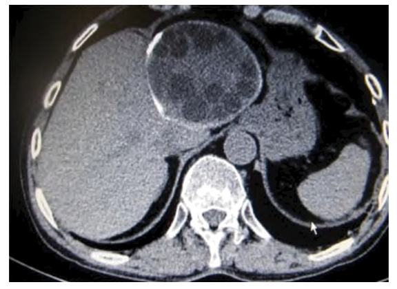 | Figure 9. CT scan of an echinococcal cyst of the left lobe of the liver with a picture of its death and corresponding changes |
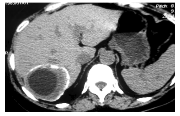 | Figure 10. CT scan of recurrent echinococcosis of the liver. Calcified fibrous capsule |
3. Conclusions
- The use of combined methods of radiation diagnostics, such as ultrasound and computed tomography, provides effective detection of liver echinococcosis and its complicated forms. The developed diagnostic strategies make it possible to determine individual surgical tactics, taking into account the location and size of echinococcal cysts, their relationship with the vascular and biliary structures of the liver, the stage of development of the parasite and the nature of the complications that have arisen.