American Journal of Medicine and Medical Sciences

p-ISSN: 2165-901X    e-ISSN: 2165-9036

2024;  14(5): 1253-1266

doi:10.5923/j.ajmms.20241405.23

Received: Mar. 13, 2024; Accepted: Apr. 20, 2024; Published: May 13, 2024


Pathomorphological and Biochemical Assessment of Its Effectiveness Using a New Topical Drug in the Complex Treatment of Acute Paraproctitis

Yakubov Davron Ruslanovich, Zokhirov Adkhamjon Rafiqovich

Assistant of Department of General Surgery No.2, Tashkent Medical Academy, Tashkent, Uzbekistan

Correspondence to: Zokhirov Adkhamjon Rafiqovich, Assistant of Department of General Surgery No.2, Tashkent Medical Academy, Tashkent, Uzbekistan.

Email:

Copyright © 2024 The Author(s). Published by Scientific & Academic Publishing.

This work is licensed under the Creative Commons Attribution International License (CC BY).
http://creativecommons.org/licenses/by/4.0/

Abstract

Acute paraproctitis (AP) is one of the most common purulent surgical diseases. It account for approximately 0,5-4% of hospitalizations and surgical hospitals in 24-50% of hospitalizations for proctological diseases. The op is not limited to loc change and tissue damage. Purulent-necrotic mass and general morphofunctional and pathophysiological cause of the change in loc. Intoxication and reduce To speed up wound healing, antiseptic and antibiotic therapy in addition to local treatment, it is advisable to use the drug detoxification. The drug suksinasol is an infusion that has considered a drug detoxification, and cell metabolism affects the case in antihypoxic effects of various cell damage. In case of acute paraproctitis, the correct choice of surgical tactics and the use of detoxification, wound-healing drug suksinasol will help prevent postoperative complications and a speedy recovery of patients.

Keywords: Acute paraproctitis, Reosorbylact, Succinasol, Pelviorectal paraproctitis, Ishiorectal paraproctitis, Subcutaneus paraproctitis

Cite this paper: Yakubov Davron Ruslanovich, Zokhirov Adkhamjon Rafiqovich, Pathomorphological and Biochemical Assessment of Its Effectiveness Using a New Topical Drug in the Complex Treatment of Acute Paraproctitis, American Journal of Medicine and Medical Sciences, Vol. 14 No. 5, 2024, pp. 1253-1266. doi: 10.5923/j.ajmms.20241405.23.

1. Introduction

Intruduction. Acute paraproctitis (op) is one of the most common purulent surgical diseases. Patients in this group account for about 0.5-4% of hospitalizations in various surgical hospitals and 24-50% of those with proctological diseases.The issues of the treatment of acute paraproctitis, despite their long history, are constantly discussed in the domestic and foreign literature and still remain relevant. The prolonged course of the disease in the postoperative period, the formation of a wound that cannot be treated for a long time, a leak of the rectum causes discomfort for the patient and reduces his performance. Acute paraproctitis is not limited to local changes and tissue damage. Purulent-necrotic masses are local and general multifaceted and cause specific morphofunctional, pathophysiological changes.
Errors in the diagnosis of the clinical picture of acute paraproctitis, improper selection of the operating method, failure to eliminate the internal opening of the primary "leak" in the rectum wall, as well as insufficient conduct of complex treatment measures in the postoperative period lead to the development of various complications. Recurrence of the disease in the postoperative period was noted in 8-12%, the formation of rectum leaks - 5-7.5%, postoperative insufficiency of the anal sphincter - 8-16%.
In acute paraproctitis, the formation of postoperative complications and the observation of recessive paraproctitis require optimal surgical tactics for the treatment of the disease.
In severe forms of acute paraproctitis, the wound is cleaned of purulent-necrotic tissues and regeneration is slow, general intoxication is observed in the ham. In order to reduce intoxication and accelerate the completion of jaroxat, it will be advisable to use disinfecting drugs in addition to maxillary antiseptic processing and antibiotic therapy. The drug succinasol is an infusion drug that has a detoxifying, antitipoxic effect and affects cell metabolism in various cell lesions. The yantaric acid contained in succinazole is a tabic metabolite of the Krebs cycle in cell metabolism, increasing the production of ATF. For this reason, it increases microcirculation in the tissue, enhances regeneration, promotes the rapid recovery of hemodynamics.
Today, large-scale measures have been implemented in our country to provide the population with affordable and high-quality medicines and imported substitute medicines from domestic raw materials. “...in the field of further development of the pharmaceutical network, improvement of the supply of affordable quality medicines to the population and medical institutions”, important tasks are set out. In this regard, on the basis of domestic raw materials, it is important to satisfy the need of the population for cheap pharmaceutical products due to the creation of new drugs, the activity of which is not inferior to foreign analogues.

2. Material and Research Methods

Characteristics of experimental materials.
Experimental studies were conducted in 84 male-sex bats with a weight of 220-250 g, kept in the TTA vivarium. To create a model of acute paraproctitis in the ecperiment, S.V.The SHaxray method was used.
To do this, the rat fresh feces were mixed with distilled water in a 1:4 ratio, passed through 5 layers of gauze and prepared a suspension. The assistant held the labaratory rat behind him under ether narcosis, and 0.5 cm from the anal sac was injected into the mucous membrane from the inside with a solution of 0.1-0.2 ml of 10% calcium chlorine. With the same nina, a ready-made autocal suspension was injected into the tissue of the subcutaneous and rectum in an amount of 0.5-0.7 ml.
In Stage 1, a model of acute paraproctitis was performed in 6 bats, and the model was confirmed. In Phase 2, acute paraproctitis was induced in 54 rats for the study, and on Day 5 of the study, a purulent cavity was cut open. Treatment-research work was conducted in equal 2 groups:
1- after cutting and opening the acute paraproctite in the examining guru, the maxillary processing (washed with 3% hydrogen peroxide, levomecol maz was put on), no detoxification treatment was carried out;
2- after cutting and opening acute paraproctitis in rats in the main group, the drug Reosorbylact was taken 1 ml/100 g of body weight per day for the purpose of detoxifying treatment.
3- after cutting and opening acute paraproctitis in rats in the main group, the drug Succinasol was taken to a body weight of 1 maxal 1 ml/100 g per day for the purpose of detoxifying treatment.
Studies were carried out in dynamics from 1, 3, 6 and 10 days after the start of treatment. In all groups, the fatal outcome was not recorded until the end of the experiment (10 days). At the appointed time, the general condition of the animals was assessed, the injury area was measured, the injury was photographed and the degree of regeneration was determined. At the appointed time, the rats were removed from the experiment by decapitation, the blood for hematological and biochemical studies, the rectum for morphological studies, and the surrounding tissue were cut Whole.
AP overview with pain from patient.
Tashkent Medical Academy 2-Department of general surgery, TTB surgical stripes clinical base and private clinic "healthy life" in the period 2019-2022 op various clinical forms bop 162 students were presented with the results of the analysis on the examination and treatment of the patient. Anaerobic paraproctitis patients did not receive scientific research work.
Patients with a large part of the acute diagnosis of primary paraproctitis thethe year - acute recurrent paraproctitis (Figure 1) - without 121 (74.7% was), only 41 (25.3%).
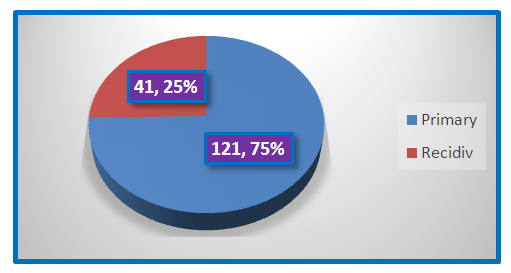
Figure 1. Types of acute paraproctitis
According to the purulent process for localization, many are recorded in the cell cavity under the skin-68 (41.9%) patients have superficial (under the skin) lungs. For 62 (38.2%) who are patients with purulent flare when the ishiorectal is full, doctors in patients with advanced pelvic-thoracic edema - 32 (19.7%) (Figure 2).
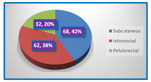
Figure 2. The clinical form of acute paraproctitis is frequency
It turns out that the disease is more common in men. Thus, 101 (69.2%) of the 162 acute paraproctitis patients we observed were male. Among the patients who underwent surgery, there were 61 (30.8%) women. The incidence rate of acute paraproctitis in men and women was 2.25: 1, which corresponds to the statistics of the whole of Uzbekistan.
The spread of the inflammatory process, the lengthening of the duration of treatment and the development of complications occur due to the late referral and late hospitalization of patients with acute paraproctitis, the inability to perform surgical intervention in a timely manner. The periods of hospitalization of patients with various clinical forms of acute paraproctitis were analyzed (Table 1).
Table 1. Op various clinical forms provide hospital construction patients during the bull period
     
Acute parapructitis information for relapse surgery is presented in Table 2.
Table 2. Acute paraproctitis for relapse operations
     
Recurrent forms of acute paraproctitis were observed in 41 (25.3%) patients. The highest faizi of relapses were found in 11 (26.8%) patients with ishiorectal 24 (58.5%) and pelviorectal paraproctitis. In superficial forms of acute paraproctitis, 14.6% recidivism has been recorded. Analyzing the data in Table 4, we can conclude that in the absolute sense, superficial acute paraproctitis repeats almost the same as ischiorectal acute paraproctitis. However, when focusing the number of relapses on the number of patients in the form of acute paraproctitis, it turned out that subcutaneous paraproctitis gives the least relapses. The clinical picture of various forms of acute paraproctitis has many general manifestations, each of which is characterized by certain local manifestations. To some extent, common signs characteristic of acute paraproctitis have been identified in all its clinical manifestations (Table 3).
Table 3. Common symptoms in patients with acute paraproctitis
     
Table 4. Local manifestations of acute paraproctitis
     
Characteristic local manifestations of acute paraproctitis are pain in the anus, rectum, perineum, defecation, and impaired pus separation act (Table 4).
When examining the rectum with a finger, valuable diagnostic information was obtained in patients with 158 (97.5%) acute paraproctitis: pain, infiltration, thickening, smoothness of the folds of the mucous wall of the rectum, the presence of infiltrates in pararectal tissues, its size and boundaries, bulging into the intestine, varonkasimon-shaped location in one or another anal crypt socket. Due to severe pain in the Perianal area, this study was not carried out in 4 (2.5%) patients.
In 4 (2.5%) patients with acute pelviorectal paraproctitis, a bimanual examination helped determine the localization of the foci of inflammation.
All patients underwent surgery under spinal anesthesia. In the treatment of patients, three types of surgical assistance were carried out: opening the purulent cavity, opening the purulent cavity into the intestinal cavity, opening paraproctitis with ligature through the inner hole of the purulent path, and opening the abscess later with delay of the inner hole.
All patients are divided into 2 groups: control and basic. The control group included 92 (56.8%) patients with acute paraproctitis, with the use of traditional antiseptics (3% washed with peroxide and levomecol mazi applied) in the treatment of perianal wounds in the postoperative period. The drug Reosorbylact in intoxication reduction maxadi was infused intravenously for 3-5 days, 400 ml (6-7 ml/1 kg of body weight) per day. The main group consisted of 80 (43.2%) patients, to whom traditional antiseptics were used in the treatment of perianal wounds, and the drug Succinasol in intoxication reduction maxad was infused intravenously for 3-5 days, 400 ml (6-7 ml/1 kg of body weight) per day.
Properties of the drugs rheosorbylact and Succinasol.
1 liter of Reosorbylact contains 6 g of sodium chloride, 0.3 g of potassium chloride, 0.1 g of calcium chloride, 0.2 g of magnesium chloride, 19.7 g of sodium lactate and 60 g of sorbitol. The balanced cationic composition of the blood, ions K+, Ca2+, Mg2+ have a positive effect on the normalization of cardiac activity and nerve conduction. A large amount of lactate contributes to a significant increase in the alkaline reserve of the carbon buffer in the blood plasma, which is very important for the prevention and elimination of acidosis. The isotonic concentration of sorbitol promotes the recovery of energy reserves, improves microcirculation and tissue perfusion, stimulates the development of collateral circulation and reduces blood hypercoagulation.
Succinasol is a complex drug with antigypoxic, antioxidant, rheological, anti-shock, detoxifying, metabolic effects (see "REKA-MED FARM" LLC QK). The main pharmacologically active ingredient is yantaric acid. Yantaric acid is a natural metabolite of the Krebs cycle and has the ability to oxidize in tissues when oxygen pressure decreases in tissues and oxygen oxidation of NAD-dependent substrates ends. Considering that the rate of phosphorylation of yantaric acid is much higher than that of NAD-dependent substrates, more ATF is synthesized per unit time due to oxidation of yantaric acid.
The drug has an effect that regulates water-salt metabolism and activates energy metabolism, normalizes the acid-base state not only due to passive neutralization of fully oxidized products, but also due to the normalization of metabolic processes in cells. Improves microcirculation during various bleeding, strokes, physical movements, restores hemodynamic parameters and heart muscle activity. The effect of the drug appears 20-30 minutes after infusion. Restores hemodynamic indicators and activity of the heart muscle during various bleeding, strokes, physical movements.

3. Clinical Research Methods

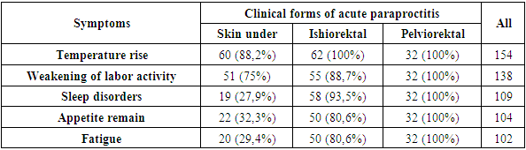
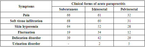
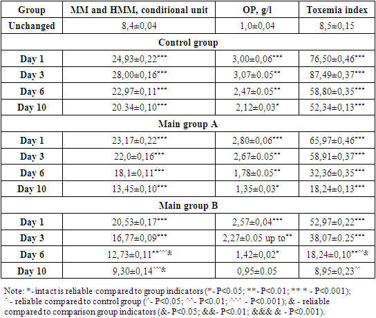
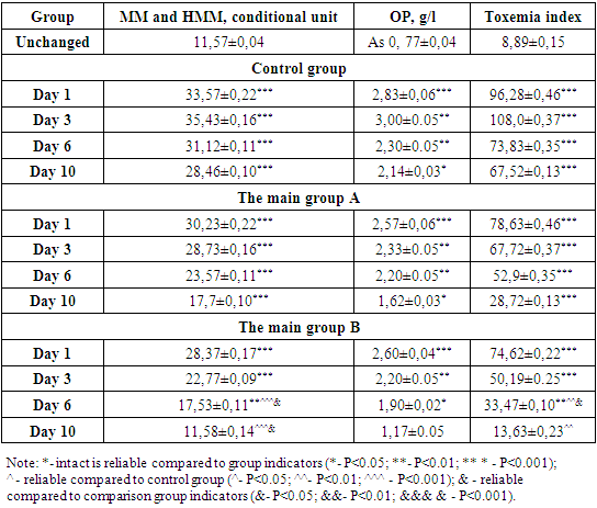
Assessment of the general condition of experimental animals. The development of the disease was assessed according to the condition of the animals, clinical manifestations of op (general depletion, increased level of malaise) were noted. Animal wool usually has a specific brightness, and changes in color characterize their condition.
Assessment of the condition of the wound and the area of purulent jarochat. Analysis of the effectiveness of the drug was carried out on the basis of a visual examination of animals and their wounds. The criteria for the effectiveness of the drug for the wound were:
Ø degree and duration of inflammatory manifestations in the wound area (edema, hyperemia, wound exudate);
Ø cleansing jarokhat from pus;
Ø condition of the wound bottom;
Ø formation of granulation tissue;
Ø reducing the area of the wound defect;
Ø marginal epithelial appearance;
Ø accelerate wound healing;
Examination of patients. Symptoms of acute paraproctitis were identified by begging, complaints and Anamnesis collection, examination, palpation. In the Perianal area, the time, nature, dynamics of development, temperature reaction, stool and urination disorders of pain in the anus were studied, as well as microtrauma of the rectum during defecation, previous diseases of the rectum and anus, hypothermia, etc. The examination of patients revealed the general condition of the skin, turgor and color, gait, shape of the anus, the presence of signs of inflammation in the perianal soxa. Palpation of the anus has been used to determine tissue pain, infiltrate size, and fluctation. Local manifestations characteristic of acute paraproctitis have been identified in the rectum (pain, swelling, infiltration, smoothness of mucous membrane folds, the presence of Infiltrate in pararectal tissues, its borders, bulging into the intestinal cavity, etc.).
Hematological research methods. The study of peripheral blood was carried out in a hematological analyzer with the calculation of the leukoformula. Based on the results obtained, the leukocyte index of intoxication was calculated in accordance with the recommendations of the Kalf-Kalif. On the basis of hematological parameters, the sliding index (SI), the leukocyte intoxication index (LII) and the neutrophil reactive response (NRR) were calculated.
Biochemical research. The activity of common protein, glucose, creatinine, urea, ALT, and AST enzymes was determined using a set of chemical reagents produced by Human (Germany) in the Mindray BA-88A (China) biochemical analyzer photometer.
Specific research methods. According to modern concepts, endogenous intoxication (EI) is a measure of the body's metabolic reaction to any aggressive factor. However, it should be noted that the variety of toxic substances does not allow creating an optimal method of clinical laboratory analysis. The severity of endogenous intoxication syndrome was assessed by integral blood parameters displacement index (DI), leukocyte intoxication index (LII), and neutrophil reactive reaction (NRR) high mass molecules (HMM) and moderate mass (MM), oligopeptides (OP) in erythrocytes, blood plasma, and erythrocyte sorption capacity (ESC).
Histological research methods. The work used the research methods described in the classical guides to histomorphology. Rats are trapped in a solution of rectum and surrounding tissue Carnoy (fixative composition: icy acetic acid, 10 parts; chloroform, 30 parts; ethyl alcohol, 60 parts). Samples are stained with hematoxylin and eosin. Microscopic studies were conducted under the Nikon Etslipse E-400 light microscope (Japan).
Method of statistical analysis. Statistical processing of digital data was carried out for practical applications using SPSS 16.0 and Windows Statistitsa 6.0. Mean values and standard deviations, medians and quartile ranges, as well as non-parametric methods (Mann-Whitney, Wilcohon, Kruskal-Wallis tests) were identified.

4. Results

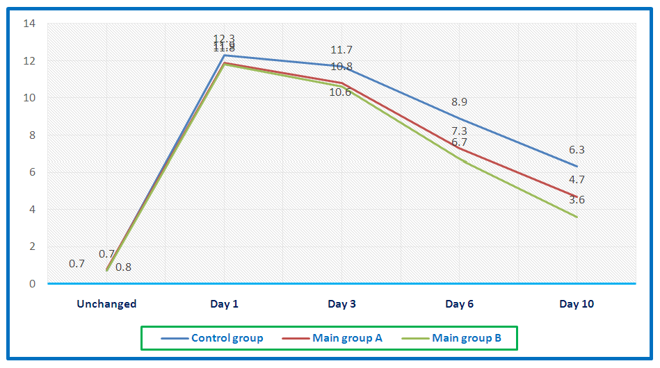
Assessment of the effectiveness of succinasol in rat acute paraproctitis model on injury termination, morphological changes and laboratory results.
Study of the effect of succinasol on the general condition of rats and the end of injury.
From the 3rd day of the experiment, when the acute paraproctitis model was induced in rats, lethargy, apathy, low activity, dullness and shedding of their wool, weight loss, blurred eyebrows and sclera were noted in animals. On the 5th day, the kiss developed vividly in rats, and a purulent cavity was cut open. Jaroxat was treated daily with an antiseptic, and as a detoxifying treatment, the prepaates succinazole (in the main rice) and rheosorbylact (in the control rice) were kept in the injection. In rats, the above symptoms increased due to increased purulent jarochate and general intoxication.
It must be said that animal hair is usually of a specific tone and usually lying on the skin. In dynamic observation in their rats in the main group, by the 6th day, the condition and appetite of animals gradually improved, they became more active, a little more aggressive, the appearance of skin wool improved, wounds on the surface of the skin healed. In animals in the control group, these indicators began to be observed on the 7-8th day. by the 10th day in rats, against the background of detoxification therapy with succinazole, the purity of their wool was gradually restored, erosion on the body disappeared. By 12-13 days, indifference and lethargy remained in the control group; they sat more in the corner of the cage; when taken, the animals became aggressive.
In the study of the leukocyte formula of experimental animals in acute paraproctitis, the development of leukocytosis was found, especially in the early stages of the experiment (see Table 5). Thus, leukocyte levels in peripheral blood increased statistically significantly by 1.9 (P<0.001) and 1.84 (P<0.001) times in rats on Days 1 and 3 respectively.
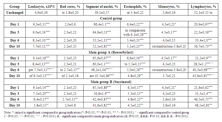
Table 5. Dynamics of changes in hematological indicators of peripheral blood of experimental animals, m±m
On the 3rd, 6th, and 10th days of the experiment, leukocytosis rates of 1.57 (P<0.001) and 1.39 (P<0.001) times decreased gradually, respectively. Leukocytosis was mainly associated with an increase in the relative content of segmented nuclear neutrophils (P<0.001) and eosinophils (P<0.001), which was due to the presence of a purulent-inflammatory process. At the same time, we observed relative lymphopenia (P<0.001), especially in the early stages of the study.
Study of Succinasol's effect on endogenous intoxication indicators in rats in acute paraproctitis.
Due to the fact that acute paraproctitis is a purulent-inflammatory process, there is an increase in lipid peroxidation in tissues, the intensity of which can be determined by the content of active compounds. In this regard, studies have shown that serum MDA intact in rats is statistically significantly higher than in animals (see Figure 3).
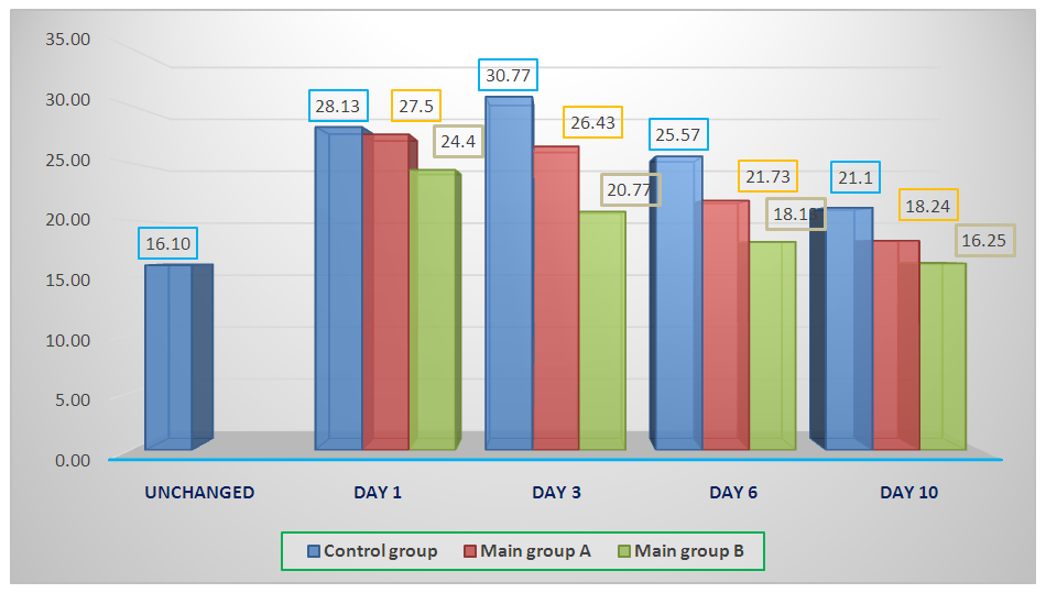
Figure 3. Dynamics of changes in the content of MDA (nmol/ml) in the blood serum of experimental animals
Thus, on Day 1 of the experiment, MDA levels exceeded standard values and were 19.6±0.02 nmol/ml, while in intact rats the value of this indicator was 2.6±0.02 nmol/ml. On the 3rd day of the experiment, the intensity of LPO decreased slightly as a result of detoxification therapy to 16.6±0.02 nmol/ml. Next, we observed its gradual decrease. The development of a purulent inflammatory process leads to an increase in lipid peroxidation, which indicates the destruction of biomembranes. In rats in the main cluster, the MDA indicator decreased 1.22 (P<0.001) times faster than in the control cluster.
Consequently, the National detoxifying drug succinasol not only reduces the level of toxicosis, but also has an antioxidant effect associated with the presence of succinate in its composition. In its activities, it outperforms Rheosorbylact.
Analysis of the index of Leukocyte displacement in rats with op showed its increase on the 1st day of the experiment (Figure 4). The next day of the experiment, we observed a gradual decrease in this indicator. In this, control remained relatively high in rats in the guru.
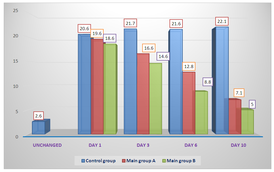
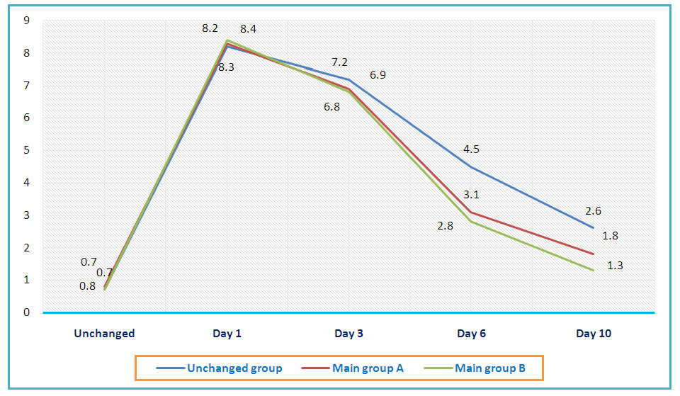
Figure 4. The effect of hemocorrectors on the dynamics of changes in the leukocyte displacement index in rats with acute paraproctitis
The use of Reosorbylact and Succinasol in rats with acute paraproctitis for 1-3 days of the experiment did not significantly affect the leukocyte displacement index. On Day 6, we observed that this indicator decreased by 1.45 and 1.6 (p<0.01) times compared to the values of the control group, respectively, and exceeded the standard values. Apparently, this was due to the activation of bone marrow hematopoiesis (immune system) in response to a decrease in toxemia.
Analysis of the dynamics of changes in the reactive response of neutrophils in rats with acute paraproctitis showed a sharp increase of 2.24 (p<0.001) times on Day 1 of the experiment compared to the values of rats in the Intact Group (Figure 5). This was due to the presence of an inflammatory-infectious process in experimental animals, the development of neutrophilia aimed at activating the macrophage-reticular system. Subsequently, this indicator gradually decreased and normalized until the 10th day. In rats in the main group, this indicator decreased faster than in the control group.
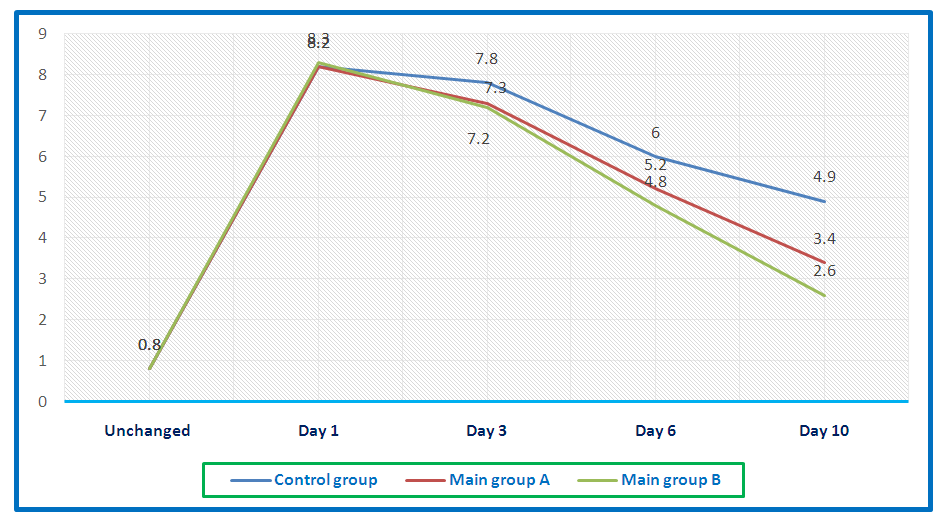
Figure 5. The effect of hemocorrectors on the dynamics of changes in neutrophil reactive response in rats with acute paraproctitis
The same dynamics was noted in the LII indicators calculated on Kalf-Kalif (Figure 6) and Ostrovsky (Figure 7). In rats with acute paraproctitis, their values increased within 1-3 days of the experiment, and then gradually decreased to the norm until the 10th day of the experiment. The use of succinasolnin against the background of local treatment of acute paraproctitis contributed to a faster decrease in LII values compared to the control group.
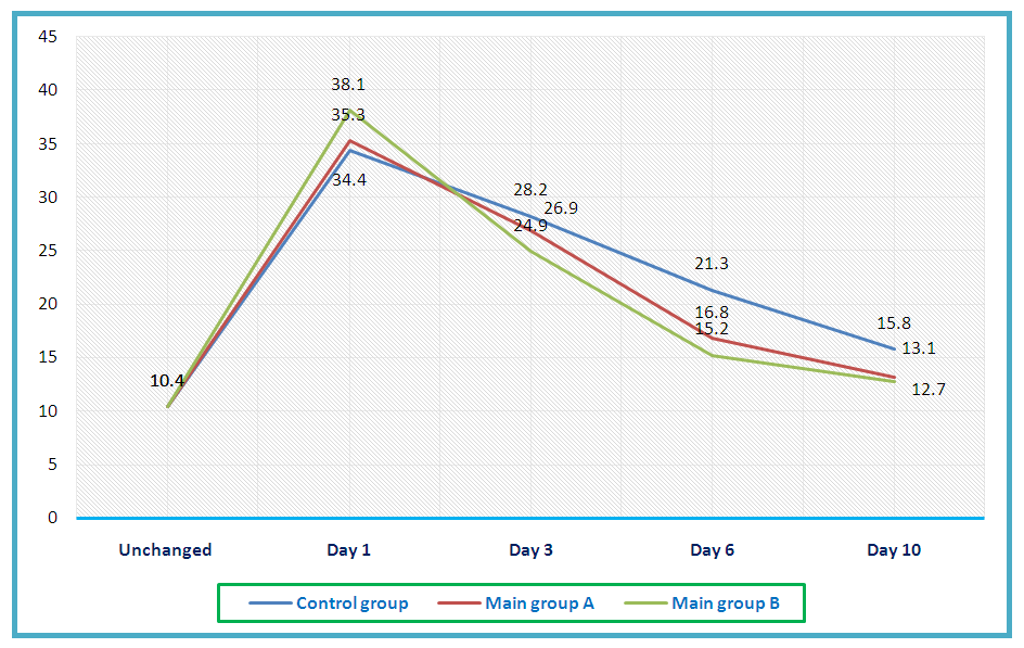
Figure 6. The effect of hemocorrectors on the dynamics of LII changes in calf-Calyph in rats with acute paraproctitis
Figure 7. Endogenous intoxication is an integral part of the overall inflammatory response syndrome
The generally accepted sign of endogenous intoxication, which allows you to assess the severity and prognosis of the disease and the effectiveness of treatment, is characterized by the level in the blood of low and medium molecular proteins, as well as their components - oligopeptides (OP). For this reason, the parameters of low to moderate molecular mass, oligopeptides, were studied against the background of the use of Rheosorbylact and Succinazole in the op.
Studies have shown that in rats with acute paraproctitis, on Day 1 of the experiment, the low to moderate Molecular Mass in the blood plasma increased statistically significantly by 2.8 (p<0.001) times compared to the values of rats in the Intact Group (Table 6). In later periods, this indicator gradually decreases compared to the values of the previous period. By the 10th day of the experiment, it was found that the rate decreased 1.5 (P<0.001) faster than the control guru in rats in the basif guru.
Table 6. Dynamics of changes in blood plasma toxicity indicators during treatment of rats with acute paraproctitis, m±m
     
The dynamics of changes in the composition of oligopeptides in the blood plasma of rats with acute paraproctitis was of such a character as ham. On Days 1, 3, and 6 of the experiment, the levels of this compound were statistically significantly higher in the control guru to rats in the intact guru at 2.8 (P<0.001); 2.6 (P<0.01); and 1.7 (P<0.05). On Day 10 of the experiment, we observed that oligopeptides in the blood plasma of rats in the main rat were 0.95 (P<0.05) and 1.35 (P<0.05) in the control rat. The results obtained indicate the development of endogenous intoxication in the body of rats with acute paraproctitis. In our opinion, this is due to metabolic disorders as a result of purulent-inflammatory, manifested by an increase in low and moderate Molecular Mass in the blood plasma, and this leads to the accumulation of oligopeptides. The detected changes affect the course of the wound process in rats with naturally acute paraproctitis. Confirmation of our opinion indicates the need for an increased index of plasma toxicity of experimental animals for disintoxic therapy. A 1.4 (P<0.05) fold faster decrease in the indicators in the main group than in the control group indicates the effectiveness of Succinasol against Reosorbylact.
It must be said that toxins of different molecular weights can accumulate not only in liquid biological moxites of the body, but also in cells, especially in erythrocytes. For this reason, we have studied low to moderate molecular mass levels in acute paraproctite dynamics, as well as oligopeptides and erythrocyte toxicity and its treatment with detoxifying drugs (see Table 7).
Table 7. Dynamics of changes in indicators of erythrocyte toxicity during treatment of rats with acute paraproctitis, m±m
     
Studies conducted have shown that rats with acute paraproctitis in erythrocytes have a statistically significant low to moderate molecular mass content of 2.6 (P<0.001) times the values of the Intact Group on Day 1. This was due to a clear violation of metabolic processes and the presence of a purulent-inflammatory process in the body of rats. In later periods, we observed a gradual decrease in the amount of these compounds in the blood erythrocytes of experimental animals. However, it was observed that its values decreased 3.6 and 10 days after the control group in the main groups by 1.26 (P<0.001) and 1.34 (P<0.001) times faster than in the 1.52 (P<0.001). By the 10th day, we observed that the values of rats in the main group almost approached those in the Intact Group.
The same dynamics was characteristic of the levels of oligopeptides in blood erythrocytes of rats with acute paraproctitis. Thus, on Day 1 of the experiment, oligopeptide levels increased statistically significantly by 3.3 (p<0.001) times compared to the values of intact rats. In later periods, oligopeptide levels gradually decrease. On the 3rd, 6th and 10th days of the experiment, we observed that the values of oligopeptides in blood erythrocytes decreased in the main group by 1.26 (P<0.001) and 1.34 (P<0.001) times faster than 1.52 (p<0.001), respectively, and did not differ significantly from the standard values on the 10th.
The most obvious changes in rats with acute paraproctitis were found in the erythrocyte toxicity index. The largest changes are characteristic of the first day of study, with the erythrocyte toxicity index exceeding the value of rats in the Intact Group by 8.94 (P<0.001) times. In later periods, the values of the erythrocyte toxicity index gradually decreased in rats with acute paraproctitis. Although gradually, the indicators in the main group decreased faster than in the control group. At the same time, unlike reosorbylact, succinazole had an effect that not only reduces the formation of toxic metabolites, but also their excretion from the body. This is confirmed by the fact that the standard values of the toxicity index of blood plasma and erythrocytes are reached faster.
According to the literature, the presence of a high concentration of toxins in the blood leads to the activation of lipid peroxidation processes, an increase in the permeability of erythrocyte membranes and the accumulation of toxins in erythrocytes. Indeed, previous studies have shown an increase in lipid peroxidation, manifested by the high production of active metabolites. On the other hand, we also observed an increase in the amount of low, medium and high molecular toxins in the blood erythrocytes of experimental animals. It must be said that the redistribution of toxic load between plasma and blood erythrocytes is a necessary part of the body's natural detoxification [14,52]. Endotoxins bind to the transmembrane protein of erythrocytes - glycophorin, and in this form pass to the detoxifying organs.
In this regard, we also studied the sorption capacity (ESQ) of erythrocytes in the blood of experimental animals (Figure 8). Studies have shown that in rats with acute paraproctitis, the index of erythrocyte sorption capacity increased by 1.64 (p<0.001) times compared to normal values in 1 day of the experiment. Over the next period, this rate gradually decreased, and in the main group it decreased faster than in the control group. However, these indicators are still significantly higher than the values of intact rats, which indicates the presence of endogenous intoxication in their body.
Figure 8. In rats with acute paraproctitis, the sorption capacity of erythrocytes is the effect of hemocorrectors on the dynamics of changes
According to the literature, the presence of a high concentration of toxins in the blood leads to the activation of lipid peroxidation processes, an increase in the permeability of erythrocyte membranes and the accumulation of toxins in erythrocytes. Indeed, previous studies have shown an increase in lipid peroxidation, manifested by the high production of active metabolites. On the other hand, we also observed an increase in the amount of low, medium and high molecular toxins in the blood erythrocytes of experimental animals. It must be said that the redistribution of toxic load between plasma and blood erythrocytes is a necessary part of the body's natural detoxification. Endotoxins bind to the transmembrane protein of erythrocytes - glycophorin, and in this form pass to the detoxifying organs.
Morphological changes of tissues in the prarectal area in 1,3,6,10 days during the treatment of acute paraproctitis, called in rats in the experiment.
Isolated rectum and pararectal soft tissue in rats were studied histologically using the hematoxylin-eosin method. When acute paraproctitis was initially called in rats, pararectal kletchatka, subcutaneous fat layer, and rectum circumference in soft tissues initially microscopically showed vascular reactions: fullness, tumors in intermediate tissue, dystrophic and necrotic changes, and acute infiltration of resident macrophages in the same Sox. Mainly in the area around the pararectal ketchatka, foci of increased foci steatonecrosis in adipose tissue, in serous curtains, foci of acute fullness and interstitial edema are detected. The damage to the cell components of the factors that lead to the inflammatory process continues with the development of the process of paranecrosis, necrobiosis and necrosis. It is in damaged cells that a violation of the metabolism (dystrophy) occurs, when the activity of enzymes involved in the Krebs cycle deepens the hypoxia process, which is caused by a violation of blood circulation.
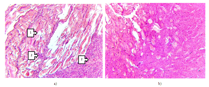
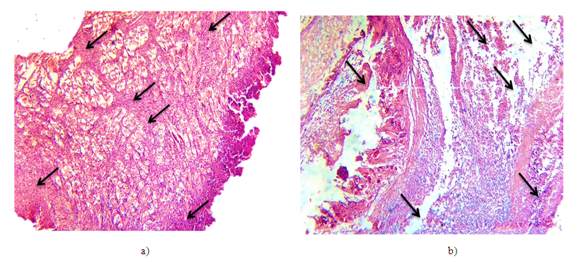
Figure 9. a - Control group. Day 1. Around the pararectal kletchatka, sharply developed fullness, interstitial tumors and destructively altered tissue components are identified. In the tangle of most venous vessels, fullness is clearly described (indicated by arrows). Paint G-E.10x4. b - Control group. Day 3. Destructive and degenerative changes that abound in the rectum area. Formation of acute interstitial edema. In the area of muscle fibers, many necrosis and tissue detritus are vividly described against a general background. Paint G-E.10x4
Figure 10. a - Main group A. Day 3. Foci of massive defragmentation and necrosis are detected between the muscle floors of the pararectal Area (1). Muscle fibers have lost their cross section; many foci of Leukocyte infiltration are detected in the area of muscle fascia (2). Paint G-E.10x10. b - Main group B. Day 3. Changes in muscle tissue around the pararectal area. Most of the myocytes are in the state of paranecrosis and of the same size, and stroma is found to have no obvious manifestation of signs of fullness. Paint G-E.10x10.
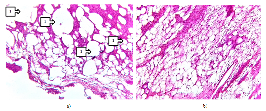
Figure 11. a - Main group A. Day 6. Pararectal area adipose tissue is large in size. Cystic dilated foci are detected as a result of changes in different sizes of lipocytes, large-cell appearance and steatonecrosis (1). Paint G-E.10x40. b - Main group B. Day 6. Pararectal area adipose tissue. Granular structures accumulated in the cytoplasm of lipocytes are identified. Lipocytes Dearness are the same size. Cystic dilated foci are not detected. Leukocytes located singly are detected in adipose tissue stroma. Paint G-E.10x10
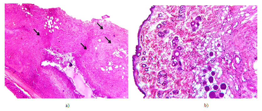
Figure 12. a - Main group A. Day 10. The general appearance of a part of the rectum is determined by the uneven orientation of the inflammatory infiltrate and fibrous structures that multiply on the muscle floor. Paint G-E.10x4. b - Main group B. Day 10. The appearance of the skin around the Anus on a general background. All structural components appear to be normative, and the post-inflammatory reparative regeneration process is found to be fully restored. The capsules are fully preserved around the hair follicles. Intermediate tumors of different levels are partially identified (indicated by arrows). Paint G-E. 10x10
Group A rats were observed to develop focal coagulation necrosis of the pararectal area tissue resulting in acute paraproctitis, phlegmanosis inflammation, and drastic development of dissirculatory changes around the same area, development of necrotic processes in adipose tissue, necrobiosis, ultimately deep-defected necrosis, and phlegmanosis inflammation.
When treated with the Drug Group B Succinasol, the processes of dystrophy and necrobiosis in the tissues of the pararectal sphere were stabilized in relation to the control group, regenerated in the subepidermal sphere, a sharp increase in reparative regeneration in soft tissues, a sharp proliferation of forming connective cells.

5. Discussion

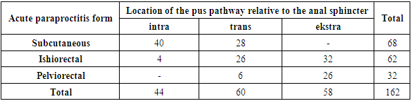
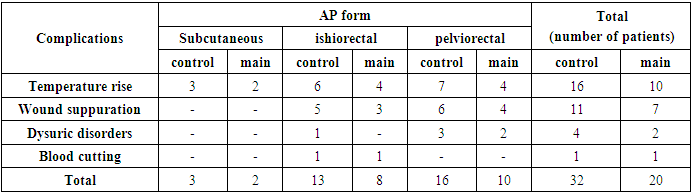
The results of the study of the course of the postoperative period and the completion of the surgical wound with the traditional and complex method of treatment in patients with acute paraproctitis.
Patients underwent 3 different types of surgery:
1-opening of the purulent cavity (palliative).
2-opening of the purulent cavity, liquidation of the inner hole (radical).
3-opening the purulent cavity, placing a rubber ligature in the inner hole (radical).
The choice of the scope of surgical intervention is determined by the following factors: the localization of the purulent-inflammatory process and its spread to the surrounding tissues, the connection of the primary purulent tract with the fibers of the anal sphincter.
After the abscess is punctured in 122 (75.3%) patients and the pus is partially evacuated, a methylene blue or brilliant green solution in equal proportions was injected into the cavity with a 3% H2O2 solution to contrast and determine the localization of the abscess. An internal opening of the primary "fistula" was detected in one of the rectum crypts.
The location of the primary pus pathway relative to the sphincter fibers was determined by probing it (Table 8).
Table 8. Distribution of patients depending on the location of the primary purulent tract in relation to the fibers of the anal sphincter
     
All patients are divided into 2 groups: control and basic. Control group 92 (56.8%) patients with acute paraproctitis reduction of intoxication the drug Maxadida Reosorbylact was infused intravenously for 3-5 days 400 ml (6-7 ml/1 kg of body weight) per day. The main group consisted of 80 (43.2%) patients who were given intravenous infusion of the drug Succinasol for 3-5 days in 400 ml (6-7 ml/1 kg of body weight) per day in an intoxication reduction maxad.
The course of the postoperative period was assessed by such criteria as the presence and nature of postoperative complications, the time of cleansing the wound from purulent and necrotic masses, the appearance and type of granulation, the onset of epithelization and the duration of the day of hospitalization.
Table 9. The following early complications were observed in patients after opening acute paraproctitis
     
In the early postoperative period, 32 (34.8%) complications were identified and treated in 92 patients in the control group. 20 (25%) complications were identified and treated in 80 patients in the main group. The most commonly observed complication was an increase in body temperature in the early postoperative period, observed in 16 (17.4%) patients in the control group and 10 (12.5%) patients in the primary group.
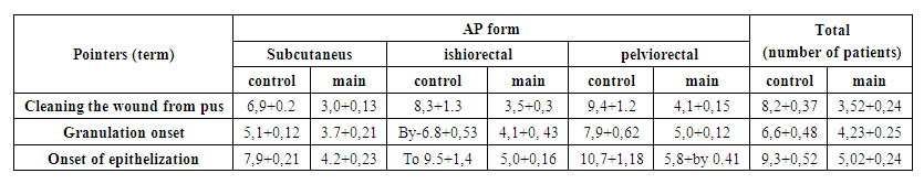
Table 10. Indications for postoperative injury termination in patients
In patients of the main group, hyperemia and loss of tissue swelling, complete cleansing of postoperative injuries from purulent-necrotic processes occurred within 3-5 days. On 4-6 days, the first granulations appeared on the surface of the wound, and on 6-9 days, the wounds became completely granular. Time of onset of local epithelization: for the subcutaneous form of the disease - 4-5 days, for the ishiorectal form - 5-6 days, for pelviorectal paraproctitis - 6-8 days. In patients in the control group, these indicators were observed to be 1-4 days late. The deterioration of the clinical course of the wound process in 6 (2.4%) patients was caused by postoperative wound suppuration.

6. Conclusions

1. Through the acute paraproctitis induction model in experimental animals, intoxication levels were found to be severely delayed when evaluated with integral blood parameters: displacement index (DI), leukocyte intoxication index (LII), and neutrophil reactive reaction (NRR) high mass molecules (HMM) and moderate mass (MM), erythrocyte oligopeptides (OP), blood plasma, and erythrocyte sorption capacity (ESC), postoperative wound termination, and pathomorphological changes in it were slow.
2. Daily injections of succinasol solution for 5 days in rats with acute paraproctitis contributed to an improvement in the condition of animals, a decrease in toxic-infectious intoxication, which was manifested by leukocytosis, an earlier decrease in neutrophilia, an increase in the low values of lymphocytes in the peripheral blood, and pathomorphological changes go more quickly to the positive side.
3. In acute paraproctitis, the formation of postoperative pararectal leaks and the observation of recessive paraproctitis require a radical method of operation. At the same time radical surgery requires a milder method of surgery to open a cisobed “purulent cavity, extend the jarochate termination for a longer period after internal cavity liquidation,” and observe anal sphincter insufficiency.
4. In the subcutaneous form of acute paraproctitis, the operation “opening of the purulent cavity, internal cavity liquidation”, the use of “opening of the purulent cavity” or “opening of the purulent cavity and installing the ligature sac” in the heavy – ischiorectal and pelviorectal types reduces the occurrence of postoperative complications.
5. The use of succinasol in acute paraproctitis leads to an early loss of signs of acute inflammation in the tissues around the wound, in the wound area, and an increase in the regeneration coefficient. Morphological, dystrophic disorders are reduced, symptoms of reparative regeneration are activated both in the epidermis and in the dermis, which leads to a complete restoration of all cellular fibrous structures and wound healing by the 10th day of treatment. The results obtained show that in the treatment of acute paroproctitis, the new topical drug Succinasol does not lag behind the classic Reosorbylact, has antioxidant properties, has a detoxifying effect of "biochemical sanitation" and restores the physiological functions of cells, which allows it to be recommended for use.

References

[1]  Akhrorova, L. B. (2023). CHOICE OF SURGICAL TACTICS FOR THE TREATMENT OF PATIENTS WITH ACUTE PARAPROCTITIS. Academia Science Repository, 4(5), 1023-1026.
[2]  Babadjanovich, K. Z., Akhrarovich, S. U., & Muradullaevich, R. I. (2023). LOOSE SET-ON IN THE TREATMENT OF ACUTE PARAPROCTITIS. JOURNAL OF BIOMEDICINE AND PRACTICE, 8(2).
[3]  Karaboyev, J. (2023). TREATMENT AND PREVENTION OF COMPLICATED FORMS OF ACUTE PARAPROCTITIS. Science and innovation, 2(D6), 21-23.
[4]  Karimov, X. Y., Ergashev, U. Y., & Yakubov, D. R. (2022). Complex treatment in severe forms of acute paraproctitis. Web of Scientist: International Scientific Research Journal, 3(9), 199-203.
[5]  Paliienko, R., & Stetsenko, M. (2022). The choice of drug treatment in patients with acute purulent paraproctitis. Perioperaciina Medicina, 5(1), 21-25.
[6]  Vasiliev, S. V., Gor, I. V., Nedozimovanii, A. I., Popov, D. E., Skuridin, G. M., & Vasilieva, E. S. (2021). Loose set-on in the treatment of paraproctitis. Ambulatornaya khirurgiya= Ambulatory Surgery (Russia), 18(2), 98-104.
[7]  Yakubov, D. R., & Dustmukhammedova, R. Z. (2023, January). ASSESSMENT OF POSTOPERATIVE PAIN LEVEL IN HEMORRHOIDS, ANAL FISSURE, PARAPROCTITIS AND PARARECTAL FISTULA. In Proceedings of International Educators Conference (Vol. 2, No. 1, pp. 217-220).
[8]  Yakubov, D. R., & Ismailov, F. M. (2023). Complex Treatment in Severe Forms of Acute Paraprocitis (Clinical Case). Genius Repository, 26, 70-72.
[9]  Yakubov, D. R., & Sagdullayeva, Y. A. (2023). GENERAL LEVEL OF INTOXICATION IN ACUTE PAROPROCTITIS. British Journal of Global Ecology and Sustainable Development, 14, 22-25.
[10]  Babich, V. A. (2010). Diagnosis and treatment of acute paraproctitis. Problems of ecological and medical genetics and clinical immunology, (3), 326-340.
[11]  Borota, A.V., Plakhotnikov, I. A., Kuhto, A. P., Borota, A. A., & Baziyan-Kuhto, N. K. (2019). MODERN METHODS OF TREATMENT OF ACUTE PARAPROCTITIS. Coloproctology, 18(S3), 19-19.
[12]  Grintsov, A. G., Antonyuk, S. M., Akhrameev, V. B., Karabak, I. S., & Matiytsiv, A. B. (2021). ON THE ISSUE OF METHODS OF TREATMENT OF ACUTE PARAPROCTITIS. Bulletin of Emergency and Reconstructive Surgery, 6(1), 56-60.
[13]  Denisenko, V. L., Drozdov, V. M., Tsyplakov, K. G., Khmelnikov, V. Ya., Sokolovsky, V. O., Smirnov, V. K., ... & Smirnova, A. O. (2022). Acute complicated anaerobic paraproctitis: a case from practice. Bulletin of the Vitebsk State Medical University, 21(6), 99-105.
[14]  Karimov, H. Ya., Ergashev, U. Yu., & Yakubov, D. R. (2021). THE RESULTS OF COMPLEX TREATMENT OF ACUTE PARAPROCTITIS.
[15]  Mustafayeva, M. F. (2014). New approaches in the surgical treatment of acute paraproctitis. Bulletin of Surgery of Kazakhstan, (3 (39)), 56-58.
[16]  Rustamov, A. E., Ismailov, F. M., Eshmurodova, D. B., Yakubov, D. R., & Eshmurodov, S. A. (2021). SURGICAL TREATMENT OF ULCERATIVE COLITIS WITH THE FORMATION OF A SMALL INTESTINAL RESERVOIR IN A PEDIATRIC PATIENT.
[17]  Sherbekov, U. A., & Rustamov, I. M. (2023). THE EFFECTIVENESS OF COMPLEX THERAPY OF ACUTE PARAPROCTITIS. Research Focus, 2(3), 220-226.
[18]  Sherkulov, K. U. (2022). ANALYSIS OF SURGICAL TREATMENT OF ACUTE PARAPROCTITIS. Scientific progress, 3(2), 793-797.
[19]  Ergashev, U. Y., Yakubov, D. R., & Mominov, A. T. (2022). SHTKIR PELVIORECTAL PARAPROCTITIS BILGAN BEMORNI DAVOLASH COMPLEX (HOLAT CLINIC). Development of pedagogical technologies in modern sciences, 1(2), 63-64.
[20]  Yakubov, D. R. (2023). A METHOD FOR MODELING ACUTE PARAPROCTITIS IN RATS.