-
Paper Information
- Next Paper
- Previous Paper
- Paper Submission
-
Journal Information
- About This Journal
- Editorial Board
- Current Issue
- Archive
- Author Guidelines
- Contact Us
American Journal of Medicine and Medical Sciences
p-ISSN: 2165-901X e-ISSN: 2165-9036
2024; 14(5): 1174-1177
doi:10.5923/j.ajmms.20241405.06
Received: Apr. 8, 2024; Accepted: May 3, 2024; Published: May 7, 2024

Assessment of Morphological Changes in the Kidneys under the Impact of Energy Drinks
 Abstract
 Reference
 Full-Text PDF
 Full-text HTML
Alimova Shakhnoza Azamatovna, Khuseynova Gulshan Khuseynovna
Bukhara State Medical Institute, Bukhara, Uzbekistan
Copyright © 2024 The Author(s). Published by Scientific & Academic Publishing.
This work is licensed under the Creative Commons Attribution International License (CC BY).
http://creativecommons.org/licenses/by/4.0/

This work is devoted to the study of the morphological state of the kidneys of rats under the influence of energy drinks on the body. The use of energy drinks can cause problems with the urinary system, the rapid depletion of the body's resources. Due to the use of energy drinks, they take a lot of energy from the body at once, which leads to the inevitable depletion of the nervous system, metabolic disorders. We have studied the morphofunctional state of the kidneys of outbred rats when exposed to an energy drink. The study was conducted on white bisexual rats weighing 125-137 g. The animals were divided into two groups: 1 - control group, 2 - experimental group of animals that received the drink in doses for 30 days. Experimental animals were on a general vivarious diet, a 12-hour light change. The slaughter was carried out by decapitation on the 30th day and the kidney was separated for histological examination. These studies were performed after standard histological examination and paraffin embedding and staining of sections with hematoxylin and eosin. Histological studies 30 days after the use of energy drinks in the experiment in the kidneys observed acellular fibrosis and hyalinosis of the stroma of the pyramid, an increase in the size of individual renal corpuscles, capillary glomeruli filled with blood, a thinned outer leaf of the capsule, and the cavity of the capsule is noticeably enlarged. In some renal bodies, processes of degenerative changes are observed. The epithelium of the proximal tubules becomes lower and the process of karyolysis is observed in some cells, dystrophic changes in the epithelium are observed in the distal tubules and collecting ducts. And, moderate ectasia of the collecting ducts, atrophy of the direct tubules and collecting ducts. Thus, the histological data obtained from the study of rat kidneys indicate that there is a high sensitivity of the kidneys to the effects of energy drinks.
Keywords: Kidney, Cortex, Medulla, Glomerulus, Morphological features, Microscopic features
Cite this paper: Alimova Shakhnoza Azamatovna, Khuseynova Gulshan Khuseynovna, Assessment of Morphological Changes in the Kidneys under the Impact of Energy Drinks, American Journal of Medicine and Medical Sciences, Vol. 14 No. 5, 2024, pp. 1174-1177. doi: 10.5923/j.ajmms.20241405.06.
Article Outline
1. Introduction
- At present, a certain “fashion” has been formed for the use of various types of energy drinks [1,2,6]. They are a new brand in a number of bad habits, that is, along with alcohol, tobacco and drugs, and it was in this historical sequence that these phenomena appeared in the life of mankind in our 21st century. Energy drinks consist of components long known to medicine [3,7,8]. So, without exception, all types of energy drinks contain caffeine and in industry it is obtained in three ways: by isolation from roasted coffee beans, which contain 0.75 - 1.5% caffeine; extraction from tea dust, which contain 1.5 - 3.5% caffeine; as well as extraction from kola nuts containing about 2% caffeine. In addition, it can be obtained chemically from uric acid or by methylation of theobromine. Of course, it is synthetic, which is cheaper caffeine, that manufacturers include in energy drinks. Another ingredient in energy shakes is taurine. Some energy drink manufacturers add mate leaf extract, damiana, far eastern magnolia vine, and ginseng. Energy drinks contain vitamins that are directly related to the body's energy metabolism. These include: ascorbic acid, B1, B2, B3, B5, B6, B12, niacin. In general, we can say that the purpose of these drinks - increases the same energy metabolism, the vitality of the body, and this succeeds by about 20-30% [4,5,7].Of course, the main consumer of energy drinks is our youth. They consume them as drinks for rest, relaxation, or to improve performance. At present, despite the growing growth in the production of various types of energy drinks, it should be noted the imperfection of the legislative framework and the level of control over their quality and safety, which can serve as a risk factor for the occurrence of various relevant diseases [3]. In the minds of our society, especially among adolescents, the psychological connection “energy is evil” has not yet been fixed, this relationship is not so pronounced. Currently, there is not enough information about the morphological changes in internal organs, especially in the kidneys during exposure to little is known about energy drinks and this requires a thorough study of the morphofunctional changes in this organ.
2. Materials and Research Methods
- For the purpose of research, laboratory white outbred rats were used: 50 individuals of three months of age, whose average weight was 125-137 g. Laboratory animals were kept in vivarium in plastic cages with small wood shavings at room temperature °C 12-hour change of light and dark. All animals of the experimental groups were divided into 2 groups, that is, the first group consisted of animals that did not consume energy drinks (control, n = 25), and the second group consisted of (experimental, n = 25) - animals that received the drink in doses for 30 days.In the course of the experiment, these animals were decapitated on the 30th day after drinking an energy drink under the influence of light ether anesthesia, the abdominal cavity was opened, and the kidneys were separated for further macro- and microscopic examination. For histopathological comparison of the control and experimental groups of rats, the kidneys were taken on the 30th day of postnatal development after the experiment, consumed energy drink. These preparations were prepared using standard histological methods. All preparations were stained with hematoxylin and eosin. The analysis of the obtained images was carried out using specialized software for medicine, using specialized software for medicine. All experiments performed on laboratory animals were carried out in accordance with the Medical Declarations adopted in 2008-2013.
3. Results and Discussions
- In 3-month-old rats of the control group, the kidneys were smooth, bean-shaped and red-brown organs, and when the weight of this organ was measured, the mass of each kidney was approximately 0.44-0.57 g, on average 0.48 g. The size of the kidneys of rats is not constant, in the form of the length of the kidneys of a 3-month-old rat, on average, they were 13-17 mm, on average, the width was 4-7.8 mm and an average thickness of 3.7-5 mm. This organ is located in the lumbar region in front of the twelfth thoracic - second lumbar segment. In these laboratory white outbred rats, the lobulation of the kidneys is weakly expressed, the cranial and caudal edges of the kidneys are blunted. In the kidney, a convex lateral and somewhat concave medial edge is distinguished, and outside the kidney is covered with a dense fibrous connective tissue and a weakly expressed fatty membrane and a serous membrane lying on ventral surface of the organ. It was found that the mass and size of the right kidney were unreliably larger than the similar parameters of the left kidney, and therefore, in the future, the right kidneys were used for morphological studies and histological preparations were made from them (1-fig.).
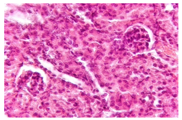 | Figure 1. Histological image of the right kidney in a 3 month old white control rat |
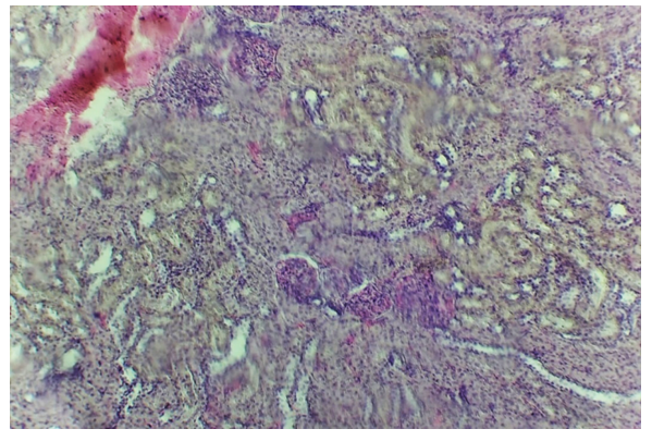 | Figure 2. Acellular fibrosis and hyalinosis of the pyramidal stroma, moderate ectasia of the collecting ducts, atrophy of the rectus tubules and collecting ducts. Hematoxylin-eosin stain. Coloring GE (magnification 20x20) |
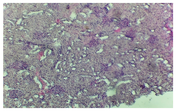 | Figure 3. There are dystrophic changes in the epithelium of the distal convoluted tubules and collecting ducts of the cortical substance of the kidney of 3-month-old albino rats. Apoptosis of groups of cells. Hematoxylin-eosin stain. Coloring GE (magnification 20x20) |
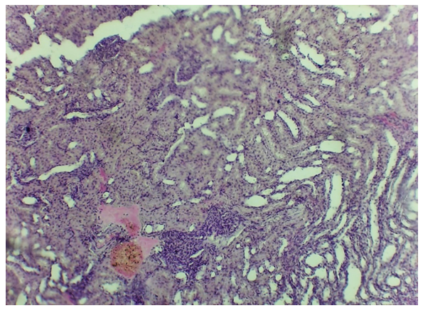 | Figure 4. There is marked polymorphocellular infiltration (neutrophilic leukocytes, lymphocytes and hemorrhage. GE coloring (magnification 20 x 20) |
|
4. Conclusions
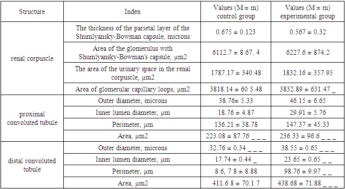
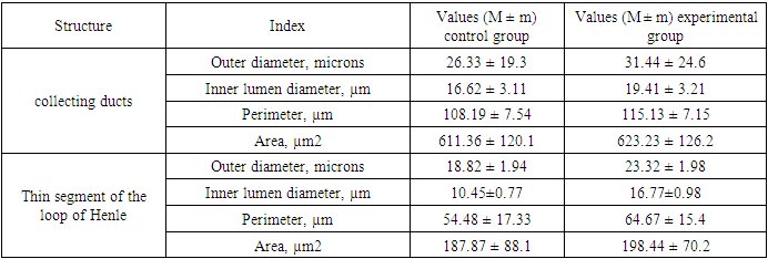
- Based on the above data, it can be concluded that histological examination after the consumption of an energy drink in the kidneys shows various changes. Histological studies 30 days after the use of energy drinks in the experiment in the kidneys observed acellular fibrosis and hyalinosis of the stroma of the pyramid, an increase in the size of individual renal corpuscles, capillary glomeruli filled with blood, a thinned outer leaf of the capsule, and the cavity of the capsule is noticeably enlarged. In some renal bodies, processes of degenerative changes are observed. The epithelium of the proximal tubules becomes lower and the process of karyolysis is observed in some cells, dystrophic changes in the epithelium are observed in the distal tubules and collecting ducts. And, moderate ectasia of the collecting ducts, atrophy of the direct tubules and collecting ducts. Thus, the histological data obtained from the study of rat kidneys indicate that there is a high sensitivity of the kidneys to the effects of energy drinks. and these changes in the end ends with organ failure. Determined that use of energy drinks the macroscopic parameters of the kidneys do not change, but at the same time it leads to an increase in the morphometric circumferential (diametrical) parameters of the renal corpuscles and all nephron tubules after the 30th day of the experiment.
References
| [1] | Kiseleva, T.F. Formation of technological and socially significant consumer properties of drinks: theoretical and practical aspects: monograph / T.F. Kiseleva; Kemerovo Technological Institute of Food Industry. – Kemerovo, 2006. – 271 p. |
| [2] | Poznyakovsky, V.M. Assortment of functional drinks on the regional market / V.M. Poznyakovsky, V.M. Kiselev, V.V. Schmidt // Beer and drinks. – 2009. – No. 5. – P. 15–17. |
| [3] | Huseynova, G. (2021). Macroscopic and microscopic characteristics of the kidneys of white outbred rats after severe traumatic brain injury. Journal of the Doctor's Bulletin, 1(1), 109-112. |
| [4] | Khuseynova, G. Kh., & Teshaev, Sh. Zh. (2020). Comparative characteristics of the morphometric parameters of the kidneys during different phases of craniocerebral infection. A New Day in Medicine, (2/1), 30. |
| [5] | Sh. A. Alimova (2022). Morphometric changes in the development of microvessels of the anal canal and the spincteric apparatus of the rectum in rats at different stages of postnatal ontogenesis. Scientific progress, 3 (4), 52-56. |
| [6] | Wide range of marketing research [Electronic resource]. – Access mode: http://www.businessanalytica.ru (date of access: 10/15/2011). Khuseynova, G. K. (2021). Macroscopic and microscopic characteristics of kidneys of white unbored rats after severe cranial injury. |
| [7] | Khuseynovna, K. G. (2022). Morphological Changes in the Kidneys in Easy Degree of Cranio-Brain Injury. Vital Annex: International Journal of Novel Research in Advanced Sciences, 1(1), 5-7. |
| [8] | Vakula T.N., Kremplevskaya S.P., Energy drinks: for or against? Journal of the Bulletin of Medical Internet Conferences, Issue No. 11. Volume 2/2012. |