-
Paper Information
- Paper Submission
-
Journal Information
- About This Journal
- Editorial Board
- Current Issue
- Archive
- Author Guidelines
- Contact Us
American Journal of Medicine and Medical Sciences
p-ISSN: 2165-901X e-ISSN: 2165-9036
2024; 14(4): 1122-1125
doi:10.5923/j.ajmms.20241404.66
Received: Feb. 27, 2024; Accepted: Mar. 23, 2024; Published: Apr. 26, 2024

Diagnostic Aspects of Changes in the Concentration of Cytokines in the Blood Serum in Patients with Chronic Uveitis
 Abstract
 Reference
 Full-Text PDF
 Full-text HTML
Muhriddin Kh. Khasanov1, 2, Nekkadam A. Nuraliev1, Jakhongir O. Safarov2, Khilola Sh. Yakhyoeva1
1Bukhara State Medical Institute, Bukhara, Republic of Uzbekistan
2Bukhara Branch of the Republican Specialized Scientific and Practical Medical Center for Eye Microsurgery, Bukhara, Republic of Uzbekistan
Correspondence to: Muhriddin Kh. Khasanov, Bukhara State Medical Institute, Bukhara, Republic of Uzbekistan.
| Email: |  |
Copyright © 2024 The Author(s). Published by Scientific & Academic Publishing.
This work is licensed under the Creative Commons Attribution International License (CC BY).
http://creativecommons.org/licenses/by/4.0/

Uveitis is a group of severe inflammatory diseases of the eyeball that can lead to such serious complications as decreased vision and even blindness. Of great importance in the pathogenesis of inflammation of the vascular layer of the eyeball is dysregulation of immune mechanisms, carried out by a large number of humoral mediators. Among them, cytokines occupy a special place. Purpose: to study the concentration of a number of cytokines in blood plasma and analyze changes in the cytokine profile depending on the severity of the inflammatory process in uveitis. Materials and methods. The subjects of the study were 45 patients (20 men and 25 women), examined and treated with a diagnosis of chronic uveitis in the hospital of the Bukhara branch of the Republican Specialized Scientific and Practical Medical Center for Eye Microsurgery (Bukhara, Uzbekistan). 22 patients who did not have inflammatory diseases in the organ of vision were selected as a control group. Quantitative determination of cytokines in blood serum was carried out by enzyme-linked immunosorbent assay using “Vector Best” kits (Russia) for tumor necrosis factor-α (TNFα) and interleukins (IL) IL-1β and IL-10. Results. Regulation of the immune response in patients with a positive result in the group of patients with a good expected outcome of uveitis occurs due to the balanced production and interaction of mediators of the acute phase of inflammation, activation of local cellular reactions. In the group of patients with uveitis that has become a chronic form of the inflammatory process of the eye or recurrent, a defect in the cytokine network is associated with an imbalance of the pro-inflammatory and anti-inflammatory effects of inflammation; the transition to a pathological process with their hyperproduction is described. Conclusions. In the blood serum of patients with varying degrees of severity of chronic uveitis, a statistically significant increase in the concentrations of IL-1β, IL-10 and TNFα was noted. The level of cytokines in the blood serum in chronic uveitis must be taken into account when assessing the severity of inflammation of the choroid.
Keywords: Uveitis, Cytokines, Interleukin, Diagnosis of uveitis, Immunoinflammatory process
Cite this paper: Muhriddin Kh. Khasanov, Nekkadam A. Nuraliev, Jakhongir O. Safarov, Khilola Sh. Yakhyoeva, Diagnostic Aspects of Changes in the Concentration of Cytokines in the Blood Serum in Patients with Chronic Uveitis, American Journal of Medicine and Medical Sciences, Vol. 14 No. 4, 2024, pp. 1122-1125. doi: 10.5923/j.ajmms.20241404.66.
Article Outline
1. Introduction
- Uveitis is an important cause of blindness in both developing and developed countries. Its prevalence in children and the elderly is estimated to be around 5–16% and 6–21%, respectively [1,2].Uveitis is a general term for inflammation of the uveal tract from any cause and usually includes a large group of diverse diseases affecting not only the uveal tract, but also the retina and vitreous. Despite a long period of study, uveitis is one of the most complex inflammatory diseases of the organ of vision; many issues of the development of uveitis remain unexplored. To date, no ideal method has been found to treat and prevent exacerbations of the disease [3,6,14].The etiology of uveitis is difficult to determine because the exact cause of uveitis is often unknown. The relationship between uveitis and systemic diseases is well known. In earlier studies, most cases were associated with autoimmune diseases, while recent reports show an association with various generalized diseases [4,5].With the development of uveitis, the lack of a clear understanding of the mechanisms of initiation of the inflammatory process in the internal structures of the eyeball and the reasons for the chronicity of the pathological process in the eye does not allow timely and effective pathogenetic therapy of the disease. This, in turn, leads to the chronic nature of the disease and a significant decrease in vision [5,6,11].According to many authors, dysregulation of immune mechanisms, carried out by a large number of humoral mediators, is of great importance in the pathogenesis of inflammation of the vascular layer of the eyeball. Among them, cytokines occupy a special place. These are low molecular weight proteins that ensure the process of intercellular interaction [7,8].Various studies have highlighted the importance of cytokines in the development and progression of various forms of infectious and non-infectious uveitis [9,10].In patients with uveitis, an increase in the level of pro-inflammatory cytokines such as IL 1 (IL-1β) and IL-6, which is similar in biological properties, was found. It was also revealed that the concentration of IL-6 in the eyes of patients with uveitis exceeds its level in the blood serum. However, IL-6 is not a specific marker of uveitis, since its level increases both in diabetic retinopathy and retinal detachment and other eye diseases [12,13]. Studies of anti-inflammatory cytokines are also described in the literature. A number of authors suggest a protective role of interleukin-10 (IL-10) in patients with uveitis, since its higher level was noted in patients with good visual functions in the outcome of the disease [15,16].
2. Purpose
- To study the concentration of a number of cytokines in blood plasma and analyze changes in the cytokine profile depending on the severity of the inflammatory process in uveitis.
3. Material and Methods
- The subjects of the study were 45 patients (20 men and 25 women), examined and treated with a diagnosis of chronic uveitis in the hospital of the Bukhara branch of the Republican Specialized Scientific and Practical Medical Center for Eye Microsurgery (Bukhara, Uzbekistan). The age group of patients ranged from 21 to 65 years. As a control group, 22 patients were selected who did not have inflammatory diseases in the organ of vision, but had refractive diseases (myopia, hyperopia, astigmatism) in the eyes, corresponding to the above comparison group by age and gender.For immunological studies, the same amount of blood was taken from the antecubital vein from all patients on an empty stomach. Quantitative determination of cytokines in blood serum was carried out by enzyme-linked immunosorbent assay using Vector Best kits (Russia) for tumor necrosis factor-α (TNFα) and interleukins (IL) IL-1β and IL-10.Intracellular cytokines are determined using modern methods based on the principles of proteomics and genomics. The cytokines produced are determined using various methods that allow them to be measured even at low concentrations [17].Statistical processing of the data obtained and collected as a result of the study was carried out using parametric analysis methods using Student's t-test. Pearson correlation analysis was performed by calculating linear and multiple correlation coefficients to determine the relationship between characteristics.
4. Results and Discussions
- Since hospitalization, all patients with chronic uveitis showed statistically significant changes in cytokine levels. The indicators of all studied groups of cytokines in patients were significantly higher than those of patients in the control group. The concentrations of IL-1β, IL-10 and TNFα in the group of patients were 3.68±0.11; 0.59±0.04; 4.65±0.29 pg/ml, respectively. The level of IL-1α, one of the main mediators responsible for the formation of a local inflammatory reaction in the eye, was 2.1 times higher than in patients in the control group. The concentration of IL-10, which is responsible for inhibiting the synthesis of anti-inflammatory cytokines, was 2.95 times higher than the values of patients in the control group. The values of TNFα, the concentration of which increases during infection and the entry of endotoxins of various natures into the body, were 2 times higher than in the control group (Fig. 1).
 | Figure 1. Cytokine profile at disease onset in patients with uveitis and controls |
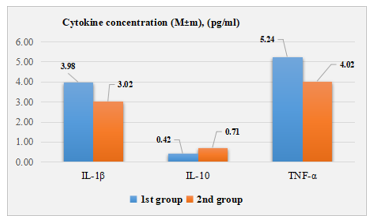 | Figure 2. Cytokine profile of uveitis patients with different disease outcomes |
5. Conclusions
- In the blood serum of patients with varying degrees of severity of chronic uveitis, a statistically significant increase in the concentrations of IL-1β, IL-10 and TNFα was noted. The level of cytokines in the blood serum in chronic uveitis must be taken into account when assessing the severity of inflammation of the choroid. Determining the concentration of a number of cytokines (IL-1b, IL-10 and TNFα) in the blood serum of patients with chronic uveitis can become a promising direction for the early diagnosis of uveitis and the development of new treatment methods.