American Journal of Medicine and Medical Sciences

p-ISSN: 2165-901X    e-ISSN: 2165-9036

2024;  14(4): 911-916

doi:10.5923/j.ajmms.20241404.24

Received: Mar. 14, 2024; Accepted: Mar. 28, 2024; Published: Apr. 10, 2024


Evaluation of the Effectiveness of PRP Therapy in Repair Processes Suture on the Uterus after Cesarean Section

J. M. Gafurov1, D. D. Saidjalilova2, B. B. Kurbanov3

1Republic Perinatal Center, Tashkent, Uzbekistan

2Tashkent Medical Academy, Uzbekistan

3Tashkent Pediatric Medical Institute, Uzbekistan

Correspondence to: J. M. Gafurov, Republic Perinatal Center, Tashkent, Uzbekistan.

Email:

Copyright © 2024 The Author(s). Published by Scientific & Academic Publishing.

This work is licensed under the Creative Commons Attribution International License (CC BY).
http://creativecommons.org/licenses/by/4.0/

Abstract

Purpose of the study: to evaluate the effect of platelet auto-plasma administered intra operatively on the processes of reparative regeneration and the formation of a full-fledged scar on the uterus after cesarean section. We studied 116 pregnant women in the period 2022-2023, who were delivered by cesarean section. We can conclude that using of platelet auto-plasma promotes the formation of a full-fledged scar on the uterus after cesarean section.

Keywords: Cesarian section, Pregnancy, Autoplasma

Cite this paper: J. M. Gafurov, D. D. Saidjalilova, B. B. Kurbanov, Evaluation of the Effectiveness of PRP Therapy in Repair Processes Suture on the Uterus after Cesarean Section, American Journal of Medicine and Medical Sciences, Vol. 14 No. 4, 2024, pp. 911-916. doi: 10.5923/j.ajmms.20241404.24.

1. Background

According to WHO, over the past thirty years, the proportion of caesarean sections (CS) has increased worldwide, reaching its highest level in the current decade, and has approached 25-30%, reaching 40-50% in large perinatal centers [1-6]. In the general population of women of reproductive age, 15-42% has a history of CS [7-9]. The CS operation, despite its technical simplicity, is a surgical procedure that has a considerable number of intra- and postoperative complications. [10-15]. The postpartum period, even with a physiological course, is favorable for the development of infectious complications; with surgical delivery, it is “superimposed” by the postoperative period and associated additional significant risk factors (blood loss, edema, ischemia, tissue destruction in the suture area, the presence of microhematomas, foreign suture material) [16,7]. Abdominal delivery causes up to 89.6% of all endometritis [17-19]. Assessment of reparative processes after CS in the body as a whole, restoration of the operated uterus and analysis of the outcomes of these processes are of great importance, including in understanding the formation of an incompetent suture/scar on the uterus after [18,20].
Along with the increase in the frequency of abdominal births and their complications, the question of the possibility of delivery of women with a uterine scar through the natural birth canal is becoming increasingly urgent, but this requires the presence of a full-fledged uterine scar [16,3,21]. The main conditions for optimal tissue regeneration and the formation of a complete scar are good blood circulation in the wound, excluding the development of ischemia and tissue hypoxia in the suture area, and a minimal inflammatory reaction, which is determined by the method of restoration of the dissected uterine wall and the type of suture material [6,22,15,18]. There are very few data in the literature on methods of influencing the reparation activity and the formation of a full-fledged scar [3,23]. Studies of the process of wound repair on the uterus have made it possible to expand the existing understanding of the course of the wound process and continue the search for technologies to activate full tissue repair [6,25]. For this purpose, such techniques as argon plasma coagulation of tissues [18], collagen membrane implantation [11], and modern cellular technologies [20] are used.
PRP therapy (plasma therapy) is the use of your own blood plasma, which is enriched with platelets (PRP - Platelet Rich Plasma). Autologous platelet-rich plasma (APRP/PRP) was first used for hemostasis during surgery and platelet transfusion in certain thrombocytopenic conditions [3]. In 1985, David Knighton first used platelet-rich plasma to treat chronic trophic ulcers [3]. As it turned out, APRP is safer than cell-based regenerative therapy. Since 1998, independently of each other, several American research groups began to conduct research on APRP to accelerate wound healing and tissue restoration in maxillofacial surgery. APRP/PRP is often used in various fields of medicine and has expanded from stimulating bone regeneration, healing wounds and ulcers, and musculoskeletal injuries to increasing the ability to engraft various types of transplants in dentistry. Due to the natural properties of platelet-enriched plasma, its introduction into the human body is one of the promising procedures in tissue repair. APRP contains α-granules, from which, after activation, many factors are released, such as transforming growth factor - beta (TGF-β), vascular endothelial growth factor (VEGF) and epidermal growth factor (EGF), as well as adhesive molecules and cytokines that stimulate reparative and anabolic processes in damaged tissues, as well as having an anti-inflammatory effect [17]. Considering the universal mechanism of action of platelet auto-plasma, it is used in various branches of medicine: dentistry, ophthalmology, traumatology, combustiology, sports medicine, and cosmetology. However, the effect of platelet autoplasma on the regeneration of damaged uterine tissue during CS surgery and the formation of a uterine scar has not currently been studied.

2. Purpose of the Study

To evaluate the effect of platelet auto-plasma administered intra-operatively on the processes of reparative regeneration and the formation of a full-fledged scar on the uterus after cesarean section.

3. Material and Methods of Research

116 women were examined in the period 2022-2023, who were delivered by cesarean section at the Republican Perinatal Center. Depending on the management, the patients were divided into groups: the main group consisted of 45 women who were injected with platelet auto-plasma during a cesarean section after suturing the incision on the uterus; the comparison group consisted of 71 women who did not receive platelet auto-plasma during surgery. Prior to the study, the voluntary informed consent of the patients was obtained. The criteria for inclusion in the study were: delivery by CS, longitudinal position of the fetus, dilation of the uterine cervix no more than 5 cm in case of emergency surgery. Exclusion criteria were: history of uterine surgery (caesarean section, myomectomy), uterine fibroids with a diameter of more than 5 cm, localized in the body of the uterus or in the isthmus, severe preeclampsia, inflammatory diseases of the female genital organs, acute infections of extra-genital localization, multiple pregnancy, premature birth. The examination of pregnant women, involved in the study was carried out in accordance with the National Standard for Management of Pregnancy and Childbirth. A comparative analysis of the course of previous and current pregnancies, childbirth and the postpartum period, as well as laboratory, instrumental and functional research methods was carried out. The direct method of using platelet auto-plasma (PAP) was as follows. To obtain PAP before surgery, 15 ml of venous blood was taken into special tubes, followed by centrifugation for five minutes at a speed of 1500 rpm. The volume of platelet-rich plasma obtained was about 12 ml. Statistical processing of the results was carried out with the determination of quantitative characteristics of the arithmetic mean (M), error of the arithmetic mean (m), and standard deviation. To assess the reliability of differences in the distribution of qualitative values, the criterion of agreement χ² (chi-square) and the magnitude of confidence intervals (p) were calculated.

4. Research Results

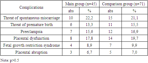
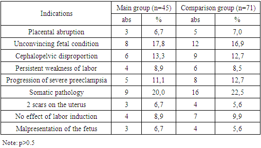
The average age of the patients who took part in the study was 25.6+5.4 years in the main group, and 26.1+5.1 years in the control comparison group (p>0.05). According to the presented results, the majority of patients were aged 21-30 years and there was no significant difference between the groups in the age of the patients (p>0.05). A study of the somatic history showed that in both groups, iron deficiency anemia was most often detected (in 20% of patients in the main group and in 15.5% in the comparison group). To a lesser extent, diseases of the gastrointestinal tract (chronic gastritis, cholecystitis in remission), diseases of the genitourinary system (chronic pyelonephritis in remission) are noted. Varicose veins were observed in 13.3% and 9.9%, respectively. Among the diseases of the nervous system, epilepsy was encountered, which was an indication for CS. Another indication was high myopia or eye surgery. A study of the anamnesis revealed that 5 patients (11.1%) of the main group and 6 women (8.5%) of the comparison group had surgical interventions on the abdominal organs for appendicitis, ectopic pregnancy, ovarian apoplexy and ovarian tumor formations.
When analyzing gynecological diseases in the groups, a history of vaginitis and bacterial vaginosis, cervical pathology, and menstrual cycle disorders were more common. Cervical pathology included ectropion (3 patients in each group) and cervical polyp (2 patients in the main group and 1 in the control group), for which surgical treatment - removal of the polyp and diathermoconization was performed. Equally often in both groups (11.1% and 7.04%, respectively), patients had a history of suffering from inflammatory diseases of the pelvic organs. In total, 26.7% of patients in the main group and 22.5% in the control group had a history of gynecological diseases.
The study of obstetric history showed that in both study groups primiparas predominated: in the main group they accounted for 93.3% (42 patients), in the control group – 91.6% (65 patients). The remaining women were multiparous. In addition, the women studied had a history of spontaneous miscarriages in 3 (6.7%) women in the main group and 7 (9.9%) women in the comparison group.
A study of the course of this pregnancy (Table 1) showed that gestation was often complicated by the threat of miscarriage, which was equally common in the study groups (22.2% and 21.1%, respectively). Preeclampsia was observed more rarely in 15.6% and 16.9% of women, respectively. Placental insufficiency was noted in 17.8% of women in the main group and in 19.7% in the comparison group. Fetal growth restriction syndrome was detected in 4 (8.9%) and 7 (9.9%) women in the study groups, respectively. Such a serious complication of pregnancy as placental abruption occurred in 3 (6.7%) women in the main group and in 5 (7.0%) women in the comparison group, which was one of the indications for cesarean section (Table 2).
Table 1. The course of this pregnancy in the women studied
     
Other more common indications for cesarean section were unconvincing fetal condition (17.8% and 16.9%, respectively), somatic pathology (20% and 22.5%) and cephaopelvic disproportion (13.3% and 12.5%). 7%).
Table 2. Indications for caesarean section
     
In addition to the above indications for emergency cesarean section, there was progression of severe preeclampsia in 11.1% and 12.7% of women. More rare indications for operative delivery were persistent weakness of labor that cannot be corrected, malpresentation of the fetus, ineffective labor induction at 41 weeks, and 2 scars on the uterus.
The stages of caesarean section were carried out traditionally. Laparotomy was performed according to Joel-Cohen, cesarean section in the lower uterine segment. An incision on the uterus was made transversely in the lower segment with a scalpel and extended with digital extension. The incision on the uterus was sutured with a double-row continuous synthetic absorbable suture material "Vicryl". The operation was performed under epidural anesthesia. For the purpose of antibiotic prophylaxis, on the basis of the woman’s voluntary informed consent, intravenous administration of a cephalosporin antibacterial drug in a dose of 2 g was carried out once during the operation. After fetal extraction, oxytocin was slowly administered intravenously at a dose of 5 units. After suturing the incision on the uterus, the suture line on the uterus was visually divided into three parts. To introduce platelet autoplasma, an injection with a thin needle was made at the border of the outer and middle third at a distance of 3-5 mm from the edge of the wound. The needle was directed towards the outer edge of the suture and inserted almost the entire length. When removing the needle, apply slow pressure to the plunger so that the plasma infiltrates the uterine tissue along the suture. After this, from the same injection site, the direction of the needle was changed to the center, and again inserted to the full length, followed by infiltration of the myometrium. Thus, from one injection it was possible to infiltrate a 5 cm long suture area (2.5 cm in each direction), saturated throughout its entire length with platelet auto-plasma. 1.5 ml of auto-plasma was injected into one infiltration zone. For 1 needle injection–3 ml. To infiltrate the seam along the entire length, 4 injections were used along the upper and lower edges of the seam. The volume of injected plasma is 12 ml. The duration of plasma administration during CS was 5-10 minutes. A comparative study of the operation parameters (technique, duration, volume of blood loss) showed that they did not differ within significantly significant limits (p>0.05). Thus, the duration of operations in the main group was 47 + 2.6 minutes, which, due to the time spent on puncturing the PAP suture, exceeded the time in the control group (41 + 1.2 minutes), but within unreliably significant limits.
The main indicators in the evaluation of surgical interventions were the nature and frequency of postoperative complications. All women in the postoperative period underwent ultrasound on days 3-4 (Table 3) and Doppler measurements of blood flow in the vessels of the uterus on days 4-5 (Table 4) after cesarean section in order to determine possible pathological processes in the uterus and the condition of the suture. When using PRP, echographic signs of hematomas were not detected in any case, which was significantly less compared to traditional suture (p<0.05). This allows us to make an assumption about the better hemostatic effect of this type of suture. Ultrasound signs of decreased uterine tone and endometritis (expansion of the cavity, gas bubbles, fibrin deposition) were found in 1 (2%) patient with a lochiometer. The results of ultrasound revealed the dependence of the rate of uterine involution on the method of restoration of the lower segment. The dimensions of the uterus according to ultrasound data on days 3-4 after surgical delivery in patients of the main and control groups are presented in Table 3. As can be seen from the table, in the control group the length of the uterus averaged 124.8±1.5 mm, width - 113, 2±2 mm, anterior-posterior size - 75.6±0.8 mm. It should be noted that the size of the uterus in the patients of the main group was significantly smaller: the length of the uterus was on average 116.8±1.4 mm, width - 105.3±1.6 mm, anteroposterior size - 70.8±1, 2 mm (p<0.05). The data obtained suggest that the use of platelet autoplasma not only does not have a negative effect on the contractile activity of the uterus, but also improves the involution of the uterus after CS.
Table 3. Ultrasound characteristics of the uterus on days 3-4 after surgery in the women studied
     
The thickness of the uterus in the area of the postoperative suture determines the so-called “edema zone,” reflecting the degree of the inflammatory reaction. The thickness of the uterus in the area of the postoperative suture (Table 3) in patients of the main group was 26.9+0.7 mm, in the control group - 26.7+0.8 mm and did not have significant differences (p>0.05). Thus, the results of the ultrasound examination confirmed the uncomplicated course of the postoperative period in women of the main group.
On days 4-5 after cesarean section, we assessed the index of blood flow resistance of the uterine vessels (Table 4), which were higher in the uterine vessels in patients in the comparison group with the traditional CS technique. Thus, the resistance index in the control group was 0.65±0.02 in the right uterine artery and 0.66±0.02 in the left uterine artery. In patients of the main group, respectively – 0.58±0.01 and 0.60±0.01 (p<0.05). The greatest difference in blood flow, and statistically significant, was determined in the radial arteries in the area of the suture on the uterus after cesarean section. The resistance index in the suture area in patients of the main group was 0.53±0.01, in the control group – 0.62±0.01 (p<0.01). Such an increase in the resistance index is associated with an increase in peripheral resistance in the vessels of the uterus. In patients of the main group who received platelet autoplasma, the resistance index was statistically significantly lower, which is associated with lower peripheral resistance, possibly due to a larger number of growing newly formed vessels in the wound area of the uterus, formed under the influence of vascular growth factors.
Table 4. Doppler characteristics of uterine vessels after surgery in the women studied
     
It is known that auto-plasma obtained using the “Plasmolifting TM” technology contains platelets and growth factors [12]. In the radial arteries of the intact posterior wall of the uterus, the resistance index was also lower in the main group (0.54±0.01), compared with the control group (0.55±0.02), but this difference was not statistically significant.
The nature of the postoperative period determined the number of bed days spent in the hospital after the operation. The average number of bed days in the main group did not differ significantly from the control group and was 5.0+1.2 and 5.2+1.4 days (p>0.05), respectively.
3 months after cesarean section, the patients were invited for an examination, which included trans-vaginal ultrasound of the pelvic organs, Doppler measurements of blood flow in the uterine vessels, and assessment of vascularization using color Doppler mapping. The size of the uterus, including the volume of the uterus, did not have significant differences between the groups. The thickness of the uterus in the area of the scar was smaller in patients of the main group and amounted to 6.1±0.7 mm, in the control group - 6.5±0.8 mm, but within unreliably significant limits. In the area of the scar, multiple hyperechoic inclusions up to 1 mm were visualized, which were regarded as unresolved suture material. Whereas, in the comparison group, in 2 patients with the traditional technique, hyperechoic formations in the scar area ranged from 8 to 10 mm. These formations may have been hematomas undergoing reverse development. In the main group, only 1 (2.2%) patient had a cystic inclusion of 4.5x2.5 mm, and in the group with the traditional technique, 3 patients had cystic inclusions in the scar area ranging in size from 3.7 mm to 7.8 mm. Another ultrasound finding was a niche-shaped defect, the depth of which was 3 mm, and the remaining myometrial tissue was 4.6 mm in the study group. In the group with the traditional technique, the “niches” had a depth of up to 8.8 mm, while the remaining part of the myometrium ranged from 1.5 to 1.8 mm, which gave us reason to classify these scars as “thin”/“incompetent”.
Blood flow in the scar area was also assessed using the resistance index of blood flow in the vessels of the uterus. It was found that the blood flow in the radial arteries in the area of the uterine scar was practically no different from the blood flow in the intact posterior wall of the uterus. Analysis of the vascularization of the scar area with color Doppler mapping revealed that the use of PRP therapy in most women (75.6%) led to moderate blood flow, in 22.2% - increased blood flow, and in only 1 patient (2.2%) blood flow was poor. Unlike the main group, in the comparison group, poor blood flow was noted in 26 women (36.6%), which was 16.6 times more often. Increased blood flow was observed in 2 patients (2.8%), which is 7.9 times less common than in the group with PRP therapy. The remaining 28 (39.4%) women in the comparison group had moderate blood flow in the scar area.
Thus, the use of platelet auto-plasma during cesarean section had no side effects or complications in the postoperative period. Statistically significant low values of the resistance index in the vessels of the uterus in patients with PRP therapy suggest more intense neovascularization of the wound on the uterus, stimulates the process of collagenization and maturation of connective tissue and the subsequent formation of a wealthy scar, which was confirmed by ultrasound and Doppler data.

5. Conclusions

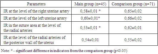
1. The introduction of platelet autoplasma into the suture area of the uterus has a positive effect on the healing of the uterine scar and makes it possible to accelerate the process of reparative regeneration.
2. The use of platelet autoplasma promotes the formation of a full-fledged scar on the uterus after cesarean section by increasing angiogenesis, improving vascularization of the uterus and the scar area.

References

[1]  Farg D., Hassan H. Surgical Repair versus Non-Surgical Management of Spontaneous Perineal Tears that Occur during Childbirth. American Journal of Nursing Research, 2019; 7(5): 781-792. doi: 10.12691/ajnr-7-5-11.
[2]  Hassan H. Evidence-Based Practice in Midwifery and Maternity Nursing for Excellent Quality of Care Outcomes. American Journal of Nursing Research, 2020; 8(6): 606-607. doi: 10.12691/ajnr-8-6-3.
[3]  Tavukcu HH, Aytaç Ö, Atuğ F, Alev B, Çevik Ö, Bülbül N, Yarat A, Çetinel Ş, Şener G, Kulaksızoğlu H. Protective effect of platelet-rich plasma on urethral injury model of male rats. // Neurourol. Urodyn. 2018; 37(4): 1286‒1293. https://doi.org/10.1002/nau.23460.
[4]  Hassan H. The Impact of Evidence-Based Nursing as The Foundation for Professional Maternity Nursing Practices. Open Access Journal of Reproductive System and Sexual Disorder, 2019; 2(2): 195-197. OAJRSD.MS.ID.000135. DOI: 10.32474/OAJRSD.2019.02.000135.
[5]  Van der Voet LF, Jordans IP, Brolmann HA et al. Changes in the uterine scar during the first year after a caesarean section: a prospective longitudinal study. // Gynecol. Obstet. Invest. 2017. https://doi.10.1159/000478046.
[6]  Hassan H., El-Sadek A., Ali L. Effect of Three Different Nursing Interventions on Intestinal Motility and Women's Satisfaction Post-Cesarean Section Birth. American Journal of Nursing Research, 2019; 7(6): 932-941. doi: 10.12691/ajnr-7-6-4.
[7]  Robson SJ, de Costa CM. Thirty years of the World Health Organization's target caesarean section rate: time to move on. // Med. J. Aust. 2017; 206:181-5.
[8]  Hassan H., EL-Kholy G., Ateya A., Hassan A. Breast Feeding Knowledge and Practices among Primiparous Women with Caesarean Section: Impact on Breast Engorgement in Upper Egypt. Communication, Society and Media, 2020; 3(2): 34- 78.
[9]  Hassan H., EL-Kholy G., Ateya A. & Hassan A. Breast Engorgement among Women with Caesarean Section: Impact of Nursing Intervention. Education, Society and Human Studies, 2020; 1(2): 22-55. doi:10.22158/eshs.v1n2p22.
[10]  Krasnopolsky V.I., Buyanova S.N., Shchukina NA, Logutova L.S. Failure of the suture (scar) on the uterus after CS: problems and solutions. // Ross. bulletin of the obstetrician-gynecologist. 2015; 3:4-8.
[11]  Makiyan Z.N., Adamyan L.V., Karabach V.V., Chuprynin V.D. A new method of surgical treatment of uterine scar failure after cesarean section using an intrauterine manipulator with a trough. // Obstetrics and Gynecology. 2020; 2: 104-10. https://dxdoi.org/1018565/aig.20202104-110.
[12]  Farag D., Bakr M., Zedan H., Hassan H., Eid S. Maternal and Newborn Outcome among Women Undergoing Elective versus Emergency Caesarean Section: A Comparative Study. Egyptian Journal of Health Care, (EJHC), 2023; 14 (3): 454-468. DOI: 10.21608/EJHC.2023.317867.
[13]  Hassan H., Zedan H., Farag D. Successful Vaginal Birth Subsequent to Cesarean Section. Mansoura Nursing Journal, 2024; 11(1): DOI: 10.21608/mnj.2024.216259.1325.
[14]  Ciebiera M, Ciebiera M, Czekanska-Rawska M, Jakiel G. Laparoscopic isthmocele treatment - single center experience. // Videosurgery Minim Invasive Gynecol. 2017; 12: 88-95.
[15]  Satpathy G, Kumar I, Matah M, Verma A. Comparative accuracy of magnetic resonance morphometry and sonography in assessment of post-cesarean uterine scar. // Indian J. Radiol Imaging. 2018; 28: 169-74.
[16]  Pomorski M, Fuchs T, Rosner-Tenerowicz A, Zimmer M. Sonographic evaluation of surgical repair of uterine cesarean scar defects. // J. Clin. Ultrasound. 2017; 45 (8): 455-60. https://doiorg/101002/jcu.22449.
[17]  Seliger G, Chaoui K, Lautenschlager C et al. Technique of sonographic assessment of lower uterine segment in women with previous cesarean delivery: a prospective, pre/intraoperative comparative ultrasound study. // Arch. Gynecol. Obstet. 2018; 298: 297-306.
[18]  Setubal A, Alves J, Osorio F et al. Treatment for uterine isthmocele, a pouch-like defect at the site of cesarean section scar. // J. Minim. Invasive Gynecol. 2017. https://doi.org/doi.-10.1016lj.jmig2017.09022.
[19]  Tang X., Wang J., Du Yet al. Caesarean scar defect: Risk factors and comparison of evaluation efficacy between transvaginal sonography and magnetic resonance imaging. // Eur. J. Obstet Gynecol. Reprod. Biol. 2019; 242: 1-6.
[20]  Tanimura S, Funamoto H, Hosono T et al. New diagnostic criteria and operative strategy for cesarean scar syndrome: endoscopic repair for secondary infertility caused by cesarean scar defect. // J. Obstet. Gynaecol. Res 2015; 41: 1363-9.
[21]  Ying-Yi Chen, Ching-Chang Tsai, Kuo-Chung Lan et al. Preliminary report on the use of a levonorgestrel intrauterine system for the treatment of intermenstrual bleeding due to previous cesarean delivery scar defect. // J. Obstet. Gynaecol. Res. 2019; 45 (10): 2015-20. https://doi.10.1111/jog.14060.
[22]  Karli P, Sahin B, Kara F. The incidence of isthmocele may be higher than reported. // J. Surg. Med. 2018; 2 (3): 283-7.
[23]  Tulandi T, Cohen A. Emerging manifestations of Cesarean scar defect in reproductive-aged women. // J. Minim. Invasive Gynecol. 2016; 23: 893-902.
[24]  Zhang Y. A comparative study of transvaginal repair and laparoscopic repair in the management of patients with previous Cesarean scar defect. // J. Minim. Invasive Gynecol. 2016; 23: 535-41.