-
Paper Information
- Next Paper
- Previous Paper
- Paper Submission
-
Journal Information
- About This Journal
- Editorial Board
- Current Issue
- Archive
- Author Guidelines
- Contact Us
American Journal of Medicine and Medical Sciences
p-ISSN: 2165-901X e-ISSN: 2165-9036
2024; 14(3): 767-770
doi:10.5923/j.ajmms.20241403.48
Received: Feb. 29, 2024; Accepted: Mar. 13, 2024; Published: Mar. 29, 2024

Determination of the Morphometric Parameters of the Epididymis of White Outbreed Rats in Age Dynamics
 Abstract
 Reference
 Full-Text PDF
 Full-text HTML
Farrukh Jumaevich Namozov
Assistant of the Department of Pathological Anatomy, Bukhara State Medical Institute, Bukhara, Uzbekistan
Correspondence to: Farrukh Jumaevich Namozov, Assistant of the Department of Pathological Anatomy, Bukhara State Medical Institute, Bukhara, Uzbekistan.
| Email: |  |
Copyright © 2024 The Author(s). Published by Scientific & Academic Publishing.
This work is licensed under the Creative Commons Attribution International License (CC BY).
http://creativecommons.org/licenses/by/4.0/

In this article, the normative morphometric parameters of the testicles in the age dynamics of white rats are presented. Epididymal weight and dimensions: Small testicular mass (0.21 g) and its dimensions (length - 0.74 cm, thickness - 0.49 cm) are observed in newborn rats. As the rats get older, the mass, length and thickness of the epididymis increase. The highest values are achieved in 12-month-old rats: weight - 0.76 g, length - 1.3 cm, thickness - 0.9 cm. Epididymal tissue volume and density: Epididymal tissue volume also increases with age, reaching the highest values in 12-month-old rats (0.64 cm3). The density of testicular tissue decreases with age, which may be related to tissue development and changes in its composition. Correlation between body weight and testicular weight: Both body weight and testicular weight increase with the age of rats, which shows some parallelism in their growth.
Keywords: Testicular hypertrophy, Purebred rats, Morphometry, Index
Cite this paper: Farrukh Jumaevich Namozov, Determination of the Morphometric Parameters of the Epididymis of White Outbreed Rats in Age Dynamics, American Journal of Medicine and Medical Sciences, Vol. 14 No. 3, 2024, pp. 767-770. doi: 10.5923/j.ajmms.20241403.48.
1. Relevance of the Study
- All over the world, special attention is being paid to research aimed at improving the early detection, treatment and prevention of diseases of the male reproductive system, including hypertrophy, caused by various physical factors. Currently, studies of the male population in different countries show an increase in spermatogenesis disorders [1-3], a decrease in the average age of men with spermogram abnormalities and various reproductive diseases [4-6]. The structure of the causes of male infertility is as follows: varicocele (14.8%), hypogonadism (10.1%), urogenital infections (9.3%), history of cryptorchidism (8.4%), previous oncological diseases (7.8 %), immunological factors (3.9%), erectile dysfunction and ejaculation (2.4%), systemic diseases (2.2%), vascular obstruction (2.2%), testicular tumours (1.2%), presence of somatic diseases (7.7%). In about 30% of cases, the true cause of male infertility cannot be determined, in which case it is diagnosed as "idiopathic infertility" [2,4-5]. Targeted measures are being implemented in our country to radically improve the healthcare system and improve the quality of medical services for the population. For this purpose, many tasks were defined, including "increasing the efficiency, quality and accessibility of medical care, implementing comprehensive measures aimed at supporting a healthy lifestyle and preventing diseases, including a healthy lifestyle the formation of a system of medical standardisation, the introduction of high-tech diagnostic methods and treatment by creating effective models of patronage and clinical examination.The purpose of the study: to study the morphometry of the testis in purebred rats.
2. Research Materials
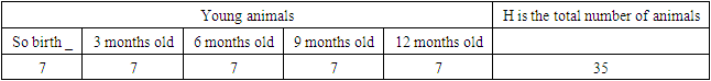
- An experimental study was conducted on the material obtained from the testicles of 35 newborn, 3, 6, 9, 12 months old male white rats.
 Animals in the morning, hungry to the stomach, either unconscious under one in the moment decapitation method slaughtered _ Belly space since opening then the testicles excesses separate received and their mass, length, width, volume and tissues density was studied. Testicle of excesses each one's mass electricity on the scales measured, length and width millimetre tape with was measured. Testicle of excesses according to the volume formula:V= 0.123×n×c2, that is where: n,c - suitable, of the testicle length and thickness 0.123 - constant coefficient. Received testicle excess Bowin in solution fixed. Raised to concentration have ethyl from alcohol from the past then they _ Hot paraffin inserted, then 6-7 μm standard thickness has been testicle excesses pieces prepared, they are oriented sagittally or frontally. Samples to van Gieson according to hematoxylin and eosin with painted _ Ready histological drugs NLCD-307B binoculars microscope under checked (Novel, China).
3. Research Results
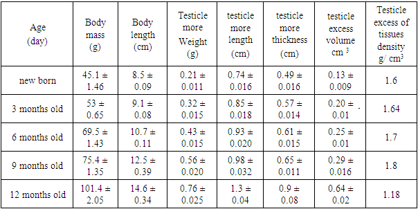
- In the process of studying the morphometry of the epididymis in non-white rats, the following data were obtained on newborn non-white rats: epididymal weight: 45.1 ± 1.46 g, body length: 8, 5 ± 0.09 cm, epididymis mass: 0.21 ± 0.011 g, epididymis length: 0.74 ± 0.016 cm, epididymis thickness: 0.49 ± 0.016 cm, epididymis thickness: volume: 0.13 ± 0.009 cm3, epididymis density: 1.6 g/cm3.These results provide insight into the characteristics of testicular hypertrophy in newborn white outbred rats.The data obtained from the study of the morphometry of the epididymis in newborn white outbred rats provide important information about the development and characteristics of this organ in this type of genotype.It is interesting to note that the weight of the testicle is an important indicator that can be related to its functional activity and the general condition of the animal. Other parameters such as the length, thickness and volume of the epididymis also play an important role in evaluating their structure and possible effects on reproductive processes.The analysis of the density of the epididymal tissue allows us to understand the structural composition of this organ. It can be useful in studying pathologies or the effect of various factors on the epididymal tissue. These results not only add important insights to the field of reproductive biology but may also have practical implications for developing strategies to treat or prevent reproductive disorders in rats and possibly other mammals.Morphometric parameters of the epididymis of 3-month-old rats: epididymis body weight: 53 ± 0.65 g, body length: 9.1 ± 0.08 cm, epididymis mass: 0.32 ± 0.015 g, the length of the epididymis: 0.85 ± 0.018 cm, the thickness of the epididymis: 0.57 ± 0.014 cm, the volume of the epididymis: 0.20 ± 0.01 cm3, the tissue of the epididymis density: 1.64 g/cm3.During the experiment with rats of different age groups, the following characteristics were revealed. In 6-month-old rats, the body weight of the testis is 69.5+1.43 g, the body length is 10.7+0.11 cm, the weight of the testis in them is 0.43+0.015 g, The testis is the length of is calculated. 0.93+0.020 cm, testicle excess thickness - 0.61+0.015 cm, testicle excess volume - 0.25+0.01 cm3, equal to g/cm3 (Figure 1). Testicle excess tissues density -1.7 _ organise did _ 9 months old in rats testicle body weight of the rest reached 75.4+1.35 g, body length reached 12.5+0.39 cm. Testicle more weight 0.56+0.020 g, testicle more length 0.98+0.032 cm, testicle more thickness - 0.65+0.011 cm, testicle more volume - 0.29+0.016 cm3, testicle more tissue density g/cm3 -1.8 (Figure 1). 12 months old in rats testicle the body weight of the rest is up to 101.4+2.05 g increased, the body length reached 14.6 + 0.34 cm. Testicle more weight is 0.76+0.025 g, testicle more length is equal to 1. 3+0.04 cm, testicle excess thickness - 0.9+0.08 cm, testicle excess volume - 0.64+0.02 cm3, testicle excess of tissues density was estimated as -1.18 in g/cm3. Above morphometric check results are given in Table 1 below.
|
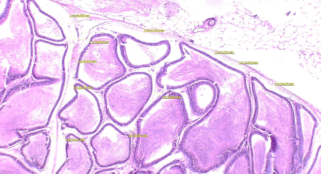 | Figure 1. Morphometric dimensions of the testis of a nonwhite male rat (hematoxylin and eosin stain, medium magnification) |
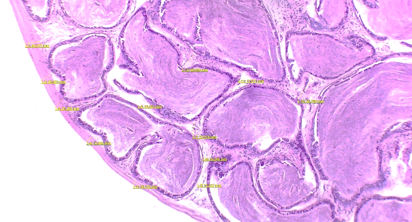 | Figure 2. Morphometric dimensions of the testis of a nonwhite male rat (hematoxylin and eosin stain, medium magnification) |
4. Conclusions
- "White without breed in rats testicle more from the study of "morphometry". the release of the following conclusion can:1. Testicle more weight and dimensions:- Newborn in rats' testicles more smaller mass (0.21 g) and his dimensions (length - 0.74 cm, thickness - 0.49 cm) are observed.- Rats ageing with Testicle excess of mass, its length and thickness increases. Most high 12 months to indicators age in rats obtained: weight - 0.76 g, length - 1.3 cm, thickness - 0.9 cm.2. Testicle excess tissue volume and density:- Testicle more size and age looking increased, 12 months in rats the highest to indicators reached (0.64 cm3).- Testicle excess tissue density of age growth decreases, this of tissue development and his of the composition change with depends to be can _3. Body weight and testicle gain weight _ between dependency:- Rats age with body weight and testicles excess the weight also increases, this they're in growth some kind of parallelism shows.This data on reproductive biology and testicles more development and of activity medical aspects in the field next studies for useful.