American Journal of Medicine and Medical Sciences

p-ISSN: 2165-901X    e-ISSN: 2165-9036

2024;  14(3): 734-738

doi:10.5923/j.ajmms.20241403.41

Received: Feb. 25, 2024; Accepted: Mar. 13, 2024; Published: Mar. 22, 2024


Indicators of the Hemostasis System in Women with Premature Birth and Obstetric Blood Loss

Аkhtamova N. A., Shavazi N. N.

Department of Obstetrics and Gynecology 1, Samarkand State Medical University, Samarkand, Uzbekistan

Correspondence to: Аkhtamova N. A., Department of Obstetrics and Gynecology 1, Samarkand State Medical University, Samarkand, Uzbekistan.

Email:

Copyright © 2024 The Author(s). Published by Scientific & Academic Publishing.

This work is licensed under the Creative Commons Attribution International License (CC BY).
http://creativecommons.org/licenses/by/4.0/

Abstract

This study investigates the hemostasis system in women with premature birth and obstetric blood loss, focusing on the role of endothelial dysfunction in these pregnancy complications. Conducted at the Samarkand State Medical University, the research involved 151 pregnant women aged 18 to 36 years, divided into three groups based on clinical presentations. The study assessed the plasma and cellular components of the hemostasis system, with a particular emphasis on platelets and biochemical markers of endothelial dysfunction, including highly sensitive von Willebrand factor (vWf) and apoptosis protein p53. Results indicated adaptive changes in the hemostatic system during pregnancy, with significant alterations in activated partial thromboplastin time, prothrombin index, fibrinogen levels, and international normalized ratio. Elevated levels of biochemical and functional endothelial markers were associated with the risk of obstetric hemorrhage during preterm labor. The study highlights the importance of monitoring endothelial function as part of prenatal care and suggests that these markers could serve as predictors of complications in pregnant women. Further research is needed to develop effective treatment strategies and improve maternal and child health outcomes.

Keywords: Hemostasis system, Premature birth, Obstetric blood loss, Endothelial dysfunction, Biochemical markers, von Willebrand factor, Apoptosis protein p53

Cite this paper: Аkhtamova N. A., Shavazi N. N., Indicators of the Hemostasis System in Women with Premature Birth and Obstetric Blood Loss, American Journal of Medicine and Medical Sciences, Vol. 14 No. 3, 2024, pp. 734-738. doi: 10.5923/j.ajmms.20241403.41.

1. Introduction

Obstetric hemorrhage is a leading cause of maternal morbidity and mortality worldwide, despite advancements in obstetric care. Understanding the etiopathogenesis of obstetric hemorrhage and identifying opportunities for improved prognosis, prevention, and treatment are crucial for addressing this persistent issue. The study presented in this article focuses on the hemostasis system in women experiencing premature birth and obstetric blood loss, aiming to shed light on the underlying mechanisms and potential interventions [1-8].
The hemostatic system undergoes adaptive changes during pregnancy, playing a vital role in gestation. Dysregulation of this system can lead to complications such as premature birth and obstetric haemorrhage. This research explores the relationship between endothelial dysfunction, a key factor in vascular health, and these pregnancy complications. By examining markers of endothelial function and hemostasis, the study seeks to identify biochemical indicators that could serve as predictors of obstetric hemorrhage and guide the development of effective treatment strategies [4,9-15].
Furthermore, the study delves into the impact of various risk factors, including maternal and fetal conditions, obstetric procedures, and socioeconomic factors, on the likelihood of obstetric hemorrhage. Early identification and management of these risk factors through regular prenatal care and appropriate interventions are emphasized as crucial steps in preventing obstetric hemorrhage [16-19].
In summary, this research contributes to the growing body of knowledge on the hemostasis system's role in pregnancy and its complications. By investigating the links between endothelial dysfunction, hemostatic changes, and obstetric hemorrhage, the study aims to inform the development of targeted interventions to improve maternal and child health outcomes.

2. Materials and Methods

The study was conducted at the branch of the Russian National Research Medical Center for Medical Research in Samarkand, specifically in the Department of Pathology of Pregnant Women. The research spanned from 2021 to 2023 and involved examining 151 pregnant women, who were subsequently divided into three distinct groups based on their clinical presentations.
The inclusion criteria for the study targeted women aged 18 to 36 years, with a particular emphasis on specific age groups. The average age of the participants was 27±2.1 years. The participants' social status varied, with 28% being homemakers and 27% students. The study also assessed the participants' Body Mass Index (BMI), which is relevant as it can influence the volume of blood loss during childbirth.
An in-depth analysis of the participants' anamnesis was conducted, encompassing their reproductive, gynecological, somatic, and infectious history. The current pregnancy status, causes of bleeding, and the postpartum period were also assessed. Additionally, the study examined the reproductive and obstetric history of the women, including their parity and gestational age—this comprehensive analysis aimed to identify potential risk factors and correlations with obstetric hemorrhage.
The study further delved into the participants' prevalence of somatic diseases and risk factors. The frequency of obesity, myopia, and varicose veins was analysed, as these conditions can potentially impact pregnancy outcomes. Additionally, the assessment covered chronic arterial hypertension (CAH) and iron deficiency anemia (IDA), both of which are significant concerns in prenatal care. The study also considered the presence of chronic pyelonephritis and the incidence of frequent acute respiratory viral infections among pregnant women.
The laboratory analyses focused on the hemostasis system, which is crucial for maintaining vascular integrity during pregnancy. The study examined the plasma component of the hemostasis system and the cellular component, with a particular emphasis on platelets. Furthermore, the research aimed to identify biochemical markers of endothelial dysfunction, such as the susceptible von Willebrand factor (vWf) and the apoptosis protein p53 in blood serum. These markers are essential for understanding the functional state of the endothelium and its role in pregnancy complications.
The Mann-Whitney test was employed to assess the significance of differences in the studied characteristics for statistical analysis. This non-parametric test is commonly used in medical research to compare two independent groups when the data are not normally distributed. Applying this statistical method allowed for a rigorous evaluation of the data collected in the study, ensuring the validity of the findings.

3. Results

The analysis was conducted to predict the risk of abnormal blood loss in preterm birth. For this purpose, various features of the anamnesis were studied, including reproductive, gynecological, somatic, and infectious, the course of the current pregnancy, causes of bleeding and the postpartum period.
The following results were obtained when analyzing the reproductive history in the compared groups. The average age at menarche was 13.1±1.21 years in the first group, 14.3±1.43 in the second study group and 12.4±1.08 in the control group. As can be seen from the presented data, menarche's age and menstruation duration were statistically comparable in the study groups.
A distinctive feature of the obstetric history of pregnant women with UPR is the high frequency of spontaneous abortions in group I (70.7%) in group II (67.6%), threat of premature birth in group I (57.8%); in group II (78.6%), history of premature birth in group I (62.4%); in group II (61.3%).
When studying the parity of women in the first group, 12 women (22%) had a 1st birth, 13 women (23.6%) had a 2nd birth, 21 (38.1%) had a 3rd birth, 4 9 women (16.3%) had 1st or more births; in the second group, eight women (14.3%) had 1st births; 11 women (19.6%) had 2nd births. 3rd birth - in 23 (41.1%) 4th or more births - in 14 women (25%) in the control group; 1st birth was in 26 women (65%), 2nd birth - in 9 women (22.5%), 3rd birth - in 4 (10%) 4th - in 1 woman (2.5%) (Fig. 1).
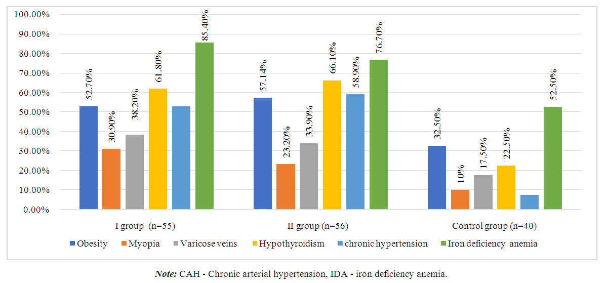
Figure 1. Analysis of somatic pathology among the study groups
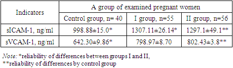
The gestational age of women in the leading 2 groups was from 28 to 34 weeks. The average gestational age of pregnancy in the main groups was, on average, 28.66±4.76 weeks, and in the control group, 38.82±2.05 weeks.
When analysing somatic diseases, it was revealed that women in clinical groups had a comparable frequency of a history of obesity of varying degrees, myopia and varicose veins (VV) of the lower extremities (Fig. 1).
Chronic arterial hypertension (CAH) and iron deficiency anemia (IDA) were more often diagnosed in women from the first group, 29 (52.7%), 47 (85.4%), and the second group, 33 (58.9%); 43 (76.7%) (p=0.0125).
Chronic pyelonephritis and frequent acute respiratory viral infections are not associated with bleeding during premature birth. A history of somatic pathology may increase the risk of pathological blood loss during childbirth, regardless of gestational age. An assessment of the medical history is necessary to prevent abnormal bleeding during preterm birth.
It should be noted that these results may be limited and cannot be generalised to the entire female population since the study was conducted only on a group of women with PC and preterm birth. To fully understand the impact of infectious and inflammatory diseases on the likelihood of early pregnancy, additional research is needed to consider a more comprehensive range of factors.
Thus, the analysis showed that women with pathological blood loss during premature and term labour were more likely to have IDA.
To fully understand the influence of the above reasons for the likelihood of PC occurring during premature birth, it is necessary to conduct additional studies that can predict this pathology.
The course of pregnancy is accompanied by adaptive changes in the hemostatic system, which plays a vital role in the gestation process. The results of the study of the plasma component of the hemostasis system are presented in Table 1.
Table 1. Parameters of the plasma link of the system hemostasis (n=151)
     
The APTT rate in the study groups with UPR was 1.3 times higher than in the control group (p<0.05). PTI: in the study groups, it was 1.1 times higher than in the control group (p<0.05). Fibrinogen: 1.1 times more than in the control group (p<0.05). INR: in the study groups, it was 1.7 times higher than in the control group. (R<0.05).
The study of the coagulation system in pregnant women also includes an analysis of the cellular component of hemostasis. The scientific literature has demonstrated the role of platelets in ensuring tropism of the vascular intima: electron microscopy confirmed the angiotrophic function of platelets [6]. This further justifies the inclusion in our study of an analysis of the state of the cellular link of the hemostasis system, which provides a complete study of the factors influencing the morphofunctional state endothelium. The results obtained are presented in Table 2.
Table 2. Platelet component of the hemostasis system in the study groups (n=151)
     
The platelet counts in patients in the first and second groups were 1.4 times higher than in the control group. The determination of the average platelet volume in the first and second groups was 1.3 times higher than in the control group (p <0.05).
Thus, when analysing some parameters of hemostasis, no significant differences were found in the groups of participants.
The study identified biochemical markers of endothelial dysfunction. Indicators that are accessible for laboratory analysis and objectively reflect changes in the functional state of the endothelium were selected. The most reliable and practical laboratory indicators of the studied pathology are: susceptible von Willebrand factor (vWf) and apoptosis protein p53 in blood serum.
The concentration of Von Willebrand factor averaged 101.6%, with a standard deviation of ±9.47% in the control group. It was revealed that the average level of von Willebrand factor was 120.0%. The standard deviation was ±20.18% in the first group in women with UPR, its concentration in group II in women with UPR was 128.0±19.05, and in the control group in women with a physiological course of pregnancy was 101.6±9.47.
The concentration of apoptosis protein p53 in peripheral blood plasma in women with UPR was 3.33 times higher than in women with a physiological pregnancy (control group).
D-dimer is a protein, a product of the destruction of the fibrin molecule, which indicates a disorder in the coagulation system. Thus, in women with UPR in the first and second groups, its value was 1.78 higher than in the control group.
Thus, the study's results made it possible to determine indicators that can be used as markers of endothelial dysfunction in women with PR. Thanks to this, it will be possible to develop more effective treatment technology and use these indicators as predictors of complications in pregnant women.
The study's results (Table 3) demonstrate that these indicators can be potential indicators of the state of the endothelium in pregnant women, especially in the context of the risk of pregnancy failure.
Table 3. Markers of endothelial function in pregnant women in the study groups
     
Endothelial dysfunction was found in women of groups I and II; their blood serum levels were higher than those of the control group. The Mann-Whitney test was used to assess the significance of differences in the studied characteristics. In summary, this study shows that women with threatened preterm labour experience endothelial dysfunction, which is manifested by elevated serum biochemical markers. This may be one of the reasons for gestational disorders and requires further study to develop promising methods for treating and preventing this pathology.
The average values of markers - von Willebrand factor and apoptosis marker protein p53 were compared between groups of women - with UPR (group I) (group II) and women with a physiological course of pregnancy (control group).
Table 4. Content of markers of endothelial dysfunction in the blood
     
The average von Willebrand factor also exceeded the level of the control group in the leading group of women with VPR - 120.0 ± 20.18%, compared to 101.6 ± 9.47% in the control group. In 49.8% of women with UPR, an increase in the D-dimer level in the blood above 300 ng/ml was detected. This indicates the processes of fibrin cross-polymerization during intravascular coagulation. The concentration of apoptosis protein p53 in the blood serum was also higher in the first group - 0.01 ± 0.011 U/ml compared to 0.003 ± 0.0013 U/ml in the control group.
Functional endothelial markers such as von Willebrand factor, soluble vascular cell adhesion molecule-1, and soluble intercellular adhesion molecule-1 are commonly used to indicate endothelial dysfunction.
The precise mechanisms underlying the association between endothelial dysfunction and obstetric hemorrhage during preterm labour are not fully understood. However, it is believed that endothelial dysfunction may lead to impaired placental blood flow and abnormal uterine contractions, which are known risk factors for obstetric hemorrhage. In addition, poor coagulation and fibrinolysis, which are also associated with endothelial dysfunction, may contribute to the development of bleeding during preterm labour.
Activation was detectedsICAM-1 by 1.3 times and sVCAM-1 by 1.1 times, on average in both groups, which was confirmed by a significant increase in blood serum levels in pregnant women compared to similar indicators in healthy pregnant women.
Thus, functional endothelial markers are valuable tools for assessing endothelial dysfunction in pregnant women and can provide important insights into the pathogenesis of obstetric hemorrhage during preterm labor. Further research is needed to fully understand the mechanisms underlying the association between endothelial dysfunction and obstetric hemorrhage and to develop effective interventions to prevent or treat this serious complication of pregnancy.
Increased expression is observed during preterm birthsICAM-1AndsVCAM-1, which is associated with increased recruitment and infiltration of leukocytes into the placenta and decidua. This infiltration contributes to inflammation, oxidative stress, and vascular damage, which may precipitate obstetric hemorrhage.
In addition to their role in attracting leukocytes, sICAM-1AndsVCAM-1 also participate in regulating vascular tone and permeability. Increased expression of these markers may lead to endothelial dysfunction and impaired vasodilation, which may further exacerbate the risk of obstetric hemorrhage.

4. Conclusions

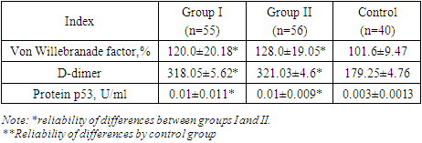
Recently, risk factors for pregnancy loss have traditionally included vascular and hemodynamic disorders in pregnant women, which are observed in various somatic diseases. Generalized endothelial dysfunction is the basis for hemodynamic and microcirculation disorders, including in the uteroplacental basin, developing in various somatic pathologies. There are several hypotheses explaining the development of endothelial dysfunction in UPR.
These changes are associated with the intensification of intravascular blood coagulation processes, including in the uteroplacental blood flow. The severity of shifts in the vascular-platelet, coagulation, fibrinolytic and anticoagulant components of hemostasis is determined by the characteristics of the course of pregnancy and the initial state of the coagulation system. Thus, the results of the study show that women with threatened preterm birth have higher values of markers (von Willebrand factor, apoptosis marker p53 protein, D-dimer and levels sICAM-1 and sVCAM-1) than in women with a physiological pregnancy. This confirms the involvement of endothelial dysfunction and apoptosis in the possible development of pathological bleeding during premature birth and the postpartum period.

References

[1]  S. Slawek-Szmyt, K. Kawka-Paciorkowska, A. Ciep\lucha, M. Lesiak, and M. Ropacka-Lesiak, “Preeclampsia and Fetal Growth Restriction as Risk Factors of Future Maternal Cardiovascular Disease—A Review,” Journal of Clinical Medicine, vol. 11, no. 20, p. 6048, 2022.
[2]  H. Roqué, E. Funai, and C. J. Lockwood, “von Willebrand Disease and Pregnancy,” J Matern Fetal Neonatal Med, vol. 9, no. 5, pp. 257–266, Jan. 2000, doi: 10.3109/14767050009053443.
[3]  M. Rick, B. Konkle, C. S. Kitchens, C. M. Kessler, and B. A. Konkle, “von Willebrand disease,” Consultative Hemostasis and Thrombosis, pp. 90–102, 2013.
[4]  M. D. Phillips and A. Santhouse, “von Willebrand disease: recent advances in pathophysiology and treatment,” The American journal of the medical sciences, vol. 316, no. 2, pp. 77–86, 1998.
[5]  G. Osmenda et al., “Treatment of denture-related stomatitis improves endothelial function assessed by flow-mediated vascular dilation,” Archives of Medical Science, vol. 13, no. 1, pp. 66–74, 2017.
[6]  H. Horiuchi, T. Doman, K. Kokame, Y. Saiki, and M. Matsumoto, “Acquired von Willebrand syndrome associated with cardiovascular diseases,” Journal of atherosclerosis and thrombosis, vol. 26, no. 4, pp. 303–314, 2019.
[7]  V. A. Holmes, “Changes in haemostasis during normal pregnancy: does homocysteine play a role in maintaining homeostasis?,” Proceedings of the Nutrition Society, vol. 62, no. 2, pp. 479–493, 2003.
[8]  J. P. Granger, B. T. Alexander, M. T. Llinas, W. A. Bennett, and R. A. Khalil, “Pathophysiology of Preeclampsia: Linking Placental Ischemia/Hypoxia with Microvascular Dysfunction,” Microcirculation, vol. 9, no. 3, pp. 147–160, Jun. 2002, doi: 10.1038/sj.mn.7800137.
[9]  A. E. Berezin, “The endothelial progenitor cell dysfunction in hypertension: the diagnostic and predictive values,” Vessel Plus, vol. 2, no. 22, pp. 2574–1209, 2018.
[10]  J. Roberts, “Endothelial Dysfunction in Preeclampsia,” Semin Reprod Med, vol. 16, no. 01, pp. 5–15, Mar. 1998, doi: 10.1055/s-2007-1016248.
[11]  R. W. Powers, J. M. Catov, L. M. Bodnar, M. J. Gallaher, K. Y. Lain, and J. M. Roberts, “Evidence of Endothelial Dysfunction in Preeclampsia and Risk of Adverse Pregnancy Outcome,” Reprod. Sci., vol. 15, no. 4, pp. 374–381, Apr. 2008, doi: 10.1177/1933719107311780.
[12]  J. S. Possomato-Vieira and R. A. Khalil, “Mechanisms of endothelial dysfunction in hypertensive pregnancy and preeclampsia,” in Advances in pharmacology, vol. 77, Elsevier, 2016, pp. 361–431. Accessed: Feb. 25, 2024. [Online]. Available: https://www.sciencedirect.com/science/article/pii/S105435891630031X.
[13]  E. Pasquier, L. De Saint Martin, C. Bohec, M. Collet, F. D. George, and D. Mottier, “Unexplained pregnancy loss: a marker of basal endothelial dysfunction?,” Fertility and sterility, vol. 100, no. 4, pp. 1013–1017, 2013.
[14]  M. Norman and H. Martin, “Preterm Birth Attenuates Association Between Low Birth Weight and Endothelial Dysfunction,” Circulation, vol. 108, no. 8, pp. 996–1001, Aug. 2003, doi: 10.1161/01.CIR.0000085069.09770.3D.
[15]  K. McLaughlin, M. C. Audette, J. D. Parker, and J. C. Kingdom, “Mechanisms and clinical significance of endothelial dysfunction in high-risk pregnancies,” Canadian Journal of Cardiology, vol. 34, no. 4, pp. 371–380, 2018.
[16]  M. Lazdam et al., “Elevated Blood Pressure in Offspring Born Premature to Hypertensive Pregnancy: Is Endothelial Dysfunction the Underlying Vascular Mechanism?,” Hypertension, vol. 56, no. 1, pp. 159–165, Jul. 2010, doi: 10.1161/HYPERTENSIONAHA.110.150235.
[17]  A. M. Germain et al., “Endothelial Dysfunction: A Link Among Preeclampsia, Recurrent Pregnancy Loss, and Future Cardiovascular Events?,” Hypertension, vol. 49, no. 1, pp. 90–95, Jan. 2007, doi: 10.1161/01.HYP.0000251522.18094.d4.
[18]  X. Chen and T. O. Scholl, “Maternal biomarkers of endothelial dysfunction and preterm delivery,” PloS one, vol. 9, no. 1, p. e85716, 2014.
[19]  Н. А. Ахтамова and Н. Н. Шавази, “PREDICTION OF OBSETRIC BLOOD LOSS IN WOMEN WITH PRETERM BIRTH (LITERATURE REVIEW),” УЗБЕКСКИЙ МЕДИЦИНСКИЙ ЖУРНАЛ, vol. 3, no. 5, 2022, Accessed: Feb. 25, 2024. [Online]. Available: https://tadqiqot.uz/index.php/medical/article/view/5509.