-
Paper Information
- Next Paper
- Previous Paper
- Paper Submission
-
Journal Information
- About This Journal
- Editorial Board
- Current Issue
- Archive
- Author Guidelines
- Contact Us
American Journal of Medicine and Medical Sciences
p-ISSN: 2165-901X e-ISSN: 2165-9036
2024; 14(3): 675-678
doi:10.5923/j.ajmms.20241403.29
Received: Jan. 25, 2024; Accepted: Feb. 22, 2024; Published: Mar. 9, 2024

Characteristics of Potomorphological Changes in the Myocardial Layer of the Heart in Young People with Chronic Ischemic Heart Disease
 Abstract
 Reference
 Full-Text PDF
 Full-text HTML
Jumanov Ziyadulla Eshmamatovich1, Butaev Sherzod Fayzulloevich2, Mamadiyarova Dilfuza Umirzakovna3
1Associate Professor of the Department of Pathological Anatomy of Samarkand State Medical University, Doctor of Medical Sciences (DSc), Uzbekistan
2Competitor of the Department of Pathological Anatomy of Samarkand State Medical University, Uzbekistan
3Associate Professor of the Department of Pedagogical-Psychology of Samarkand State Medical University, Doctor of Philosophy in Psychology (PhD), Uzbekistan
Correspondence to: Jumanov Ziyadulla Eshmamatovich, Associate Professor of the Department of Pathological Anatomy of Samarkand State Medical University, Doctor of Medical Sciences (DSc), Uzbekistan.
| Email: |  |
Copyright © 2024 The Author(s). Published by Scientific & Academic Publishing.
This work is licensed under the Creative Commons Attribution International License (CC BY).
http://creativecommons.org/licenses/by/4.0/

Microscopic examination of the hearts of 28 deceased persons aged 25-45 was conducted in order to study the specificity of photomorphological changes in the myocardial layer of the heart in young people with chronic ischemic heart disease. Morphological changes of ischemic type, hypertrophy of cardiomyocytes and growth of interstitial connective tissue to perivascular areas, quantitatively less focal sclerotic changes were noted in the myocardium. It was found that morphometric indicators differ between young people.
Keywords: Chronic ischemic heart disease, Myocardium, Youth, Coronary cardiosclerosis
Cite this paper: Jumanov Ziyadulla Eshmamatovich, Butaev Sherzod Fayzulloevich, Mamadiyarova Dilfuza Umirzakovna, Characteristics of Potomorphological Changes in the Myocardial Layer of the Heart in Young People with Chronic Ischemic Heart Disease, American Journal of Medicine and Medical Sciences, Vol. 14 No. 3, 2024, pp. 675-678. doi: 10.5923/j.ajmms.20241403.29.
Article Outline
1. Introduction
- Currently, there are about 126.5 million cases of ischemic heart disease in the world. Coronary heart disease (CHD) remains one of the leading causes of death, accounting for more than 9 million deaths per year worldwide [3,5,6].The term stable coronary artery disease is often used interchangeably as a synonym for chronic ischemic heart disease and covers a variety of conditions in which there is a recurrent mismatch between myocardial oxygen supply and demand. This is often seen when long-standing atherosclerotic obstruction in the epicardial coronary arteries results in poor flow and distal ischemia. However, this is not the only mechanism. Various pathophysiological processes, such as coronary artery vasospasm, microcirculatory dysfunction, or congenital anomalies, can lead to chronic recurrent ischemia [4,7].The characteristics of cardiovascular diseases in young people have not been sufficiently studied. Among men with coronary artery disease, those under 39 are considered young; Different age criteria are adopted for women: some authors set the same age limit for men, while others raise it to 45 years. According to various sources, the incidence of ischemic heart disease in young people is 5-10% of all cases of the disease. Risk factors for young people are generally the same as for ischemic heart disease: arterial hypertension, hypercholesterolemia, excess body weight, physical inactivity, diabetes, etc. Undoubtedly, the way of life (emotional background) plays an important role in the emergence of СHD in young people. According to a study conducted in England, the risk of developing coronary artery disease is 3 times higher in men with depression [2].
2. The Purpose of the Study
- Study of morphological and morphometric aspects of myocardial changes in chronic ischemic heart disease in young people.
3. Materials and Methods
- In order to study the morphological characteristics of the myocardial structure in chronic ischemic heart disease in young people, microscopic examination of the hearts of 28 deceased persons aged 25-45 years was performed, of which 21 (75%) were men, 7 (25%) were women. These ages were divided into the following subgroups for the purpose of in-depth study of pathomorphological changes in the myocardium: Group 1: 25-30-year-olds; Group 2: 31-35 years old and Group 3: 36-40 and Group 4: 41-45 years old. Based on the goals and objectives of the study, morphological features of myocardial structures were studied. Material for special histological examination was taken from the anterior wall of the left ventricle and the interventricular septum. The obtained tissue fragments were fixed in 10% neutral formalin, passed through an alcohol battery, and paraffin blocks were prepared. The prepared histological sections were stained with hematoxylin and eosin, according to Van-Gieson. Photomicrographic methods were conducted.
4. Results and Discussion
- 25-30-year-olds died from chronic ischemic heart disease, 6 of them were women. The average weight of the heart of the deceased was 343.3±5.1, its dimensions were 10.5x8.7x5.1 cm, the thickness of the left ventricle was 1.11±0.12, and the right ventricle was 0.33±0.02 cm. Myocardial consistency elasticity. In the micropreparations prepared from the myocardium layer of the heart of this age group, the growth of intermediate connective tissue in the perivascular areas between the muscle fibers is determined. Due to the thickening of the wall of small intramyocardial artery blood vessels, their spaces are narrowed. Hypertrophy of cardiomyocytes around foci of coronary cardiosclerosis is observed (Fig. 1).
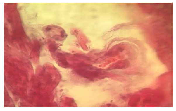 | Figure 1. 25-30-year-old patients with foci of coronary cardiosclerosis in intramyocardial blood vessels. Stained in hematoxylin-eosin. Ob.40, ok.10 |
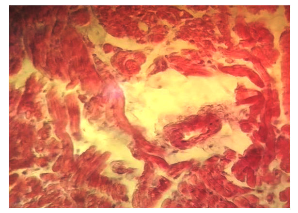 | Figure 2. Coronarocardiosclerosis caused by sclerotic changes in intramyocardial blood vessels in patients aged 31-35 years. Stained in hematoxylin-eosin. Ob.40, ok.10 |
|
|
|
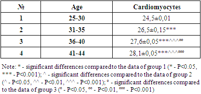
5. Conclusions
- Thus, morphological changes in the myocardial layer of the heart of young patients with chronic ischemic disease are manifested mainly in the form of coronary cardiosclerosis. The narrowing of the wall of intracardiac arteries develops more strongly. The myocardium shows morphological changes of the ischemic type, hypertrophy of cardiomyocytes and growth of intermediate connective tissue to perivascular areas, and quantitatively less focal sclerotic changes. Hypertrophy of cardiomyocytes in young people who died of chronic ischemic heart disease, 36-40 in 25-30-year-olds; Compared to 31-35-year-olds, the enlargement of cardiomyocytes is slower in 41-44-year-olds compared to younger patients.