-
Paper Information
- Paper Submission
-
Journal Information
- About This Journal
- Editorial Board
- Current Issue
- Archive
- Author Guidelines
- Contact Us
American Journal of Medicine and Medical Sciences
p-ISSN: 2165-901X e-ISSN: 2165-9036
2024; 14(3): 575-582
doi:10.5923/j.ajmms.20241403.08
Received: Feb. 13, 2024; Accepted: Feb. 28, 2024; Published: Mar. 2, 2024

Morphological Changes in the Immune System During Renal Failure in Rats and Corrections with Pomegranate Seed Oil
 Abstract
 Reference
 Full-Text PDF
 Full-text HTML
Khamdamova M. T. , Teshaev Sh. D., Khikmatova M. F.
Bukhara Medical Institute named after Abu Ali Ibn Sina, Uzbekistan
Copyright © 2024 The Author(s). Published by Scientific & Academic Publishing.
This work is licensed under the Creative Commons Attribution International License (CC BY).
http://creativecommons.org/licenses/by/4.0/

Renal failure (RF) is a condition in which the kidneys are unable to perform their functions fully. Kidney failure can be temporary or chronic and can develop gradually or suddenly. Kidney failure can be caused by a variety of causes, such as chronic kidney disease (eg, chronic glomerulonephritis or chronic pyelonephritis), acute kidney failure (eg, due to injury or infection), and systemic diseases such as diabetes mellitus or hypertension. Kidneys are one of the most important organs that ensure the stability of the internal environment. The thymus is an organ of the endocrine system located in the upper part of the chest cavity behind the sternum. Quite a lot of work has been devoted to the study of the morphology of the human thymus. It plays an important role in the development and functioning of the immune system. Morphologically, the thymus is a bilobed organ, which consists of a cortex and medulla.
Keywords: Salts, Kidney, Pressure, Pomegranate oil, Kidney stones, Sodium, Spleen, Thymus, Morphology
Cite this paper: Khamdamova M. T. , Teshaev Sh. D., Khikmatova M. F. , Morphological Changes in the Immune System During Renal Failure in Rats and Corrections with Pomegranate Seed Oil, American Journal of Medicine and Medical Sciences, Vol. 14 No. 3, 2024, pp. 575-582. doi: 10.5923/j.ajmms.20241403.08.
1. Introduction
- The thymus is the best experimental model for studying the effect of new progressive methods of treating accelerated aging of the body. The thymus capsule and connective tissue of the interlobular septa contain blood vessels, lymphatic vessels, and nerve fibers. From the connective tissue, blood vessels enter the thymus lobule. In the cortical zone, capillaries form loops, go to the cortico-medullary zones and collect in venules. The cortico is richest in blood vessels medullary zone. Further cortico-medullary the venules leave the thymus together with the medullary ones. The hemocapillaries of the cortical zone of the thymus lobules are surrounded by relatively densely located epithelial cells, thus the latter participate in the formation of the hematothymic barrier that protects the differentiating thymocytes of this zone from various antigens traveling through the bloodstream. Epithelioreticulocytes differentiate and various cellular immunity appears in the body and form thymus-dependent zones (in the spleen, lymph nodes, etc.). The epithelial islets of the thymus of young adult animals secrete into the blood a secretion that contains hormones of the thymosin family. These hormones regulate humoral immunity in the animal and human body. Lymphoid cells of the outer part of the cortical zone are represented predominantly densely arranged lymphoblasts. Their diameter is about 7-8 microns, they contain a rounded nucleus with nucleoli [1,2,3,4,5,6,7,8,9,10,11,12]. Quite often, cells are detected at various stages of mitotic division. In the inner part of the cortical zone, lymphocytes are located less frequently compared to the outer part. Lymphocytes in this zone are smaller in diameter and contain a small number of intracellular orgenella - free ribosomes, mitochondria, tubules of the granular endoplasmic reticulum. (65) The shape of the thymus in 68.8% of cases is leaf-shaped, in 9.6% - cylindrical, in 7.2% - indeterminate, in 4.8% - truncated cone, in 2.4% - cone-shaped, and in the rest (7.2%) other shapes (double cylinder, double cone, quadrangle, bean, oval and trapezoid) [1,13,14,15,16,17,18,19,20,21,22,23,24]. 3 options for the shape of the organ and its lobes: lepto-, meso- and brachymorphic types, as well as the arrangement of the lobes according to the ratio of the width of the lobe to its thickness; 3 more options: compact, laminar and convolute. It is noted that with age the lobes become more elongated. In fetuses and newborns, 60-70% of the lobes are compact, and with age they turn (up to 80%) into laminar and convolute [25,26,27,28,29,30,31,32]. The lobes of the human thymus, like the organ itself, often have a spindle-shaped shape, and therefore authors describing its macrostructure often introduce a certain terminological confusion in the name of the upper and lower parts of the organ. In our opinion, the most suitable terms for the macroscopic description of the thymus are: the cranial and caudal ends of the lobes, the upper and lower poles of the thymus. The thymus in rats, as well as in other mammals, is an organ of the immune system and plays an important role in the development and maturity of T lymphocytes. Typically, the thymus in rats is a bilobed organ located in the upper part of the chest cavity, behind the sternum and in front of the spinal column. It consists of the cortex and medulla, with the cortex located closer to the organ capsule, and the medulla located inside. The thymus cortex in rats consists of a network of epithelial cells that form structures known as thymocytes. These cells play a role in the process of maturation of T lymphocytes and training them to recognize self and foreign antigens. The thymus medulla in rats also contains epithelial cells, fibrous tissue, capillaries, and immunocompetent cells such as T lymphocytes and macrophages. Here the final differentiation and maturation of T lymphocytes occurs. The size of the thymus in rats, as in other mammals, varies with age. Typically, the thymus in rats goes through a series of morphological changes during their life, which are associated with its functional activity and changes in the immune system. In newborn rats, the thymus is usually larger than in adult rats. This is due to the active process of development and migration of immunocompetent cells, such as T lymphocytes, into the thymus in the period immediately after birth. During the first weeks of life, the size of the thymus in rats can increase significantly, since the organ actively functions in the process of forming the immune system and training immunocompetent cells.The spleen has the shape of a flattened and elongated hemisphere with pointed ends. It has two surfaces: diaphragmatic and visceral. The hilum of the spleen is located on the visceral surface. There are two edges: the upper (anterior) edge, separating the visceral surface from the diaphragmatic one, is sharp and the lower (posterior) edge is blunt. There are also two ends (poles): the rear one - it is rounded and faces up and back, and the front one is sharper. The mass of the spleen according to Yu.I. Borodina et al. (1987) for men aged 20 to 40 years is 192g, for women - 153g, and according to M.R. Sapin and L.E. Etingen (1996) its average weight in men is 140 g, in women - 130 g. The length of the spleen is approximately 13-14cm, width 6-10cm, and thickness 3-4cm. The relative weight of the spleen of adults is 0.25 - 0.30% (N.P. Bisenkov, 1972) [1,2,33,34]. The spleen is covered on all sides by peritoneum, which is firmly fused with its fibrous capsule. ME AND. Fedonyuk et al. (1997) identified 3 layers in the spleen capsule: superficial, middle and deep. They differ from each other not only in thickness and architectonics, but also in the degree of development of fibrous structures. Due to the fact that the collagen fibers of the superficial and deep layers pass through the middle layer, all three layers are firmly connected to each other.
2. Methods
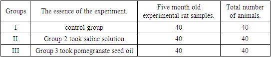
- This experiment confirms the possibility of a negative effect of excess salt intake on kidney function and the development of various diseases. Therefore, controlling salt intake is considered an important aspect of maintaining healthy renal function. When it comes to the potential effects of salt on the kidneys and the possible formation of stones through excess salt and drinking water, this is an area of interest for research.In this case, the animals are divided into three groups, and the effects of salt on the kidneys, as well as the potential effects of these factors through excess nutrition, were potentially investigated. The first group of animals is a control group, in which the scientists used a standard diet with normal salt content to compare the results with other groups. The second group of animals consumed an average amount of salt water in their diet, approximately 12-14 ml per day for every 100 grams of body weight. This amount of water was provided for one month. This can lead to various aspects such as increased blood pressure and kidney damage related to the retention of water and sodium in the body.The third group of animals also consumed salt in the specified amount, but they took pomegranate oil (according to Mohammad Tacher, B., Delnia, A., Hamidekh Zhalili, R., Elham, A., & Azar, H. 2013) at a dose of 5 ml once a day for a month in combination with food. For 10 days, the control group of shameless rats, from the 141st day to the 150th day, was injected with 0.5 ml of distilled water through a metal probe through the oral cavity. In the experiment, but only one of them did not complete the experiment.
|
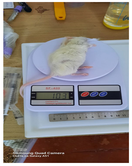 | Figure 1 |
3. Results
- Kidney damage from salt can lead to various diseases such as kidney stones, kidney failure and other genitourinary problems. The main mechanism of salt's effect on the kidneys is an increase in blood volume. Increasing the amount of salt causes the body to retain water, which increases blood volume. Increased blood volume puts pressure on the walls of blood vessels and increases stress on the kidneys, the organs responsible for filtering the blood and removing excess water and waste. Chronic salt consumption can lead to the development of a number of kidney problems. First, high blood pressure can damage the blood vessels in the kidneys, leading to poor kidney function. Secondly, excess salt promotes the formation of kidney stones. Increasing the amount of sodium in the urine can lead to a buildup of salts, which can form kidney stones.In conclusion, excessive salt intake can negatively affect the kidneys, leading to increased blood pressure and damage to blood vessels, as well as the formation of kidney stones. Therefore, limiting your salt intake and drinking enough water are important to maintain kidney health.
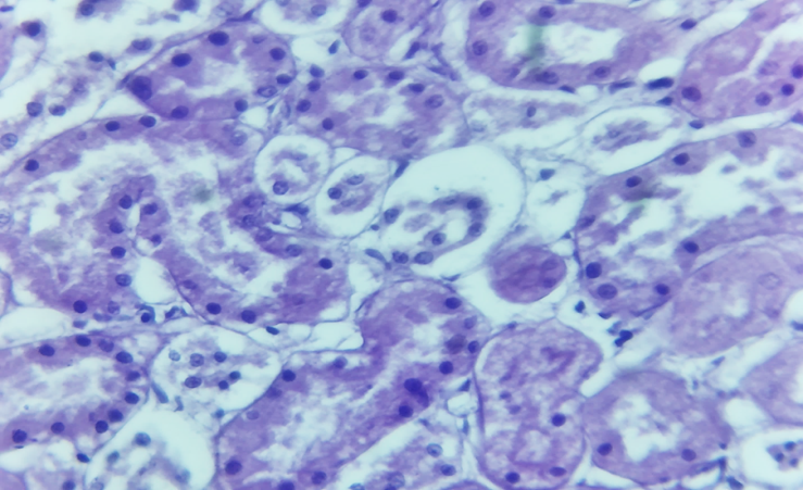 | Figure 2. Microscopic picture of renal failure ( hydropic degeneration and nuclear necrosis are detected in the proximal and distal tubules (1). Vacuolar degeneration and foci of karyolysis are detected (2). Staining with hematoxylin-eosin, 10x10) |
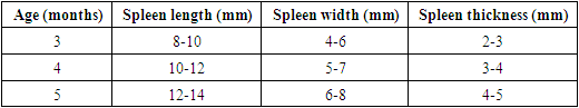
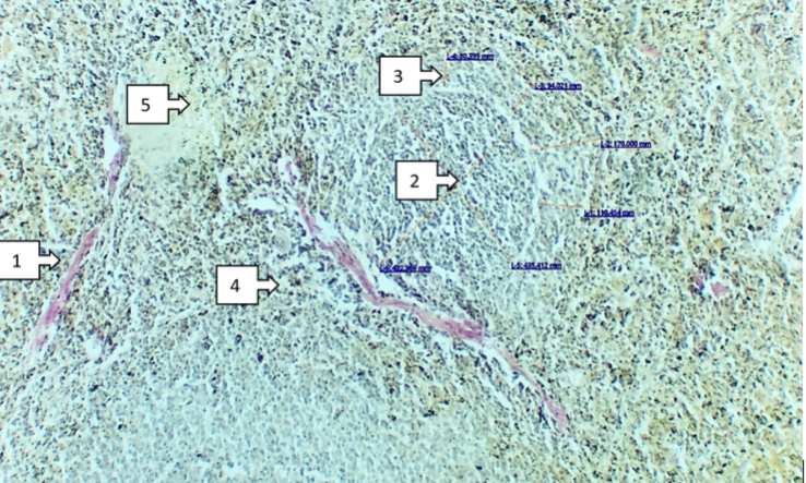
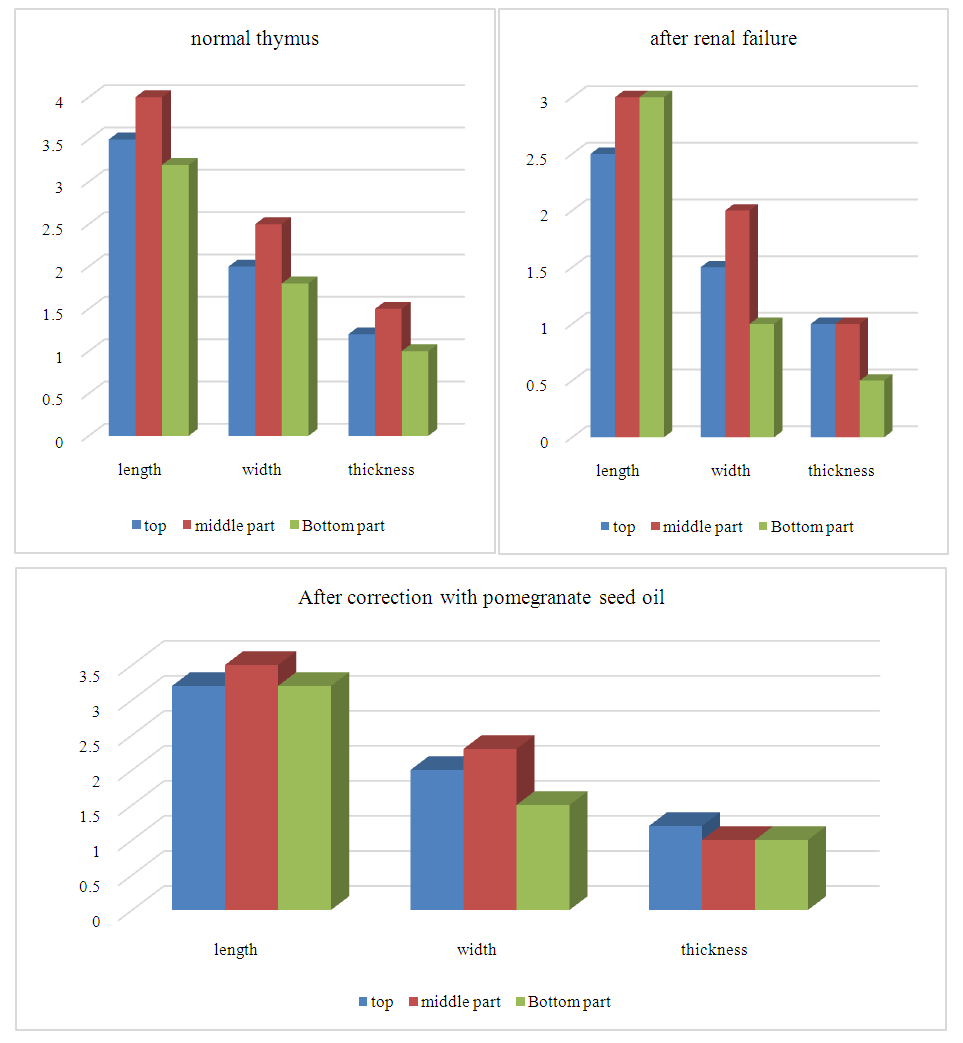 | Figure 3. Information on the sizes of the parts of the thymus |
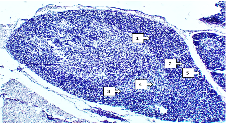 | Figure 4. Morphological structure of the thymus Dye heme-eosin about 10x20 approx. Decreased T-lymphocytes with foci in the subcapsular part of the cortex (Cortex) (1) Increased epithelial (reticuloepithelial) thymic cells (2) Epithelial (reticuloepithelial) thymic cells in the medulla, located in the middle. There are very few lymphocytes (3), among the epithelial (reticuloepithelial) cells of the thymus, demasosomes are thickened, and the number of macrophages is increased (4). The interlobular connective tissue of the thymus is thickened (5) |
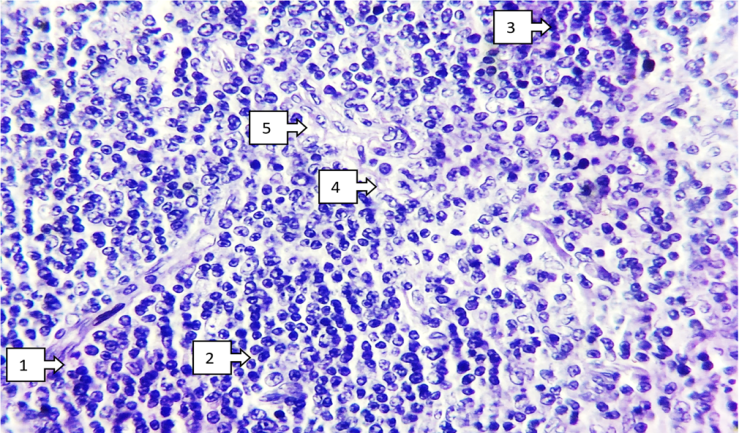 | Figure 5. Morphological structure of the thymus after correction. Chem -eosin dye about 10x20 approx. The thin connective tissue of the thymus is smooth, there are no fat cells around it, and it is rich in blood vessels (1). The number of prethymocytes, T lymphocytes, increases through mitosis (2). The nucleus of reticuloepithelial "Nanny" cells is enlarged, and there is an increase in the number of lymphocytes around mitotic division (3). In the medulla, the number of reticuloepithelial cells increased, the number of mature lymphocytes increased, and stromal edema disappeared (4). Desmosomes between reticuloepithelial cells are smoothed, the number of macrophages is slightly reduced (5) |
|
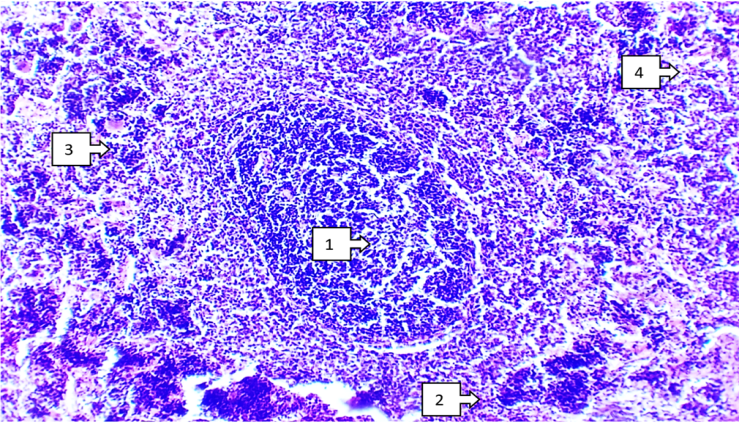 | Figure 6. Morphological structure of the spleen. Heme -eosin dye about 10x20 approx. White pulp area: Central part of the lymphatic follicle - swelling in the reactive center, hyperplasia of B-lymphocytes (1). The mantle and marginal areas are reduced (2). Red pulp area: the splenic band (Chordaelienalis) is reduced, B lymphocytes, plasma cells and macrophages are increased (3). Splenic sinusoids (sinuslienalis) dilate, aggregate red blood cells, breakdown of red blood cells, hemosiderins of different sizes (4). Expansion of the trabecular venous space (in size), stasis of blood-like elements in it (4) |
 | Figure 7. Morphometric and morphological appearance of the spleen after correction. Van paint - painted in Gizon about 10x20 approx. The trabeculae of the spleen are elastic connective tissue of pale pink color, smooth and thinned (1). White pulp area: The central part of the lymphatic follicle is in the reactive center, B lymphocytes multiply and increase (in size) (2). The mantle and marginal areas (in size) are enlarged and thickened (3). The red pulp area: enlarged in the splenic cords (Chordaelienalis), increased B-lymphocytes, plasma cells and macrophages (4) |
|
4. Discussion
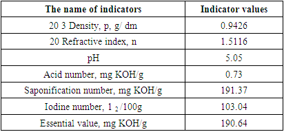
- Pomegranate oil is extracted from pomegranate seeds, which are a rich source of antioxidants, vitamins and minerals. It contains high amounts of polyphenols, including flavonoids and ellagic acid, which have powerful anti-inflammatory and antioxidant properties. Research shows that pomegranate oil may have a positive effect on kidney health. It helps reduce inflammation, prevent the formation of kidney stones, and improve kidney function. In addition, pomegranate oil helps reduce the amount of protein in the urine, which is a sign of kidney damage. Thanks to its powerful antioxidant properties, pomegranate oil helps protect the kidneys from damage caused by oxidative stress. This makes it a potentially useful agent for the prevention and treatment of various kidney diseases. According to the World Health Organization, the recommended daily intake of salt for adults is less than 5 g (less than a teaspoon). Medical effectiveness.Among vegetable fats, oil from the seeds of fruit plants is of particular interest, as it has a unique chemical composition and, due to its phytochemical and antioxidant properties, has practical applications in the food and pharmaceutical industries. In many countries around the world, seed oils are included in various food diets. The presence of all classes of glycolipids in many seed oils makes them an excellent source of unsaturated fatty acids in the diet. They are a rich source of fatty acids and bioactive fat-soluble elements. The high levels of polar lipids indicate that these oils may be a suitable and valuable source for obtaining appropriate concentrations of polar lipids. The presence of all glycolipid classes in these oils makes them an excellent product for formulating nutritional diets. If we take into account the literature data on the significant oil content in dry pomegranate seeds, then approximately 72 tons of pomegranate oil per year can be obtained from the available seed waste. All this served as the basis for conducting relevant research. Raw samples of pomegranate fruit pomace were dried at room temperature with occasional ventilation to bring the moisture content to a constant level. A certain amount of dry seeds was transported to Germany, where, through the German international scientific organization DAAD, one of the authors (N.G. Kurbanov) was on a scientific trip to the Institute of Food Technology and Food Chemistry of the Technical University of Berlin. For research about 2 kg of dry pomegranate seeds were selected for uniformity, weighed, their moisture content was brought to a constant level, after which their total lipids were extracted by the use of hexane. Samples in an amount of 5 g were first homogenized in methanol (50 ml) for 1 min in a blender, then hexane was added in an amount of 100 ml and homogenized for 2 min. Subsequently, the mixture was filtered, and the liquid was dissolved in a hexane /methanol mixture in a volume ratio of 2:1 (100 ml + 50 ml) and further homogenization was carried out for 3 minutes. The mixture was then filtered again and washed with fresh solvent (2:1, w/w1, 150 ml). The mixed filtrates were purified by repeated addition of 0.2 volumes of 0.75% sodium chloride solution. Everything was mixed without shaking, and the layers were separated so that the hexane completely covered them. The purified lipids were collected in a vial and treated with sodium sulfate to remove any remaining moisture. After filtration, the extract was dried on a rotary evaporator at a temperature of 40°C. The resulting oil (total lipids) was weighed and stored in hexane at 20°C. The fatty acid composition of the oil was analyzed as a methyl ester prepared according to Mohamed F. Ramadan and Jorg -T. Mursel. To do this, each 0.1 g oil sample was esterified with 10 ml of a mixture made from 25 ml of sulfuric acid (98%) and 500 ml of methanol (94.8%) for 120 minutes at 80°C. Next, the methyl esters were extracted with 10 ml of hexane and the amount of extract was reduced to 0.5 ml in a nitrogen environment to avoid oxidation of unsaturated fatty acids. After this, an analysis was carried out using the gas chromatographic method. As can be seen from the table data, the content of oil obtained from pomegranate seeds in the form of total lipids is 95% in relation to the dry weight.
5. Conclusions
- In the composition of total lipids, unsaturated fatty acids are predominant, accounting for about 63.84% of all fatty acids. Among them, 40.4% is oleic acid, 10.4% is heptadecenoic acid, 10.0% is li-zero, 1.93% – palmitoleic, 1.01% – γ-linolenic and 0.10% – ly-nolenic acid. The amount of saturated fatty acids in the total lipids of pomegranate oil is about 36.3%, among them palmitic (20.7%) and stearic (14.8%) acids predominate. Margaric (0.12%) and behenic (0.64%) acids were also found in small quantities in pomegranate seed oil, which had not previously been found in the works of Spanish, Arab and Iranian authors conducted with the fruits of various types and varieties of pomegranate.