-
Paper Information
- Next Paper
- Previous Paper
- Paper Submission
-
Journal Information
- About This Journal
- Editorial Board
- Current Issue
- Archive
- Author Guidelines
- Contact Us
American Journal of Medicine and Medical Sciences
p-ISSN: 2165-901X e-ISSN: 2165-9036
2024; 4(2): 360-364
doi:10.5923/j.ajmms.20241402.43
Received: Jan. 20, 2024; Accepted: Feb. 8, 2024; Published: Feb. 12, 2024

Congenital Malformation of the Central Nervous System - Aicardi Syndrome (Clinical Case)
 Abstract
 Reference
 Full-Text PDF
 Full-text HTML
Djurabekova Aziza Takhirovna1, Umida Khamidovna Vaseyeva2, Akmaljon Akhmatjonovich Gaibiev3
1Doctor of Medical Sciences, Professor of the Department of Neurology, Samarkand State Medical University, Uzbekistan
2Basic Doctoral Student of the Department of Neurology, Samarkand State Medical University, Uzbekistan
3Doctor of Medical Sciences, Associate Professor of the Department of Neurology, Samarkand State Medical University, Uzbekistan
Copyright © 2024 The Author(s). Published by Scientific & Academic Publishing.
This work is licensed under the Creative Commons Attribution International License (CC BY).
http://creativecommons.org/licenses/by/4.0/

Aicardi syndrome is a rare genetic disorder characterised by agenesis of the corpus callosum, epileptic seizures of infantile spasms with early onset, specific lacunar changes on the ocular fundus, typical EEG changes (split brain pattern), delayed psychomotor development, and facial dysmorphism.
Keywords: Aicardi syndrome, Genetic disease, Corpus callosum, Congenital brain malformations
Cite this paper: Djurabekova Aziza Takhirovna, Umida Khamidovna Vaseyeva, Akmaljon Akhmatjonovich Gaibiev, Congenital Malformation of the Central Nervous System - Aicardi Syndrome (Clinical Case), American Journal of Medicine and Medical Sciences, Vol. 4 No. 2, 2024, pp. 360-364. doi: 10.5923/j.ajmms.20241402.43.
1. Introduction
- The corpus callosum (the great commissure of the brain) is a layer of nerve fibres connecting the cortex of the two hemispheres of the large brain. The corpus callosum forms between 8-20 weeks of gestation. Once formed, the corpus callosum continues to grow, increasing in length and width. Crossing of fibres from one hemisphere to the other begins at 12 weeks of gestation.Agenesis of the corpus callosum refers to congenital structural disorders of neuroontogenesis. It can be isolated, but is more often combined with other congenital brain anomalies - microgyria, pachygyria, lissencephaly, hydrocephalus. Clinical manifestations of agenesis of the corpus callosum are polymorphic: there is a combination of dysraphic status, mental retardation of varying degrees, epileptic seizures, motor disorders, and abnormal development of internal organs. Agenesis of the corpus callosum may be hereditary and may be the result of spontaneous mutations. Among the variants of agenesis of the corpus callosum presented in the table, the most common and widely reported in the literature is Aicardi syndrome.The relevance of studying congenital brain malformations (CBMD) in children is associated with a high proportion of the disease in the structure of infant and perinatal mortality and childhood disability [1,3,5]. According to WHO statistics for 2024, the prevalence of CBMD varies in different countries from 3 to 17% (4-6% on average). In the pathomechanism of ontogenesis of malformation development, an anomaly (defect) of the neural tube is one of the leading places among all detected human congenital anomalies - 10-30% [2,4,6]. The actual percentage frequency of malformations of the central nervous system, in particular the brain, remains unspecified, possibly due to the clinical and diagnostic difficulties of childhood, or the difficulty of registering such patients. Accordingly, an important task for specialists in this area is to objectify the collection, management of data and confirmation of the results obtained [5,7].It is known that IUGRD display a wide polyetiological section of disorders of early and late ontogenesis with multifactorial nature of congenital anomalies. This creates prerequisites for the search for the most informative and important risk factors for their formation, which help to correctly analyse and evaluate the prognosis of the probability of congenital pathology with the subsequent use of preventive measures to prevent the birth of children with IUGRD. But as scientific literature shows, there are leading causes that appear as various combined effects and manifest themselves multifactorially. So inevitably, a significant contribution to the development of congenital malformations in children, depends on the initial state of health of parents, unfavourable ecological system in the environment, infection, hereditary predisposition [8]. Many scientists associate a high percentage of CBMD with the problem of perinatal CNS damage, which remains relevant not only among the neurological service, but also among specialists: neonatologists, ophthalmologists, paediatricians. Moreover, taking into account the possibilities of the last decades to improve neonatal care for the survival rate of perinatally affected children, it is populisation of congenital brain pathologies that falls on this period, and what is characteristic, in the majority among premature children. Pathological changes in premature infants are often accompanied by severe anomalies, congenital pathology of visual development, as a result of hypoxic-ischaemic lesions of the CNS. The dominant position among the factors of combined lesions, childhood blindness and low vision in the last few years is occupied by vasoproliferativity of eye lesions in premature infants. According to the data of Russian scientists (Ovchinnikova T.V. et al.), the second place in the structure of the identified nosologies, diseases of CBMD is occupied by periventricular leukomalacia and intraventricularhaemorrhage. The impact of these diseases on the visual tract can lead to optic atrophy, optic nerve hypoplasia or cerebral visual impairment. The direct connection of the visual tract system and brain sections lead to the fact that in multifactorial brain damage in children, different sections of the visual pathway or visual cortex may be involved in the process of pathomechanism, which is revealed by a combination of neurological and neuroophthalmological symptomatology [5]. According to many authors of scientific publications, MRI is a sensitive and specific diagnostic method for CBMD. The surge in the development and introduction of angiography, computed tomography and nuclear magnetic resonance into the clinical practice of neurologists has made new demands concerning the traditional issues related to the causes of the onset of CBMD in children, the issue of complications, which from the point of view of etiopathogenesis can significantly affect the treatment tactics up to its complete change [6]. Thanks to neuroimaging technologies, the problem of diagnostically masked diseases such as encephalopathy, cerebral palsy, oligophrenia, psychomotor developmental delays and others, under which ADHD was concealed, has been solved.The main difficulty in interpreting the clinical picture of neurological disorders in children with IUGRD lies in the high polymorphism of syndromes, in addition, there are different types of IUGRD that do not reveal themselves until a certain point, because of insufficiently clear symptoms [3,7].The issue of detection and clinical and functional manifestation of CHD in children at different ages remains debatable and has not been fully resolved. Consequently, the multiple manifestations of GDM should be used as an informative value for early detection of this pathology.The most frequent of the CBMD is considered to be the lesion of the corpus callosum, from the position of etiopathogenesis has a detailed scale of causes. Thus, this unfavourable impact of environmental factors can cause mutations and the appearance of various developmental anomalies. In the literature, the results of presumed time of exposure, intrauterine "teratogenetic - terminal period" are described [4,8], with the formation of agenesis of the corpus callosum (AMT). A congenital multisystem disease characterised by epileptic seizures, delayed psycho-speech development, chorioretinal lacunar foci on the ocular fundus, and the presence of a malformation of the corpus callosum - Aicardi syndrome, which still causes certain difficulties for specialists in this field. Dysmorphic microcephaly is not uncommon in young patients with Aicardi syndrome, which emphasises the diagnostic value of a thorough external examination. In addition, the presence of psychomotor development: spastic paraplegia or hemiplegia, delayed psycho-speech development of varying degrees, mask the disease, and increase the difficulty of differentiating with cerebral palsy. The main sign of the disease is epileptic seizures in the form of infantile spasms, manifesting in the first year of life, reaching a frequency of up to 10-25 times a day, with an early debut of seizures, manifested by severe disorders of psychomotor development. Diagnosis of ophthalmological changes is considered equally important, as there is a correlation between the degree of involvement of the visual analyser and the central nervous system in the pathological process [7]. Neuroimaging, in turn, describes in some cases, a combination of CBMD: atrophy of the brain substance (deepening and widening of the convexital furrows, widening of the interhemispheric and lateral gaps); structural changes in the projection of the optic radius; Dandy-Walker anomaly; pachygyria in various combinations [2].Clinical case description.Patient A., 11 months old. He was admitted for inpatient treatment to the paediatric neurology department of MC SamSMU 12/2023. Diagnosis on admission: G80.0 Spastic cerebral palsy. Infantile cerebral palsy, spastic tetraparesis, GMFCS V level. G40.2 Localised (focal) (partial) symptomatic epilepsy and epileptic syndromes with complex partial convulsive seizures. Focal epilepsy, probably structural, with tonic left-sided seizures G96.8. Other specified lesions of the central nervous system. Delayed psychomotor development.Complaints from the mother about paroxysmal states in the form of disturbance of consciousness, tonic tension with fixation of gaze to the left, small amplitude clonks of distal musculature from 2 to 10 times a day, a single hemiconvulsive right-sided seizure, delayed psycho-motor development.According to the mother: a child from a close marriage (twin brother and sister on the mother's side), on the father's side aggravation, drug use. First pregnancy, first delivery: premature operative delivery at 36 weeks, Apgar score 7\8b, Caesarean section. Emergency. Indications: foetal distress. Pregnancy course: without peculiarities. In the 3rd trimester, fetal fetal GM abnormalities were detected by ultrasound. Duration: 36 weeks. Condition of the newborn Birth weight: 1970 grams Height: 44 cm Baby: premature. Transfer at the second stage to the Department of Neonatal Pathology, with the diagnosis: PEP. Dysgenesis of the corpus callosum. Congenital clubfoot. Oppression syndrome. Retinopathy of prematurity. The child has paroxysmal states in the form of disturbance of consciousness, tonic tension, small amplitude clonus of distal musculature, axial myoclonias. Observed by an orthopedist with the diagnosis: congenital multiple arthrogryposis (q 74.3), congenital hip deformities (q 65.8), severe atypical clubfoot. Conservative treatment was carried out - stage plastering, percutaneous ponsetiAchillotomy on both sides. Receives valproic acid 120 mg 2 times a day, levetiracetam 100 mg 2 times a day. Left-sided tonic seizures lasting up to 1 minute, 1-2 times a day persist. Before admission, a right-sided hemiconvulsive seizure lasting up to 1 minute was noted; it did not recur again and was spontaneously resolved. He was admitted to the DN department for the first time with the purpose of pre-examination, correction of therapy and rehabilitation treatment. Somatic status: Height/body length: 78 cm Body weight: 8.9 kg. Temperature: 36.5°C BMI: 14.6 kg/sq.m. Body surface area: 0.44 square metres. General condition on admission: moderately severe. Consciousness: clear. Nutrition: satisfactory. Constitution: normosthenic. Condition of skin, visible mucous membranes, lymph nodes Skin colour: swarthy. Development of FML: moderately. Distribution of FML: uniform. Rash: on the anterior surface of the right thigh a papular element with hyperaemia. Skin moisture: normal. Turgor: preserved. Cyanosis: absent. Presence of oedema: absent. Lymph nodes: not enlarged. Oral mucosa: clean. Oropharyngeal mucosa: mucous pink. Degree of tonsils enlargement: not enlarged. Condition of musculoskeletal system Damage: none. The degree of muscular development: satisfactory. Muscle tone: increased. Description of the state of the joints: deformed. Movement of joints: limited. Description: in knee and ankle joints. Spinal pain: none. Deformity of the spine: none. Description of the shape of the thorax: normal shape. Additional information: postoperative scars on the lower extremities without signs of inflammation. Respiratory status Respiratory rate: 39/min Respiratory rhythm: regular. Breathing: normal. Independent breathing: natural. Nature of breathing: puerile. no. Nasal breathing: free. Condition of the cardiovascular system HR: 122 /min. Pulse filling: moderate filling. Pulse tension: moderate tension. Heart rhythm: not disturbed. Heart tones: clear. Presence of a heart murmur: yes. Characteristics of the noise: along the left edge of the sternum. Description of the noise: systolic. Heart area: not changed. Boundaries of relative cardiac bluntness: within the normal limits. Boundaries of the heart: not changed. Additional information: BP is not measured, negative reaction. Condition of gastrointestinal tract organs Tongue colour: pink. Plaque on the tongue: absent. Size of the tongue: normal. Size of the abdomen: not enlarged. Symmetry of the abdomen: symmetrical. Shape of the abdomen: rounded. Bloating: none. Abdomen on palpation: soft. Abdominal pain on palpation: none. Symptoms of peritoneal irritation: negative. Stool: without pathological changes. Stool character: tendency to constipation. Vomiting: none. Liver: palpated at the edge of the rib arch. Spleen: does not protrude from under the edge of the rib arch. State of the genitourinary system Urination: not disturbed. Character of urination: painless. Area of the kidneys: not changed. Puncture symptoms: not determined by age.Neurological status: Multiple stigmas of dysembryogenesis: low oblique forehead, microcrania, low-lying dysplastic auricles, wide bridge of nose, long back of nose, transverse palmar crease. No general cerebral, meningeal symptoms. No seizures at the time of examination. Olfaction is not impaired. Eye slits D=S. Pupils OD=OS. Photoreaction: alive. Eyeballs - convergent strabismus, D>S. Movement of eyeballs in full volume. Sensitivity on the face is not disturbed. The trigeminal nerve exit points are painless on palpation. No nystagmus. Hearing. The face is symmetrical at rest, facial muscle strength is sufficient. Swallowing and phonation are not disturbed. Tongue in the mouth along the midline. Turns of the head in full volume. Volume of movements is limited, muscle strength of arms and legs 2 points, muscle tone is increased D>S, tendon reflexes in arms and legs are high, D>S, pathological reflexes of Babinski on 2 sides. Does not group, holds his head, turns from stomach to side. Spastic tetraparesis. There are no disorders of superficial and deep sensitivity. There are no disorders of pelvic organ functions. Psychomotor development does not correspond to the age: does not sit up, does not turn over. He does not follow the toy with his eyes. No babbling or humming (Fig. 1).
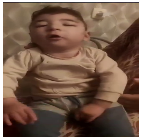 | Figure 1. Patient 2 years old with Aicardi syndrome |
 | Figure 2. CT scan of patient T.B., Diagnosis: Brain malformation-agnesia of the corpus callosum. Aicardi syndrome. Infantile spasms. Delayed psychomotor development. Enlargement of the lateral ventricles. Cystic formation of the optic nerve on the left (choreoreoretinal cyst) |
2. Conclusions
- Thus, the interrelationship of dysembryogenetic syndromes is characterised by a combination of combinations of clinical and neurological features, in the form of epileptic manifestations, non-paroxysmal neurological disorders, ophthalmological, cardiological, neurophysiological and neuroradiological disorders. Unlike many other neurogenetic syndromes with a known molecular defect, DNA testing in Aicardi syndrome is difficult to diagnose; accordingly, more attention should be paid to the diagnosis of the disease and a comprehensive clinical and neuroimaging study.