-
Paper Information
- Next Paper
- Previous Paper
- Paper Submission
-
Journal Information
- About This Journal
- Editorial Board
- Current Issue
- Archive
- Author Guidelines
- Contact Us
American Journal of Medicine and Medical Sciences
p-ISSN: 2165-901X e-ISSN: 2165-9036
2024; 4(2): 336-339
doi:10.5923/j.ajmms.20241402.37
Received: Jan. 15, 2024; Accepted: Feb. 2, 2024; Published: Feb. 12, 2024

Polyhydramnios: Features of the Course of Gestation, Birth and Methods of Delivery
 Abstract
 Reference
 Full-Text PDF
 Full-text HTML
Yusupbaev R. B., Igamberdieva D. U., Dauletova M. Zh.
Republican Specialized Scientific and Practical Medical Center for Obstetrics and Gynecology, Uzbekistan
Copyright © 2024 The Author(s). Published by Scientific & Academic Publishing.
This work is licensed under the Creative Commons Attribution International License (CC BY).
http://creativecommons.org/licenses/by/4.0/

The article presents the data of a retrospective analysis of the history of childbirth in pregnant women with polyhydramnios, delivered in RSSPMCOG. The frequency of occurrence, features of the course of gestation, childbirth and methods of delivery were studied.
Keywords: Polyhydramnios, Pregnancy, Childbirth, Amniotic fluid
Cite this paper: Yusupbaev R. B., Igamberdieva D. U., Dauletova M. Zh., Polyhydramnios: Features of the Course of Gestation, Birth and Methods of Delivery, American Journal of Medicine and Medical Sciences, Vol. 4 No. 2, 2024, pp. 336-339. doi: 10.5923/j.ajmms.20241402.37.
Article Outline
1. Introduction
- Today, one of the pressing problems of obstetrics is the pathology of amniotic fluid. In the development of pregnancy progression, amniotic fluid is one of the important components. Currently, available data on the functional interdependence between the condition of the fetus and the composition of the amniotic fluid indicate the diagnostic value of studying amniotic fluid [1,2,5].Polyhydramnios has a different etiology, the causes may be related to diseases of the mother or fetus and placenta. Idiopathic polyhydramnios occurs in 20.1-66.7% of cases and no pathology on the part of the mother and fetus is detected [5].The most common dysfunction of the amniotic membranes is polyhydramnios, which is associated with an ever-increasing infectious index in pregnant women. Polyhydramnios is one of the clinical manifestations of intrauterine infection, which is a pressing problem in obstetrics and modern perinatology. Polyhydramnios is one of the specific signs of intrauterine infection, the frequency of which has now increased to 3-5% [2,7,10]. At the same time, the course of pregnancy with polyhydramnios is often complicated by preeclampsia, premature abruption of a normally located placenta, premature birth, fetal hypoxia and asphyxia [4,5,8].Polyhydramnios is one of the predictors of fetal malformations. Excessive accumulation of amniotic fluid occurs with many chromosomal disorders, such as Down syndrome. Poorly managed gestational diabetes associated with fetal macrosomia may be one of the causes of polyhydramnios, but its pathogenesis is not fully studied. One possible explanation is polyuria due to fetal hyperglycemia, leading to increased osmotic diuresis [4,8,9,10].The occurrence frequency of polyhydramnios, depending on the duration of pregnancy, ranges from 0.12 to 3% and varies greatly within different periods of gestation: 16–19 weeks – 1.5%; 20–23 weeks – 8.9%; 24–27 weeks – 12.2%; 28–32 weeks – 28.4%; 33–38 weeks – 19.6%.In addition to the above, the following are of great importance:- degree of polyhydramnios, which depends on the amount of amniotic fluid (mild, moderate, severe);- rate of increase of amniotic fluid (acute, chronic);- gestational age in which polyhydramnios occurs (primary, secondary).Despite numerous studies of amniotic fluid, polyhydramnios is one of the most serious complications of pregnancy and is a serious problem leading to placental dysfunction and causing a high risk of perinatal pathology [2,3,5,6].Purpose of the study. To study the frequency and characteristics of pregnancy in women with polyhydramnios.
2. Materials and Methods of the Research
- A retrospective analysis of the birth history of pregnant women with polyhydramnios was carried out at the Republican Specialized Scientific and Practical Medical Center for Obstetrics and Gynecology. According to the birth history record, clinical and anamnestic data, the course of pregnancy, childbirth and the condition of the newborns were carefully analyzed. Records of clinical, laboratory and instrumental data were studied. The study of amniotic fluid, utero-fetal-placental circulation was carried out at the Republican Specialized Scientific and Practical Medical Center for Obstetrics and Gynecology of the Ministry of Health of the Republic of Uzbekistan using the method of Dopplerography with color mapping and Doppler ultrasound with the help of the Mindray DC-70 apparatus. The study was conducted in the second and third trimesters. An objective measurement of the amount of fluid is performed using mathematical values obtained after scanning the uterine cavity in quadrants (pockets).
3. Results of the Research
- A retrospective analysis of 3315 histories of births that occurred at the Republican Specialized Scientific and Practical Medical Center for Obstetrics and Gynecologywas carried out. The results of a retrospective analysis showed that among the general population of pregnant women admitted for delivery at the RSSPMCOG in 2020, 6% (n=199) of women had polyhydramnios.According to a retrospective analysis, the age of the examined pregnant women with polyhydramnios ranged from 21 to 40 years, the average age was 28+2.64 years. The proportion of women under the age of 25 was 38.80%; 55.60% of women were aged 26-35 and 5.60% were at the age of 36 and older (Fig. 1).
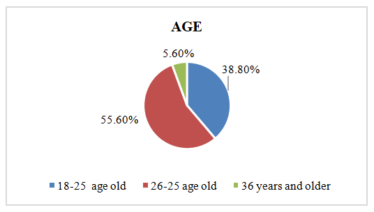 | Figure 1. Age of pregnant women with polyhydramnios (according to retrospective analysis) |
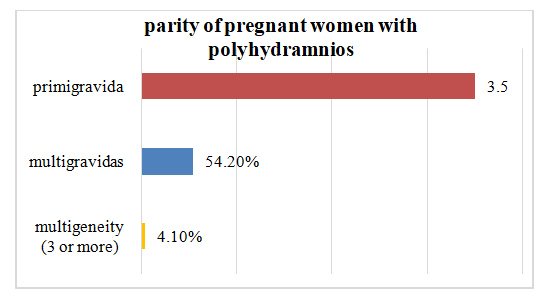 | Figure 2. Parity of pregnant women with polyhydramnios |
|
|
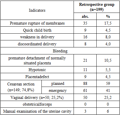
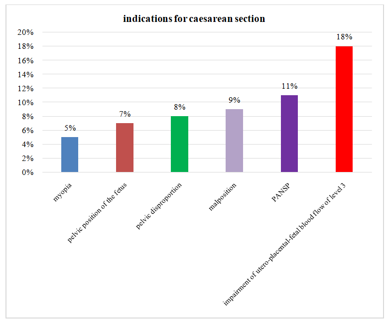 | Figure 3. Indications for abdominal delivery in pregnant women with polyhydramnios |
|
4. Conclusions
- Thus, our retrospective analysis showed that polyhydramnios occurs in 6% of cases among all births according to the clinic of the Republican Specialized Scientific and Practical Medical Center for Obstetrics and Gynecology. Polyhydramnios plays a leading role in the development of obstetric and perinatal complications. The timing of delivery was between 25 and 37 weeks of gestation. Perinatal mortality in a retrospective analysis was 15.5% due to early delivery due to acute polyhydramnios. Implementing timely diagnostic and therapeutic measures such as amnioreduction can improve perinatal outcomes.