-
Paper Information
- Previous Paper
- Paper Submission
-
Journal Information
- About This Journal
- Editorial Board
- Current Issue
- Archive
- Author Guidelines
- Contact Us
American Journal of Medicine and Medical Sciences
p-ISSN: 2165-901X e-ISSN: 2165-9036
2024; 14(1): 33-36
doi:10.5923/j.ajmms.20241401.08
Received: Dec. 14, 2023; Accepted: Dec. 30, 2023; Published: Jan. 3, 2024

Markers of Osteogenesis in Oral Fluid in Children with Musculosketal Disorders
 Abstract
 Reference
 Full-Text PDF
 Full-text HTML
Abdullaev J. R.
Tashkent State Dental Institute, Uzbekistan
Correspondence to: Abdullaev J. R., Tashkent State Dental Institute, Uzbekistan.
Copyright © 2024 The Author(s). Published by Scientific & Academic Publishing.
This work is licensed under the Creative Commons Attribution International License (CC BY).
http://creativecommons.org/licenses/by/4.0/

The article presents data from a study conducted to study the indicators of osteogenesis markers in oral fluid in children with disorders of the musculoskeletal system. Analysis of the results obtained indicates that when assessing the cariogenic situation in children with disorders of the musculoskeletal system, it is advisable to take into account the multifactorial nature of the carious process.
Keywords: Factors in the development of caries, Disorders of the musculoskeletal system, Markers of osteogenesis in the oral fluid
Cite this paper: Abdullaev J. R., Markers of Osteogenesis in Oral Fluid in Children with Musculosketal Disorders, American Journal of Medicine and Medical Sciences, Vol. 14 No. 1, 2024, pp. 33-36. doi: 10.5923/j.ajmms.20241401.08.
Article Outline
1. Introduction
- As is known, the influence of the general condition of the body on the possible development of dental caries was actively discussed by domestic and foreign cariologists back in the 70s of the last centuries. In the studies of Leus P.A. (2007), the results of experimental analytical and epidemiological studies confirmed the indirect effect of general factors (through blood and saliva) on the condition of dental tissues). Meanwhile, the concept of providing dental (including preventive) care for damage to hard dental tissues in patients with musculoskeletal disorders should take into account etiopathogenetic features, including the endogenous factor in the development of the disease. It should be noted that diseases of the musculoskeletal system today occupy a leading position in the structure of the general morbidity of the population. In recent years, in research and clinical work, osteoprotegerin (OPG), which belongs to the tumor necrosis factor family, has been identified as diagnostically significant markers of bone metabolism, along with proven markers of bone tissue remodeling. Osteoprotegerin is synthesized by osteoblastic cells (on the surface of the cell membranes of osteoblasts, T-lymphocytes and stromal cells). M. Sakata, H. Shiba, H. Komatsuzawa et al [1999] made a presumable conclusion that OPG, synthesized by dental mesenchymal cells, locally regulates the resorptive processes of dental hard tissues with the help of cytokines. In more recent work, the pulp of intact and carious teeth was studied. Intact tooth samples showed OPG immunoreactivity only in the odontoblastic layer. There is no information about the features of reactive changes in tooth enamel (its physicochemical and structural characteristics, physiological properties) with changes in the RANKL - OPG system in the literature available to us.Meanwhile, the condition of teeth is largely determined by the characteristics of the environment surrounding the tooth - oral fluid. It is with the properties of oral fluid that the processes of natural secondary maturation of enamel are associated, i.e. postoperative increase in its caries resistance.Saliva is an important element of the body’s resistance to caries throughout life. Literary sources indicate that ARVI, infectious hepatitis, hypertension and musculoskeletal disorders can inevitably lead to the formation of a low level of dental resistance to caries. Meanwhile, at present, the prevalence of caries in permanent teeth in adolescents has a slight downward trend: in 6 years - from 22% to 13%, 12 - from 78% to 73%, 15 - from 88% to 82%. It should be noted that knowledge of etiopathogenetic concepts and epidemiological features of the carious process in adolescents with musculoskeletal disorders allows the attending physician to draw up an optimal plan for the treatment and prevention of diseases of the hard dental tissues in adolescents, especially when combined with various pathologies.
2. Purpose of the Study
- To study the indicators of osteogenesis markers in the oral fluid in children with musculoskeletal disorders.
3. Material and Research Methods
- During the entire observation period, under our control and constant dynamic observation there were 326 children of both sexes with pathologies of the musculoskeletal system, aged from 7 to 18 years, permanent residents of special (correctional) educational institutions for pupils with disabilities. The control group consisted of 30 practically healthy children, without pathology of the musculoskeletal system.Before collecting oral fluid (ORF), children and parents (guardians) are explained in detail the methodology and purpose of collecting the biosubstrate. The collection of gastric fluid was carried out in a children's dental clinic from 8 to 9 o'clock (the period of maximum secretion) on an empty stomach into sterile tubes. Patients were advised not to carry out procedures that stimulate salivation: chewing gum; eating food, water; mouth rinse; teeth cleaning. It is recommended to eliminate emotional and physical stress, and rinse your mouth with distilled water (t-22-24°) for 5-10 minutes, followed by removing any remaining water with a clean napkin. Method of collecting gastric fluid: the patient sits comfortably, lowers his head and sits in this position without moving, and does not swallow saliva. After 2 minutes, the patient spits the contents into a calibrated sterile cylinder. The total number of procedures is 5, the total collection time is 10 minutes (recommendations of TsNIIS, 1991). Next, the GC was centrifuged (speed 8000 rpm, time 15 min), aliquoted into 200 μl plastic tubes and stored frozen at t=-76°C until the start of the study.telopeptides in the blood plasma, which were determined by enzyme-linked immunosorbent assay using the Cross Laps reagent kit ELISA" from Nordic Bioscience Diagnostic A/S" respectively. Determination and calculation procedures were carried out according to the attached instructions. The Cross Laps method is based on the use of antibodies against a synthetic octapeptide, identical to the C- telopeptide region of type I collagen, and characterizes the level of CTX formed during the degradation of collagen under the action of cathepsin K of osteoclasts and matrix metalloprotein. The content of serum calcitonin, parathyroid hormone, 25-hydroxyvitamin D3 osteocalcin was determined by enzyme-linked immunosorbent assay using commercial test systems from Vector-Best (Russia). Biochemical methods for studying oral fluid.The state of bone metabolism in oral fluid was studied using the level of basic markers: P-Cross Laps (a specific degradation product of type I collagen, a marker of bone resorption) and osteocalcin (a marker of osteosynthesis). Excretion of free hydroxyproline in daily urine was carried out according to the method of Neumann and Logan, modified by P.N. Sharaeva (2002). The content of total glycosaminoglycans (GAG) based on the level of hexuronic acids (L.I. Slutsky, 1969; modified by P.N. Sharaeva et al., 1987); The content of GAGs in the blood was expressed in µmol hexuronic acids per 1 liter of plasma (µmol /l).The results were processed using the STATISTICA 10.0 program (StatSoft, Inc., USA). If the characteristic distribution law in the studied samples corresponds to the normal law, the hypothesis about the equality of average sample values was tested using Student's t-test.
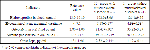
4. Results and Its Discussion
- One of the markers that allow us to evaluate the processes of bone tissue regeneration is the levels of alkaline and acid phosphatase. Alkaline phosphatase plays a key role in mineralization by breaking down inorganic pyrophosphate and releasing free inorganic phosphate. It is believed that this enzyme can also be used as a biochemical marker to determine the activity of osteoblasts, since it is present on fragments of their plasma membranes. One of the important markers of bone metabolism is osteocalcin, which is a non-covalent calcium-ion binding protein, also known as γ- carboxyglutamic acid protein, produced by both osteoblasts and odontoblasts. Osteocalcin binds hydroxyapatite and calcium during mineralization of the organic matrix. To date, there are limited studies examining caries in combination with musculoskeletal pathologies, and most of these studies are experimental in nature. Based on the above, the purpose of this study was to study the dynamics of osteogenesis markers in adolescents with caries with and without musculoskeletal disorders.One of the indicators that allows us to evaluate the processes of bone tissue regeneration is the levels of alkaline phosphatase. It has been established that this enzyme is involved in the regulation of phosphorus-calcium metabolism. Alkaline phosphatase plays a key role in mineralization by breaking down inorganic pyrophosphate and releasing free inorganic phosphate. It is believed that this enzyme can also be used as a biochemical marker to determine the activity of osteoblasts, since it is present on fragments of their plasma membranes. The data we obtained, presented in Table 1, indicates the activation of enzyme activity in the subjects examined, especially with disorders of the musculoskeletal system, relative to the indicators of the comparison group. As can be seen from the presented research results, when comparing indicators between groups, an increase in alkaline phosphatase activity by 37% was noted.
|