-
Paper Information
- Next Paper
- Previous Paper
- Paper Submission
-
Journal Information
- About This Journal
- Editorial Board
- Current Issue
- Archive
- Author Guidelines
- Contact Us
American Journal of Medicine and Medical Sciences
p-ISSN: 2165-901X e-ISSN: 2165-9036
2024; 14(1): 29-32
doi:10.5923/j.ajmms.20241401.07
Received: Dec. 5, 2023; Accepted: Dec. 27, 2023; Published: Jan. 3, 2024

Macroanatomical Features of the Heart in Chronic Alcohol Intoxication and Ways of Its Correction
 Abstract
 Reference
 Full-Text PDF
 Full-text HTML
M. M. Ziyodullaev1, A. S. Ilyasov2, D. A. Khasanova3
1Assistant of the Department of Internal Diseases, Bukhara State Medical Institute named after. Abu Ali ibn Sino, Republic of Uzbekistan
2Doctor of Biological Sciences, Associate Professor of the Department of Anatomy, Clinical Anatomy (OCTA), Bukhara State Medical Institute named after. Abu Ali ibn Sino, Republic of Uzbekistan
3Doctor of Medical Science, Associate Professor, Professor of the Department Of anatomy, Clinical Anatomy (OKTA), Bukharsk State Medical Institute im. Abu Ali Ibn Sina, Republic of Uzbekistan
Copyright © 2024 The Author(s). Published by Scientific & Academic Publishing.
This work is licensed under the Creative Commons Attribution International License (CC BY).
http://creativecommons.org/licenses/by/4.0/

In the ECG examination of six-month-old (adult) rats poisoned with ethyl spit, the average number of heart contractions increased from 513 to 800, and according to the echocardiographic examination, the blood pumping volume of the heart decreased from 43,9% to 37,9%. Electrophysiological changes of the heart occur due to a decrease in the amount of microelements (sodium, potassium, zinc, calcium, magnesium) important for excitable tissues under the influence of ethyl alcohol. Ethanol has a dyslipidemic effect on the body, causes fatty dystrophy and atrophy of cardiomyocytes, and causes a decrease in the blood pumping capacity of the heart.
Keywords: Heart, Rats, Alcohol, Olive oil
Cite this paper: M. M. Ziyodullaev, A. S. Ilyasov, D. A. Khasanova, Macroanatomical Features of the Heart in Chronic Alcohol Intoxication and Ways of Its Correction, American Journal of Medicine and Medical Sciences, Vol. 14 No. 1, 2024, pp. 29-32. doi: 10.5923/j.ajmms.20241401.07.
Article Outline
1. Introduction
- Alcohol abuse is an important medical problem of our time, which is still a social problem. According to the World Health Organization, 1.5 million people die every year from alcoholism. Alcohol consumption reduces people's ability to work by 15-30%. Alcoholism is a disease caused by chronic consumption of alcoholic products, causing disruption of most vital functions of the body [1]. According to many authors, chronic alcohol consumption leads to the development of a number of cardiovascular diseases, including: arterial hypertension, heart rhythm disturbances, coronary heart disease, dilated cardiomyopathy. Researchers have discovered that ethyl alcohol and its metabolites have a direct effect on the heart muscle. At the same time, drinking 80 grams of alcohol per day for ten years leads to alcoholic cardiomyopathy, which is associated with the esterification of alcohol in fatty acids [2]. The toxic effect of alcohol on the heart is divided into such types as: arrhythmic, cardiomyopathic, coronary spastic. The arrhythmic type is the most common and fatal consequence of alcohol among those listed [3]. Alcohol-induced toxicity leads to alcohol-induced cardiomyopathy, characterized by loss of contractile function and dilatation of the myocardial ventricles [4]. With dilated cardiomyopathy, not only the apex of the organ is rounded, but all the chambers of the heart also expand. On the contrary, alcoholic cardiomyopathy is characterized only by an enlargement of the left ventricle [5]. Among the complications of chronic alcohol intoxication, myocardial infarction is of particular importance for study. According to pathological anatomical studies, the most significant is the development of a large focal infarction with atherosclerotic lesions of many coronary vessels [6]. However, there is also data that states the opposite opinion, about the absence of a negative effect of alcohol consumption in persons with coronary heart disease [7]. In the cardiac muscle of patients who died as a result of ethanol poisoning, filling of blood vessels, aggregation of red blood cells, swelling of the vascular endothelium, local narrowing of the vascular wall and cardiomyolysis are observed [8]. Plants were one of the first natural sources used by humanity for the prevention and treatment of diseases. According to WHO, medicinal preparations of plant origin are the most important source of adaptive biologically active compounds that are necessary for treatment and strengthening the body’s defenses for various diseases, and a large number breadth of therapeutic action, low toxicity and the possibility of long-term course use for chronic diseases, low risk of side effects, complex action, accessibility allows herbal medicines to successfully compete with synthetic ones [9]. These factors contribute to an increase in the consumption of medicines obtained from medicinal plants, both in the Russian Federation and in China, the USA and Germany [10]. It should be noted that olive oil is of particular importance in the prevention of cardiovascular diseases. This is due to the fact that olive oil reduces the amount of unsaturated fatty acids in the blood, reduces fat oxidation, and increases the sensitivity of receptors to insulin. Olive oil has antioxidant, anti-inflammatory, and endothelial-strengthening properties. Also, it prevents cardiac remodeling [11].Purpose: to study the macroanatomical features of the heart in chronic alcohol intoxication and correction with olive oil.Tasks:1. Determine the volume of the heart affected by alcohol and its changes under the influence of olive oil.2. Identify the weight of the heart damaged by alcohol and its changes under the influence of olive oil.3. Study the number of heart contractions under the influence of alcohol and its changes under the influence of olive oil.4. Determine the weight of alcoholic rats and its changes under the influence of olive oil.5. To identify the cardiac ejection fraction of alcoholic rats and its changes under the influence of olive oil.
2. Materials and Methods of Research
- Experimental studies were carried out on 6-month-old rats. At the beginning of the experiment, all sexually mature rats were placed in a week-long quarantine, and after somatic or infectious diseases were excluded, they were transferred to the normal vivarium regime. During the experiment, the behavior and physiological state of animals in the control and experimental groups were monitored.The rats were divided into 3 groups: control group I (n = 13); Rats of group II received 40% ethyl alcohol in an amount of 7.0 g/kg body weight for 30 days (n = 13). Group III - rats received olive oil in an amount of 2 ml/kg for 30 days (n = 53).All animals were kept in a vivarium at the same temperature 15-20°C, humidity 65-70% and lighting for 14-17 hours. They were also kept under standard conditions in a metal cage with a mat, a feeder, a drinking bowl and its tube. White mongrel rats were obtained from the research center of the Bukhara State Medical Institute named after Abu Ali ibn Sina. Feeding the rats, cleaning the cages, disinfecting equipment, dividing into groups was carried out in accordance with the recommendation of the ethical committee (extract from minutes No. 2 of the meeting of the ethical committee M3 of the Republic of Uzbekistan dated March 31, 20 23 years old). According to the recommendations of the ethical committee, rats that died during the study were treated with a 20% solution of bleach, buried and included in the report.To model and simulate chronic alcohol intoxication, rats from 2 months of age were administered 40% alcohol in an amount of 7 g/kg for 30 days [12].Ethyl alcohol was administered intragastrically through a metal gastric tube.Rats in the control group were intragastrically injected with 0.5 ml of distilled water through a metal gastric tube for 30 days.Considering that the amount of olive oil that reduces the harmful effects of ethyl alcohol on the body is 2 ml/kg [13].Detoxification of rats with chronic alcohol intoxication was carried out intragastrically through a metal gastric tube.Organometric methodTo measure the morphometric parameters of the heart, an anatomical ruler and 0.05 mm calipers were used.When measuring thickness, length, width, the following parts of the heart were used: thickness of the heart (the largest anterior-posterior dimension in relation to the ventricles), length (from the place of exit from the aorta to the apex of the heart) and width of the heart (between the sides external surfaces of the heart at the level of the base of the ventricles) [14].The shape of the heart was determined using the Jilin formula: (Width/Length of the organ) ×100%. According to the formula, the shape of the heart will be as follows:Conical up to 65%, 65% to 75% ellipsoidal, more than 75% have a spherical shape [15]. Heart mass was measured on laboratory scales with an accuracy of 0.01 g (TBE - 0.50 - 0.01, made in South Korea) and recorded in the examination journal.Electrocardiographic research methodAn ECG study allows us to obtain complete information about the electrophysiological state of the heart of rats. Rats of the same age were divided into 3 groups: control group, experimental group and detoxification group. According to the rules for conducting ECG on animals, the rats were placed in special cages and were not fed for two hours. The rats were placed on a wooden table for electrocardiographic examination. Before the study, rats were anesthetized with ether anesthesia. We made sure that the body of the rats did not touch metal objects. For electrocardiography, a three-channel electrocardiograph was used. (BLT – 1203 SN: E066A000412. EC Representative Name: Shanghai international Trading Corp. GmbH (Hamburg). The study was carried out for 5-10 minutes. The electrocardiogram calibration signal was set to 50 mV, and the tape speed was set to 50 mm /s. During the study, a standard lead was used (according to Willem Uythoven). Before connecting the leads, a special ECG gel was applied to the areas where the sensors will be placed. This improves the ability of the sensors to transmit electric current into the body of the rats. The rats were placed on their backs, and the metal parts of the colored sensors were placed on the medial surface of the legs of rats using an adhesive plaster according to the instructions.I standard abduction - front right and left legs,II standard abduction - front right leg and back left leg,III standard lead - the electrical impulse passes between the front and back left legs. The ECG was recorded automatically on thermal paper 50 mm wide.EchocardiographyAn echocardiographic study was carried out on an anatomical device M-mode SSI-5000 (serial number S/N1407392) from Sono Scape (China) using a sector sensor S 2.5 MGS, designed for important for ultrasound cardiography. Echocardiography parameters were determined by measuring the main hemodynamic parameters in a standard way. Ultrasound cardiography was performed without sedation. Hair was shaved from the right and left parasternal area to the abdominal area. The animals were placed on the table either with their left or right side so that the sensor could be placed. A special gel was applied to the device sensor. Depending on the position of the rats, the sensor was located between the third and sixth ribs in the right parasternal space, and when using the left cranial parasternal space, the sensor was located or between the third or fourth ribs [16].
3. Statistical Research Methods
- Statistical analysis was carried out using well-known statistical methods and application programs. The collected data was corrected, after systematization of the initial materials, the spreadsheet program Microsoft Office Excel 2010 was used to obtain significant results. All data was subjected to statistical analysis using the IBM SPSS Statistics v.23 program (developed by IBM Corporation, USA).In order to describe the sample distribution of quantitative characteristics that do not obey the normal distribution, we used the arithmetic mean (M) and standard error (m), in the format M±m and 95% confidence limits ogo interval (95% CI).When comparing average values in normally distributed sets of quantitative data, Student's t-test was calculated. The obtained Student's t-test values were assessed by comparison with the critical values. Differences in indicators were considered statistically significant at a significance level of p<0.05. The Pearson χ2 criterion was also used, which allows us to assess the significance of the difference between the actual number of outcomes or qualitative characteristics of the sample falling into each category and the theoretical number m, which can be expected in the studied groups if the null hypothesis is valid. When organizing and conducting research, we used the principles of evidence-based medicine.
4. Research Results
- Studies have shown that the weight of six-month-old rats ranges from 101.0 to 125.0 mg, with an average of 112.98 ± 2.09 mg. Chest volume from 8.5 to 14.5 cm3, on average 11.61±0.52 cm3, organ volume from 2.5 to 2.9 cm3, on average 2.767±0.035 cm3. And the heart mass in six-month-old rats ranges from 0.59 to 0.69 mg, on average - 0.65±0.01 mg. At this age, the length of the heart ranges from 16.0 to 18.0 mm, on average 16.67 ± 0.17 mm, width from 11.0 to 13.0 mm, on average 12.4 ± 0.17 mm. In addition, the heart shape was conical in 8%, elliptical in 30% and spherical in 62% of cases.Studies have shown that six-month-old rats with alcohol poisoning have a body weight from 85.85 to 106.25 mg, on average 96.04 ± 1.78 mg. The volume of the chest is from 7.23 to 12.33 cm3, the average is 9.87±0.44 cm3, and the volume of the organs is from 2.13 to 2.47 cm3, the average is 2.35±0.03 cm3. Heart weight in six-month-old rats ranges from 0.50 to 0.59 mg, on average - 0.55±0.01 mg. At this age, the length of the heart ranges from 13.6 to 15.3 mm, on average 14.17 ± 0.15 mm, width from 9.35 to 11.05 mm, on average 10.54 ± 0.15 mm. At the same time, the shape of the heart was elliptical in 38%, spherical in 62%, conical was not determined.Studies have shown that the body weight of six-month-old rats receiving olive oil detoxification ranges from 92.92 to 115.0 mg, with an average of 103.39 ± 1.92 mg. The volume of the chest is from 7.82 to 13.34 cm3, on average - 10.68±0.48 cm3, and the volume of the organ is from 2.30 to 2.67 cm3, on average 2.55±0.03 cm3. Heart weight in six-month-old rats ranges from 0.54 to 0.64 mg, with an average of 0.59 ± 0.01 mg. At this age, the length of the heart is from 14.72 to 16.56 mm, on average 15.33 ± 0.16 mm, width from 10.12 to 11.96 mm, on average 11.35 ± 0.16 mm. In addition, the heart shape was conical in 7%, elliptical in 30% and spherical in 63% of cases.In six-month-old rats, the number of heartbeats and echocardiographic changes are shown in Table 1.
|
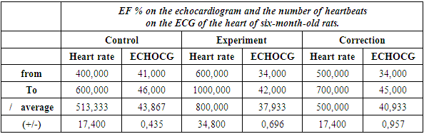
5. Conclusions
- 1. The experiment established that the heart volume in the control group decreased from 2.767±0.035 cm3 to 2.35±0.03 cm3 as a result of alcohol poisoning and increased to 2.55±0.03 cm3 as a result of correction.2. It was established that the mass of the organ decreased from 0.65±0.01 mg to 0.55±0.01 mg, and as a result of detoxification the mass amounted to 0.59±0.01 mg.3. With alcohol intoxication, ECG studies showed an increase in the number of heart contractions, and echocardiographic studies showed a decrease in the volume of heart contractions.4. During detoxification with olive oil, it was found that the number of heartbeats in rats decreased from 800±34.800 to 500±17.400, and cardiac output increased from 37.933±0.696 to 40.933±0.957.5. The experiment established that the weight of six-month-old rats decreased from 112.98±20.52 to 96.04±1.78 mg during alcohol poisoning, and as a result of detoxification the weight was 103.39±1.92 mg. It was established that the volume of the chest in the control group decreased from 11.61±0.52 cm3 to 9.87±0.44 cm3 as a result of alcohol poisoning and increased to 10.68±0.48 cm3 as a result of correction.