American Journal of Medicine and Medical Sciences

p-ISSN: 2165-901X    e-ISSN: 2165-9036

2024;  14(1): 16-23

doi:10.5923/j.ajmms.20241401.04

Received: Dec. 5, 2023; Accepted: Dec. 21, 2023; Published: Jan. 3, 2024


Clinical and Immunological Characteristics of Patients with Type 2 Diabetes Mellitus Complicated by Chronic Kidney Disease that Underwent COVID-19

Najmutdinova D. K.1, Mirakhmedova Kh. T.1, Khusanov A. M.2, Kudaybergenova D. Kh.1, Nurmatov A. Kh.2

1Tashkent Medical Academy, Uzbekistan

2Zangiata Infectious Diseases Hospital №1, Uzbekistan

Copyright © 2024 The Author(s). Published by Scientific & Academic Publishing.

This work is licensed under the Creative Commons Attribution International License (CC BY).
http://creativecommons.org/licenses/by/4.0/

Abstract

Coronavirus disease—COVID-19 (coronavirus disease 2019) has become the cause of the global pandemic in the last three years. Its etiological factor is SARS-CoV-2 (Severe Acute Respiratory Syndrome Coronavirus type 2). Patients with diabetes (DM—diabetes mellitus), in contrast to healthy people not suffering from chronic diseases, are characterised by higher morbidity and mortality due to COVID-19. Patients who test positive for SARCoV-2 are at higher risk of developing hyperglycaemia. In this paper, we present, analyse and summarize the data on possible mechanisms underlying the increased susceptibility and mortality of patients with diabetes mellitus in the case of SARS-CoV-2 infection. However, further research is required to determine the optimal therapeutic management of patients with diabetes and COVID-19. Kidney impairment in hospitalized patients with SARS-CoV-2 infection is associated with increased in-hospital mortality and worse clinical evolution, raising concerns towards patients with chronic kidney disease (CKD). From a pathophysiological perspective, COVID-19 is characterized by an overproduction of inflammatory cytokines (IL-6, TNF-alpha), causing systemic inflammation and hypercoagulability, and multiple organ dysfunction syndrome.

Keywords: COVID-19, Type 2 diabetes mellitus, IL11, IL 17A, TGF-β1

Cite this paper: Najmutdinova D. K., Mirakhmedova Kh. T., Khusanov A. M., Kudaybergenova D. Kh., Nurmatov A. Kh., Clinical and Immunological Characteristics of Patients with Type 2 Diabetes Mellitus Complicated by Chronic Kidney Disease that Underwent COVID-19, American Journal of Medicine and Medical Sciences, Vol. 14 No. 1, 2024, pp. 16-23. doi: 10.5923/j.ajmms.20241401.04.

1. Introduction

The World Health Organization (WHO) has classified the 2019 Coronavirus pandemic (COVID-19) as a global health emergency [1]. The virus causes acute respiratory distress syndrome. So far our knowledge of this disease has been limited. The new beta-coronavirus was first introduced in Wuhan, China in 2019 and then spread worldwide. SARS-CoV-2 is an RNA virus with genomes ranging from 26,000 to 32,000 base pairs [2]. The capsid contains four structural proteins: spike (S), nucleocapsid (N), membrane (M) and envelope (E). In their research, Walls et al. showed that the nucleic acid and developmental N acid are under the formed lipid novel solution of the SARS-CoV-2 virion [3]. Under an electron microscope, the S looks like a corona and thus its name: coronavirus. The S protein consists of two domains. The binding of the receptor for angiotenin-converting enzyme-type 2 (ACE-2) takes place in the upper lobular domain, which initiates a viral entry into cells. The downstream domain of the S protein is involved in the virus attachment to the host-cell membrane [4]. The function of the M protein is to bind the viral genome to the inner surface of the host-cell membrane. The C-terminal domain of the transmembrane proteins contacts the viral N protein, playing an important role in its life cycle [5]. The scheme showing the model of COVID-19 virus is presented in Figure 1.
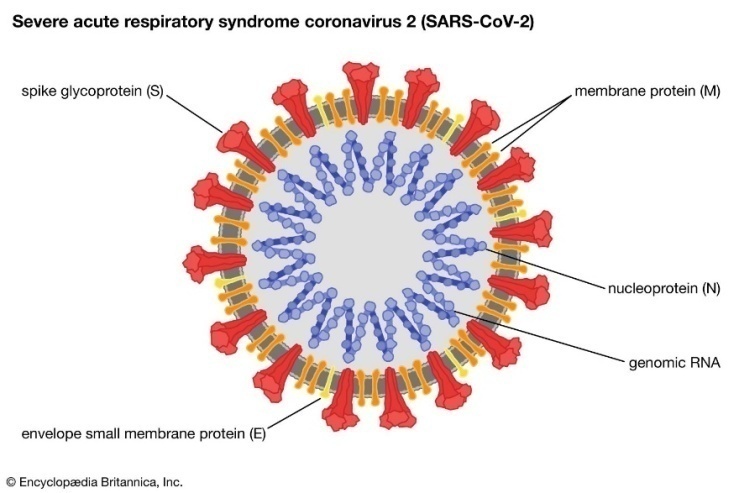
Figure 1. A model of COVID-19 depicting structural elements of the virion
In the SARS-CoV-2 replication cycle, several key stages can be distinguished: attachment and entry into the cell, transcription of the viral replicase, genome replication, translation of structural proteins and release of new virions [2]. The analysis of over 300,000 SARS-CoV-2 genomes, performed less than a year ago by Rochman et al., showed some kind of an adaptive evolution of this virus which affects not only the nucleocapsid itself, but also many regions in the viral S protein [6]. Therefore, based on mutations in the RBD region of the viral S protein, a total of seven mutant variants of SARS-CoV-2 have been described: alpha (B.1.1.7), beta (B.1.351), gamma (P.1), delta (B.1.617.2), epsilon (B.1.427), kappa (B.1.617.1), and the Omicron variant (B.1.1.529). Based on microscale thermophoresis analyses on the S-protein in SARS-CoV-2, it has been demonstrated that the alpha, beta, gamma and delta variants are characterised by the strongest interaction with the ACE2 receptor [7]. It is also worth emphasizing that the Omicron variant is typical due to its ability to avoid the human immune system [8]. The mortality rate for COVID-19 infection has been estimated at 0.5–1.0% [9,10,11]. The proportion of diabetics among COVID-19-positive patients varies according to the regions in the world. For example, in Italy as many as 36% of those seriously ill, having a positive result from a COVID-19 test, were burdened with diabetes [12], and in the United States, this same phenomenon was noted in as many as 58% of patients [13]. The vast majority of infected patients present a mild form of COVID-19, but some develop a severe form of infection that can be fatal. Factors that influence the development of severe disease are: older age, comorbidities, difficult socio-economic conditions and occupational risk (increased epidemiological treat). The most frequently quoted age limit above which there is an increased risk of serious complications is 60 years and above [14,15]. Regardless of age, patients with comorbid chronic conditions that compromise the immune system are at a higher risk of becoming infected with SARS CoV-2. These diseases mainly include: diabetes [16], renal failure [17], chronic heart and brain diseases [18,19], malignant neoplasms [14], immune disorders [15] and obesity [20]. In recent studies, a significantly higher risk of in-hospital deaths has been noted due to COVID-19 in diabetic patients than in those without glucose tolerance impairment (HR = 2.36) [14]. Interestingly, proper glycaemic control before admission to hospital, confirmed by a glycosylated haemoglobin test (HbA1c), was not associated with improved outcomes in the treatment of infections among these patients [21].
Scientists of Uzbekistan have carried out a number of studies on the specific course and consequences of COVID-19 in patients with diabetes, including the structure of death among patients with diabetes in the Republic of Uzbekistan during the COVID-19 pandemic (Haydarova F.A. and others), COVID- The state of hyperglycemia in patients hospitalized due to pneumonia associated with 19 (Alieva A.V. and others).

2. Mechanism of SARS-CoV-2 Morbidity in Diabetes

Diabetes is the seventh leading cause of death in the world and is associated with vascular complications, severely impacting quality of life(63) Diabetes predisposes patients to acquiring the infection due to an impaired immune system function. The following are responsible for this fact: the inhibition of phagocytosis, lower macrophage activity and decreased ability of neutrophil chemotaxis resulting from the reduced amount of interferon (IFN-γ) [42]. The role of glycaemic control in diabetic patients is emphasized. Although, as mentioned earlier [21], it does not seem to affect the efficacy of infection treatment, it reduces the likelihood of virus proliferation in cells [43,44]. Another possible cause of increased susceptibility to COVID-19 infection is an increase in the levels of furin, a type 1 protease bound to the cell membrane. It has been shown that higher levels of furin in diabetic patients facilitates the entry of SARS-CoV-2 into cells [33,45]. Furins mediate a viral entry into a human cell by cleaving and stimulating the SARS-CoV2 S1 and S2 proteins [3,46]. The scheme showing the pathophysiological mechanisms including an increased susceptibility of diabetic patients to COVID-19 is presented in Figure 2.
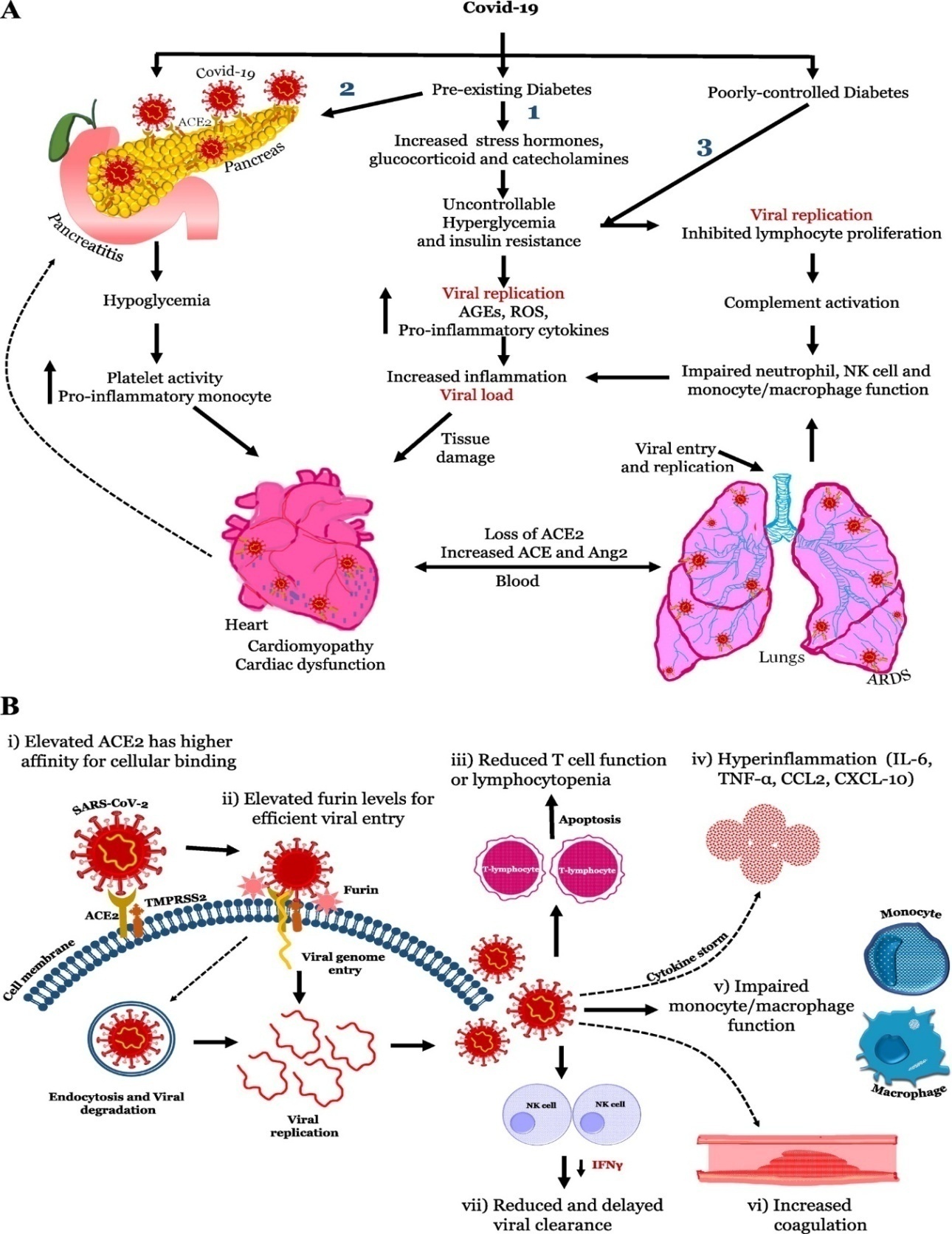
Figure 2. Patomechanism inducing an increased possibility of COVID-19 infection in diabetic patients
Thus, besides lung involvement, other organ complications are observed in patients with SARS-CoV-2 infection such as kidney damage leading to acute kidney injury (AKI)11, raising concerns regarding the clinical outcomes and prognosis of patients with preexisting comorbidities such as chronic kidney disease (CKD), end-stage kidney disease (ESKD), and kidney transplant recipients under immunosuppression therapy. Recipients under immunosuppression therapy. A meta-analysis including 73 studies evaluating the association between multi-organ dysfunction and COVID-19 development revealed that patients with CKD were more likely to develop severe SARS-CoV-2 infection (OR 1.84 [95%CI 1.47–2.30]) 12. Hence, besides disease severity, it is imperative to evaluate the clinical outcomes, prognosis, and mortality associated with COVID-19 infection in patients with history of CKD.
As 2023 approaches, the COVID-19 pandemic has killed millions. While vaccines have been a crucial intervention, only a few effective medications exist for prevention and treatment of COVID-19 in breakthrough cases or in unvaccinated or immunocompromised patients. SARS-CoV-2 displays early and unusual features of micro-thrombosis and immune dysregulation that target endothelial beds of the lungs, skin, and other organs. Notably, anticoagulation improves outcomes in some COVID-19 patients. The protein transforming growth factor-beta (TGF-β1) has constitutive roles in maintaining a healthy microvasculature through its roles in regulating inflammation, clotting, and wound healing. However, after infection (including viral infection) TGF-β1 activation may augment coagulation, cause immune dysregulation, and direct a path toward tissue fibrosis. Dysregulation of TGF-β signaling in immune cells and its localization in areas of microvascular injury are now well-described in COVID-19, and such events may contribute to the acute respiratory distress syndrome and skin micro-thrombosis outcomes frequently seen in severe COVID-19. The high concentration of TGF-β in platelets and in other cells within microvascular thrombi, its ability to activate the clotting cascade and dysregulate immune pathways, and its pro-fibrotic properties all contribute to a unique milieu in the COVID-19 microvasculature. This unique environment allows for propagation of microvascular clotting and immune dysregulation. In this review we summarize the physiological functions of TGF-β and detail the evidence for its effects on the microvasculature in COVID-19. In addition, we explore the potential role.

3. Inflammatory Cytokines in the Pathophysiology of Diabetic Nephropathy

Cytokines are a group of pharmacologically active, low molecular weight polypeptides with autocrine, paracrine, and juxtacrine effects which, in a coordinated manner, regulate inflammatory and immune responses with the participation of different cytokine-associated signalling pathways. Cytokines are produced throughout the body by cells of varied embryological origin and, in addition to their immune response regulatory role, exert important pleiotropic actions as cardinal effectors of injury [36]. At present time, it is recognized that chronic low-grade inflammation and activation of the innate immune system are closely involved in the pathogenesis of diabetes mellitus [37-39]. Plasma concentrations of diverse inflammatory parameters are elevated in diabetic patients [8-10,40,41] being strong predictors of the development of this disease [42-44]. The pathogenesis of severe COVID-19 reflects an inefficient immune reaction to SARS-CoV-2. Here we analyze, at the single cell level, plasmablasts egressed into the blood to study the dynamics of adaptive immune response in COVID-19 patients requiring intensive care. Before seroconversion in response to SARS-CoV-2 spike protein, peripheral plasmablasts display a type 1 interferon-induced gene expression signature; however, following seroconversion, plasmablasts lose this signature, express instead gene signatures induced by IL-21 and TGF-β, and produce mostly IgG1 and IgA1. In the sustained immune reaction from COVID-19 patients, plasmablasts shift to the expression of IgA2, thereby reflecting an instruction by TGF-β. Despite their continued presence in the blood, plasmablasts are not found in the lungs of deceased COVID-19 patients, nor does patient IgA2 binds to the dominant antigens of SARS-CoV-2. Our results thus suggest that, in severe COVID-19, SARS-CoV-2 triggers a chronic immune reaction that is instructed by TGF-β, and is distracted from itself.of existing TGF-β inhibitors for the prevention and treatment of COVID-19 associated microvascular thrombosis and immune dysregulation.

4. TGF-β1 Promotes Renal Fibrosis

Diabetic kidney disease pathology is characterized by thickening of the glomerular basement membrane, mesangial expansion, segmental glomerulosclerosis or global glomerulosclerosis, tubulointerstitial fibrosis, and afferent and efferent arteriole hyalinosis (Najafian et al., 2015). TGF-β is also known as a prominent regulator of immune reactions12, and it causes fibrosis13, a comorbidity of severe COVID-19 patients14,15 For a cohort of patients with severe COVID-19 requiring intensive medical care for up to 60 days, the continuous egress of plasmablasts into the blood, reflecting a continued immune reaction. Initially, within the first week of ICU admission, this immune reaction is directed against SARS-CoV-2, as all patients acquired IgG antibodies against the S and N proteins. Later, IgA-expressing plasmablasts are generated predominantly, reflecting continued instruction of the B lymphocytes by TGF-β. However, regarding their specificity, only one-third of the patients express S protein-specific IgA, and only 1 of those expressed S protein-specific IgA2, the terminal antibody class targeted by TGF-β. Circulating plasmablasts are clonally expanded and their antibodies are somatically mutated, but not specific for SARS-CoV-2 S or N protein. Taken together, these results point to TGF-β as a key cytokine regulating a chronic immune reaction in severe COVID-19, an immune reaction which is no longer directed to SARS-CoV-2.

5. IL-17A as a Proinflammatory Mediator in DN

IL-17A has been described as a circulating inflammatory protein associated with increased risk of renal damage progression [26]. Interestingly, circulating IL-17A levels are related to the severity of kidney disease and progressively decrease from subjects with normal glucose tolerance to subjects with type 2 diabetes with and without DN [40]. In agreement with this, patients with advanced DN present lower levels of IL-17A in both plasma and urine [41]. However, Zhang et al. showed an increase in CD4+ CXCR5+ PD-1+ T follicular helper cells and plasma values of IL-6 and IL-17 in patients with DN compared to healthy controls [42]. Among cirrhotic hepatitis C virus infected patients, serum IL-17A levels were higher in those that were type 2 diabetic than in non-diabetic patients and controls [43]. Although these studies have addressed circulating or urinary IL-17A levels in DN patients, local renal levels of IL-17A have not been investigated yet. Importantly, infiltration of immune cells is a key feature of DN [17]. Activated T cells (CD4+ and CD8+) are mainly located in the renal interstitium of diabetic kidneys [44,45,46]. Although CD4+ IL-17+ cells are the main source of IL-17A production, other cells, including macrophages, neutrophils, natural killer, dendritic, and mast cells, have also been described to produce this cytokine.
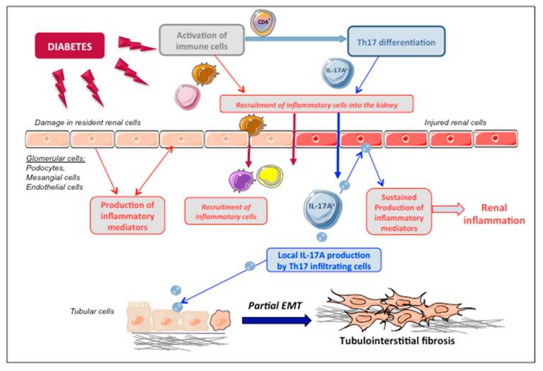
Figure 3. Proposed mechanism of IL-17A-induced renal damage in diabetic nephropathy. Under diabetic conditions, renal resident cells are activated and can produce different mediators that could contribute to recruit immune cells into the kidney. Infiltrating Th17 cells can locally produce IL-17A in the diabetic kidney. Then, IL-17A acting on IL-17R on resident renal cells can produce additional proinflammatory mediators, contributing to sustained inflammation. Moreover, IL-17A acting on tubular epithelial cells can induce phenotype changes, such as partial epithelial-to-mesenchymal transition (EMT) and secretome changes. By these mechanisms, IL-17A participates in the amplification of the inflammatory response and the progression of renal damage, finally leading to tubulointerstitial fibrosis
In this sense, in a pioneer study in renal biopsies of DN patients, we described the local activation of inflammatory pathways, specifically NF-κB activation linked to an upregulation of proinflammatory factors, such as the chemokine MCP-1 [14]. Since then, many preclinical studies have demonstrated that MCP-1 can be a therapeutic target and potential biomarker for DN. In this regard, clinical trials targeting MCP-1 or its receptor have shown promising results [48]. Nevertheless, studies evaluating the kidney expression of IL-17A in human DN are needed to further define its role in DN progression.
Providing diabetic patients with protection from the development and progression of renal injury remains a challenge for nephrologists. In this context, the need to identify new therapeutic targets and additional strategies for treating DN is clearly evident, especially since current treatments do not completely stop the development and progression of renal injury in the diabetic patient. Diabetic nephropathy is considered an inflammatory disease, and several reports recently demonstrated inflammasome activation in association with diabetic nephropathy [119]. The modulation of inflammatory processes might be useful in the prevention or therapy of DN. Inflammatory cytokines exert an important diversity of actions implicated in this disease, from development to the initial stages of diabetes to progression and to late stages of renal failure. The recognition of these molecules as significant pathogenic factors and the development of new techniques for examining changes in the expression of pathogenic genes involved in inflammatory pathways in this complication will provide new therapeutic targets.

6. Cellular and Molecular Mechanisms of Fibrosis

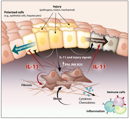
Interleukin (IL)-11 is a member of the IL-6 family of cytokines. Interleukin (IL)-11 is upregulated in a wide variety of fibro-inflammatory diseases such as systemic sclerosis, rheumatoid arthritis, pulmonary fibrosis, inflammatory bowel disease, kidney disease, drug-induced liver injury, and nonalcoholic steatohepatitis. IL-11 is a member of the IL-6 cytokine family and has several distinct properties that define its unique and nonredundant roles in disease. The IL-11 receptor is highly expressed on stromal, epithelial and polarized cells, where noncanonical IL-11 signaling drives the three pathologies common to all fibro-inflammatory diseases—myofibroblast activation, parenchymal cell dysfunction, and inflammation—while also inhibiting tissue regeneration. This cytokine has been little studied, and publications on IL-11 peaked in the early 1990s, when it was largely misunderstood.
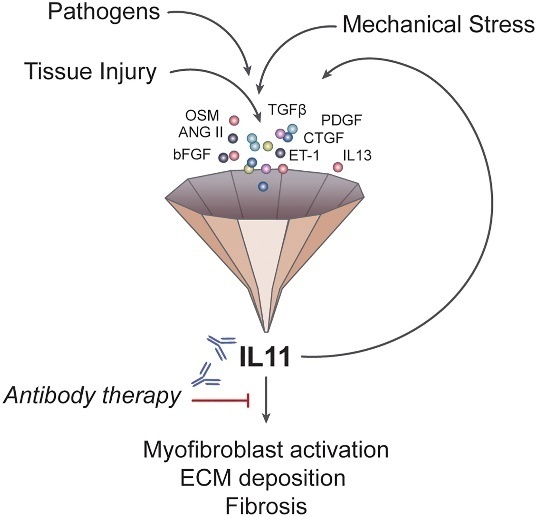
Figure 4. In fibroblasts, IL-11 acts at a point of signalling convergence downstream of multiple and diverse stimuli and is the nexus between pro-fibrotic initiating factors and organ fibrosis. ANG II, angiotensin II; bFGF, basic FGF; CTGF, connective tissue growth factor; ET-1, endothelin 1; OSM, oncostatin MAll these cells release proinflammatory and profibrotic factors such as CD40L, IL-6, transforming growth factor-β1 (TGF-β1), Rantes, and MCP-1, which act synergistically in the progression of DN [47]
IL11 is similar to IL6, and both form a GP130 heterodimer complex to initiate its downstream signaling 25–28, but their respective hexameric signaling complex formation differ 29. While IL6R is expressed most highly on immune cells, IL11RA is expressed in stromal cells, such as fibroblasts and hepatic stellate cells, and also on parenchymal cells, including hepatocytes. Hence, it may be expected that IL6 biology relates mostly to immune functions whereas IL11 activity is more closely linked to the stromal and parenchymal biology 25, 30–32. Since the nineties, high IL11 release during viral infections have been described 33, 34, and more recently, several studies have related this interleukin to fibrosis, chronic inflammation and matrix extracellular remodeling 31, 35–39. It is also known that WNT5A and IL11 have the ability of activating STAT3 signaling 40 and this ability has been postulated as a possible mechanism to link WNT5A gene with immunomodulation. WNT5A is a member of WNT family proteins which plays critical roles in a myriad of processes in both health and disease, such as embryonic morphogenesis, fibrosis, inflammation or cancer 41. Several studies have described a crosstalk between transforming growth factor-beta (TGFβ) and WNT signaling pathways during fibrotic processes 42–45, and more recently with the increase in IL11 production 46. TGFβ represents the most prominent profibrotic cytokine by upregulating production of extracellular matrix (ECM) components and multiple signaling molecules 47.L11 signaling-related genes, such as STAT3 or TGFβ, were also differentially expressed. Subsequently, bioinformatics and functional assays revealed that these four accessory proteins were implicated in both inflammatory and fibrotic responses, suggesting the involvement of ORF6, ORF8, ORF9b and ORF9c in inflammatory and/or fibrotic responses in SARS-CoV-2 infection.

7. Conclusions

In conclusion, inflammatory cytokines could play a crucial role in the pathogenesis of DN. Monitoring of cytokines helps evaluate the immune status inflammation of DN patients and identify those patients at high risk of developing diabetes to select the best therapeutic option. Taking into account 120 patients with diabetes mellitus 2 type infected with SARS-CoV-2 in Uzbekistan, as well as a review of the literature, our observations showed that among patients complicated by diabetes mellitus, severe disease and high mortality rates are observed. Therefore, we set ourselves the task of studying this situation more deeply.

References

[1]  Yuen K.S., Ye Z.W., Fung S.Y., Chan C.P., Jin D.Y. SARS-CoV-2 and COVID-19: The most important research questions. Cell Biosci. 2020; 10: 38–40. doi: 10.1186/s13578-020-00404-4. [PMC free article] [PubMed] [CrossRef] [Google Scholar]
[2]  Lu R., Zhao X., Li J., Niu P., Yang B., Wu H., Wang W., Song H., Huang B., Zhu N., et al. Genomic characterisation and epidemiology of 2019 novel coronavirus: Implications for virus origins and receptor binding. Lancet. 2020; 395: 565–574. doi: 10.1016/S0140-6736(20)30251-8. [PMC free article] [PubMed] [CrossRef] [Google Scholar]
[3]  Walls A.C., Park Y.J., Tortorici M.A., Wall A., McGuire A.T., Veesler D. Structure, Function, and Antigenicity of the SARS-CoV-2 Spike Glycoprotein. Cell. 2020;181:281–292. doi: 10.1016/j.cell.2020.02.058. [PMC free article] [PubMed] [CrossRef] [Google Scholar]
[4]  Zhang L., Lin D., Sun X., Curth U., Drosten C., Sauerhering L., Becker S., Rox K., Hilgenfeld R. Crystal structure of SARS-CoV-2 main protease provides a basis for design of improved α-ketoamide inhibitors. Science. 2020; 368: 409–412. doi: 10.1126/science.abb3405. [PMC free article] [PubMed] [CrossRef] [Google Scholar]
[5]  Rey F.A., Lok S.M. Common Features of Enveloped Viruses and Implications for Immunogen Design for Next-Generation Vaccines. Cell. 2018; 172: 1319–1334. doi: 10.1016/j.cell.2018.02.054. [PMC free article] [PubMed] [CrossRef] [Google Scholar]
[6]  Rochman N.D., Wolf Y.I., Faure G., Mutz P., Zhang F., Koonin E.V. Ongoing global and regional adaptive evolution of SARS-CoV-2. Proc. Natl. Acad. Sci. USA. 2021; 118: 116–121. doi: 10.1073/pnas.2104241118. [PMC free article] [PubMed] [CrossRef] [Google Scholar]
[7]  Kim S., Liu Y., Lei Z., Dicker J., Cao Y., Zhang X.F., Im W. Differential interactions between human ACE2 and spike RBD of SARS-CoV-2 variants of concern. J. Chem. Theory Comput. 2021; 17: 7972–7979. doi: 10.1021/acs.jctc.1c00965. [PMC free article] [PubMed] [CrossRef] [Google Scholar]
[8]  Wu L., Zhou L., Mo M., Liu T., Wu C., Gong C., Lu K., Gong L., Zhu W., Xu Z. SARS-CoV-2 Omicron RBD shows weaker binding affinity than the currently dominant Delta variant to human ACE2. Sig. Transduct. Target. Ther. 2022; 7: 8–12. doi: 10.1038/s41392-021-00863-2. [PMC free article] [PubMed] [CrossRef] [Google Scholar]
[9]  Verity R., Okell L.C., Dorigatti I., Winskill P., Whitttaker C., Imai N., Cuomo-Dannenburg G., Thompson H., Walker P., Fu H., et al. Estimates of the severity of coronavirus disease 2019: A model-based analysis. Lancet Infect. Dis. 2020; 20: 669–677. doi: 10.1016/S1473-3099(20)30243-7. [PMC free article] [PubMed] [CrossRef] [Google Scholar]
[10]  Perez-Saez J., Lauer S., Kaiser L., Regard S., Delaporte E., Guessous I., Stringhini S., Azman A.S., Serocov-POP Study Group Serology-informed estimates of SARS-CoV-2 infection fatality risk in Geneva, Switzerland. Lancet Infect. 2020; 20: E69–E70. doi: 10.1016/S1473-3099(20)30584-3. [PMC free article] [PubMed] [CrossRef] [Google Scholar]
[11]  Salje H., Tran Kiem C., Lefrancq N., Courtejoie N., Bosetti P., Paireau J., Andronico J., Hozé N., Richet J., Dubost C., et al. Estimating the burden of SARS-CoV-2 in France. Science. 2020; 369: 208–211. doi: 10.1126/science.abc3517. [PMC free article] [PubMed] [CrossRef] [Google Scholar]
[12]  Onder G., Rezza G., Brusaferro S. Case-Fatality Rate and Characteristics of Patients Dying in Relation to COVID-19 in Italy. JAMA. 2020; 323: 1775–1776. doi: 10.1001/jama.2020.4683. [PubMed] [CrossRef] [Google Scholar]
[13]  Bhatraju P.K., Ghassemieh B.J., Nichols M., Kim R., Jerome K.R., Nalla A.K., Greninger A.L., Pipavath S., Wurfel M.M., Evans L., et al. Covid-19 in Critically Ill Patients in the Seattle Region—Case Series. N. Engl. J. Med. 2020; 382: 2012–2022. doi: 10.1056/NEJMoa2004500. [PMC free article] [PubMed] [CrossRef] [Google Scholar]
[14]  Williamson E., Walker A.J., Bhaskaran K.J., Bacon S., Bates C., Morton C.E., Curtis H.J., Mehrkar A., Evans D., Inglesby P. OpenSAFELY: Factors associated with COVID-19-related hospital death in the linked electronic health records of 17 million adult NHS patients. Nature. 2020; 584: 430–436. doi: 10.1038/s41586-020-2521-4. [PMC free article] [PubMed] [CrossRef] [Google Scholar]
[15]  Petrilli C.M., Jones S.A., Yang J., Rajagopalan H., O’Donnell L., Chernyak Y., Tobin K.A., Cerfolio R.J., Francois F., Horwitz L.I. Factors associated with hospital admission and critical illness among 5279 people with coronavirus disease 2019 in New York City: Prospective cohort study. BMJ. 2020; 369: m1966. doi: 10.1136/bmj.m1966. [PMC free article] [PubMed] [CrossRef] [Google Scholar]
[16]  Richardson S., Hirsch J.S., Narasimhan M., Crawford J.M., McGinn T., Davidson K.W., the Northwell COVID-19 Research Consortium. Barnaby D.P., Becker L.B., Chelico J.D., et al. Presenting Characteristics, Comorbidities, and Outcomes Among 5700 Patients Hospitalized with COVID-19 in the New York City Area. JAMA. 2020; 323: 2052–2059. doi: 10.1001/jama.2020.6775. [PMC free article] [PubMed] [CrossRef] [Google Scholar]
[17]  Cheng Y., Luo R., Wang K., Zhang M., Wang Z., Dong L., Li J., Yao Y., Ge S., Xu G. Kidney disease is associated with in-hospital death of patients with COVID-19. Kidney Int. 2020; 97: 829–838. doi: 10.1016/j.kint.2020.03.005. [PMC free article] [PubMed] [CrossRef] [Google Scholar]
[18]  Mehra M.R., Desai S.S., Kuy S., Henry T.D., Patel A.N. Cardiovascular Disease, Drug Therapy, and Mortality in Covid-19. N. Engl. J. Med. 2020; 382: 99–102. doi: 10.1056/NEJMc2021225. [PMC free article] [PubMed] [CrossRef] [Google Scholar] Retracted
[19]  Laird J.R., Pareek G., Miner M., Sobel D.W., Balestrieri A., Sfikakis P.P., Tsoulfas G., Protogerou A., Misra D.P., Agarwal V., et al. COVID-19 pathways for brain and heart injury in comorbidity patients: A role of medical imaging and artificial intelligence-based COVID severity classification: A review. Comput. Biol. Med. 2020; 124: 103–960. [PMC free article] [PubMed] [Google Scholar]
[20]  Palaiodimos L., Kokkinidis D.G., Li W., Karamanis D., Ognibene J., Arora S., Southern W.N., Mantzoros C.S. Severe obesity, increasing age and male sex are independently associated with worse in-hospital outcomes, and higher in-hospital mortality, in a cohort of patients with COVID-19 in the Bronx, New York. Metabolism. 2020; 108: 154–262. doi: 10.1016/j.metabol.2020.154262. [PMC free article] [PubMed] [CrossRef] [Google Scholar]
[21]  Cariou B., Hadjadj S., Wargny M., Pichelin M., Al-Salameh A., Allix I. Phenotypic characteristics and prognosis of inpatients with COVID-19 and diabetes: The CORONADO study. Diabetologia. 2020; 63: 1500–1515. doi: 10.1007/s00125-020-05180-x. [PMC free article] [PubMed] [CrossRef] [Google Scholar]
[22]  Gregory J.M., Slaughter J.C., Duffus S.H., Smith T.J., LeStourgeon L.M., Jaser S.S. COVID-19 severity is tripled in the diabetes community: A prospective analysis of the pandemic’s impact in type 1 and type 2 diabetes. Diabetes Care. 2021; 44: 526–532. doi: 10.2337/dc20-2260. [PMC free article] [PubMed] [CrossRef] [Google Scholar]
[23]  Barron E., Bakhai C., Kar P., Weaver A., Bradley D., Ismail H., Knighton P., Holman N., Khunti K., Sattar N., et al. Associations of Type 1 and Type 2 Diabetes With COVID-19-Related Mortality in England: A Whole-Population Study. Lancet Diabetes Endocrinol. 2020; 8: 813–822. doi: 10.1016/S2213-8587(20)30272-2. [PMC free article] [PubMed] [CrossRef] [Google Scholar]
[24]  Holman N., Knighton P., Kar P., O’Keefe J., Curley M., Weaver A., Barron E., Bakhai C., Khunti K., Wareham N., et al. Risk Factors for COVID-19-Related Mortality in People with Type 1 and Type 2 Diabetes in England: A Population-Based Cohort Study. Lancet Diabetes Endocrinol. 2020; 8: 823–833. doi: 10.1016/S2213-8587(20)30271-0. [PMC free article] [PubMed] [CrossRef] [Google Scholar]
[25]  Wang S., Ma P., Zhang S., Song S., Wang Z., Ma Y., Xu J., Wu F., Duan L., Yin Z., et al. Fasting Blood Glucose at Admission is an Independent Predictor for 28-Day Mortality in Patients With COVID-19 Without Previous Diagnosis of Diabetes: A Multi-Centre Retrospective Study. Diabetologia. 2020; 63: 2102–2111. doi: 10.1007/s00125-020-05209-1. [PMC free article] [PubMed] [CrossRef] [Google Scholar]
[26]  Wu J., Huang J., Zhu G., Wang Q., Lv Q., Huang Y., Yu Y., Si X., Wang C., Liu Y., et al. Elevation of Blood Glucose Level Predicts Worse Outcomes in Hospitalized Patients With COVID-19: A Retrospective Cohort Study. BMJ Open Diabetes Res. Care. 2020; 8: 14–76. doi: 10.1136/bmjdrc-2020-001476. [PMC free article] [PubMed] [CrossRef] [Google Scholar]
[27]  Coppelli A., Giannarelli R., Aragona M., Penno G., Falcone M., Tiseo G., Ghiadoni Z., Greta Barbieri L., Monzani F., Virdis A., et al. Hyperglycemia at Hospital Admission is Associated with Severity of the Prognosis in Patients Hospitalized for COVID-19: The Pisa COVID-19 Study. Diabetes Care. 2020; 43: 2345–2348. doi: 10.2337/dc20-1380. [PubMed] [CrossRef] [Google Scholar]
[28]  Bode B., Garrett V., Messler J., McFarland R., Crowe J., Booth R., Klonoff D. Glycemic Characteristics and Clinical Outcomes of COVID-19 Patients Hospitalized in the United States. J. Diabetes Sci. Technol. 2020; 14: 813–821. doi: 10.1177/1932296820924469. [PMC free article] [PubMed] [CrossRef] [Google Scholar]
[29]  Zhu L., She Z.G., Cheng X., Qin J., Zhang X.J., Cai J., Lei F., Wang H., Xie J., Wang W., et al. Association of Blood Glucose Control and Outcomes in Patients with COVID-19 and Pre-Existing Type 2 Diabetes. Cell Metab. 2020; 31: 1068–1077. doi: 10.1016/j.cmet.2020.04.021. [PMC free article] [PubMed] [CrossRef] [Google Scholar]
[30]  Filippatos T.D., Liontos A., Papakitsou I., Elisaf M.S. SGLT2 inhibitors and cardioprotection: A matter of debate and multiple hypotheses. Postgrad. Med. 2019; 131: 82–88. doi: 10.1080/00325481.2019.1581971. [PubMed] [CrossRef] [Google Scholar]
[31]  Bornstein S.R., Rubino F., Khunti K., Mingrone G., Hopkins D., Birkenfeld A.L., Boehm B., Amiel S., Holt R.I., Skyler J.S. Practical recommendations for the management of diabetes in patients with COVID-19. Lancet Diabetes Endocrinol. 2020; 8: 546–550. doi: 10.1016/S2213-8587(20)30152-2. [PMC free article] [PubMed] [CrossRef] [Google Scholar]
[32]  Hampp C., Swain R.S., Horgan C., Dee E., Qiang Y., Dutcher S.K., Petrone A., Chen Tilney R., Maro J.C., Panozzo C.A. Use of Sodium-Glucose Cotransporter 2 Inhibitors in Patients with Type 1 Diabetes and Rates of Diabetic Ketoacidosis. Diabetes Care. 2020; 43: 90–97. doi: 10.2337/dc19-1481. [PubMed] [CrossRef] [Google Scholar]
[33]  Mota M., Stefan A. Covid-19 and Diabetes-A Bidirectional Relationship? Rom. J. Diabetes Nutr. Metab. Dis. 2020; 27: 77–79. [Google Scholar]
[34]  Wan Y., Shang J., Graham R., Baric R.S., Li F. Receptor Recognition by the Novel Coronavirus from Wuhan: An Analysis Based on Decade-Long Structural Studies of SARS Coronavirus. J. Virol. 2020; 94: 120–127. doi: 10.1128/JVI.00127-20. [PMC free article] [PubMed] [CrossRef] [Google Scholar]
[35]  Erkan C., Medine C.C. Comment on ‘Can angiotensin receptor-blocking drugs perhaps be harmful in the COVID-19 pandemic? J. Hypertens. 2020; 38: 1189–1198. [PMC free article] [PubMed] [Google Scholar]
[36]  Pal R., Bhansali A. COVID-19, diabetes mellitus and ACE2: The conundrum. Diabetes Res. Clin. Pract. 2020; 162: 108132. doi: 10.1016/j.diabres.2020.108132. [PMC free article] [PubMed] [CrossRef] [Google Scholar]
[37]  Esler M., Esler D. Can angiotensin receptor-blocking drugs perhaps be harmful in the COVID-19 pandemic? J. Hypertens. 2020; 38: 781–782. doi: 10.1097/HJH.0000000000002450. [PubMed] [CrossRef] [Google Scholar]
[38]  Romaní-Pérez M., Outeiriño-Iglesias V., Moya C.M., Santisteban P., González-Matías L.C., Vigo E., Mallo F. Activation of the GLP-1 Receptor by Liraglutide Increases ACE2 Expression, Reversing Right Ventricle Hypertrophy, and Improving the Production of SP-A and SP-B in the Lungs of Type 1 Diabetes Rats. Endocrinology. 2015; 156: 3559-3569. doi: 10.1210/en.2014-1685. [PubMed] [CrossRef] [Google Scholar]
[39]  Zhang W., Li C., Liu B., Wu R., Zou N., Xu Y.-Z., Yang Y.-Y., Zhang F., Zhou H.-M., Wan K.-Q. Pioglitazone upregulates hepatic angiotensin converting enzyme 2 expression in rats with steatohepatitis. Ann. Hepatol. 2013; 12: 892–900. doi: 10.1016/S1665-2681(19)31294-3. [PubMed] [CrossRef] [Google Scholar]
[40]  Ali R.M., Al-Shorbagy M.Y., Helmy M.W., El-Abhar H.S. Role of Wnt4/β-catenin, Ang II/TGFβ, ACE2, NF-κB, and IL-18 in attenuating renal ischemia/reperfusion-induced injury in rats treated with Vit D and pioglitazone. Eur. J. Pharmacol. 2018; 831: 68–76. doi: 10.1016/j.ejphar.2018.04.032. [PubMed] [CrossRef] [Google Scholar]
[41]  Viswanathan V., Puvvula A., Jamthikar A.D., Saba L., MJohri A., Kotsis V., Khanna N.N., KDhanjil S., Majhail M., Prasanna Misra D., et al. Bidirectional link between diabetes mellitus and coronavirus disease 2019 leading to cardiovascular disease: A narrative review. World J. Diabetes. 2021; 12: 215–237. doi: 10.4239/wjd.v12.i3.215. [PMC free article] [PubMed] [CrossRef] [Google Scholar]
[42]  Knapp S. Diabetes and infection: Is there a link?—A mini-review. Gerontology. 2013; 59: 99–104. doi: 10.1159/000345107. [PubMed] [CrossRef] [Google Scholar]
[43]  Moutschen M., Scheen A., Lefebvre P. Impaired immune responses in diabetes mellitus: Analysis of the factors and mechanisms involved. Relevance to the increased susceptibility of diabetic patients to specific infections. Diabete Metab. 1992; 18: 187. [PubMed] [Google Scholar]
[44]  Geerlings S.E., Hoepelman A.I. Immune dysfunction in patients with diabetes mellitus (DM) FEMS Immunol. Med. Microbiol. 1999; 26: 259–265. doi: 10.1111/j.1574-695X.1999.tb01397.x. [PubMed] [CrossRef] [Google Scholar]
[45]  Fernandez C., Rysä J., Almgren P., Nilsson J., Engström G., Orho-Melander M., Ruskoaho H., Melander O. Plasma levels of the proprotein convertase furin and incidence of diabetes and mortality. J. Intern. Med. 2018; 284: 377–387. doi: 10.1111/joim.12783. [PMC free article] [PubMed] [CrossRef] [Google Scholar]
[46]  Hoffmann M., Kleine-Weber H., Schroeder S., Krüger N., Herrler T., Erichsen S., Schiergens T.S., Herrler G., Wu N.H., Nitsche A., et al. SARS-CoV-2 Cell Entry Depends on ACE2 and TMPRSS2 and Is Blocked by a Clinically Proven Protease Inhibitor. Cell. 2020; 181: 271–280. doi: 10.1016/j.cell.2020.02.052. [PMC free article] [PubMed] [CrossRef] [Google Scholar]
[47]  Huang J., Xiao Y., Zheng P., Zhou W., Wang Y., Huang G., Xu A., Zhou Z. Distinct neutrophil counts and functions in newly diagnosed type 1 diabetes, latent autoimmune diabetes in adults, and type 2 diabetes. Diabetes Metab. Res. Rev. 2019; 35: 30–64. doi: 10.1002/dmrr.3064. [PubMed] [CrossRef] [Google Scholar].