American Journal of Medicine and Medical Sciences

p-ISSN: 2165-901X    e-ISSN: 2165-9036

2023;  13(12): 1980-1984

doi:10.5923/j.ajmms.20231312.34

Received: Nov. 25, 2023; Accepted: Dec. 13, 2023; Published: Dec. 20, 2023


Effects of Dapagliflozin on Cardiac Functional Status in Patients with Chronic Heart Failure

Abdigaffar G. Gadaev1, Guzel B. Shamsutdinova2, Oybek Z. Abdukholikov1

1Tashkent Medical Academy, Uzbekistan

2Fergana Public Health Medical Institute, Uzbekistan

Copyright © 2023 The Author(s). Published by Scientific & Academic Publishing.

This work is licensed under the Creative Commons Attribution International License (CC BY).
http://creativecommons.org/licenses/by/4.0/

Abstract

In the article, the effects of standard medical treatments with different content on intracardiac hemodynamics and N-pro natriuretic peptide in patients with chronic heart failure were studied by comparing the subjects in three groups. A highly reliable increase in left ventricular end-diastolic size and diastolic volume and ejection fraction was found in the third group of patients. This confirms that sacubitril+valsartan and dapagliflozin have a high cardioprotective effect.

Keywords: Chronic heart failure, N-pro natriuretic peptide, Succubitril+valsartan, Dapagliflozin

Cite this paper: Abdigaffar G. Gadaev, Guzel B. Shamsutdinova, Oybek Z. Abdukholikov, Effects of Dapagliflozin on Cardiac Functional Status in Patients with Chronic Heart Failure, American Journal of Medicine and Medical Sciences, Vol. 13 No. 12, 2023, pp. 1980-1984. doi: 10.5923/j.ajmms.20231312.34.

1. Introduction

Chronic heart failure (CHF) is one of the urgent medical and social problems of modern medicine [19,12,15]. This is due to its prevalence, severe consequences, and high cost of treatment [14,4,10].
Mortality due to CHF is 4-8 times higher than in the general population, and half of patients die within 5 years of diagnosis. Its IV functional class (FC) has a half-year mortality rate of 44% [21,24,1,11,17,16].
According to epidemiological data, in the Russian Federation and European countries, in most cases, CHF develops as a result of arterial hypertension (95%) and ischemic heart disease (IHD) (69.7%). In our republic, the main cause of this serious complication is often the two diseases listed above [18,2].
Due to the increase in the life expectancy of the population, the positive results achieved in the treatment of cardiovascular diseases, and the prevalence of risk factors that cause IHD and hypertension (HK), which are the main diseases that cause CHF, this serious complication is becoming more and more common among the world’s population [3,5,22,23]. Despite the progress made in recent years, this confirms that CHF still remains a heavy financial burden on the health economy of all countries around the world.
Systemic changes are observed in all organs of CHF, and remodeling processes in the heart are of particular importance [20].
It is known that a number of examination methods are used in the diagnosis and evaluation of the effectiveness of treatment of CHF. Among them, natriuretic hormones are of particular importance as a biological marker. Currently, there are a number of its representatives, among which brain and N-pro brain sodium uretic peptides are widely used in the diagnosis and evaluation of CHF.
A.M. Richards was the first to show the use of concentration of N-prosodium uretic peptide in blood to control the effect of treatment in patients suffering from CHF. In it, monitoring was carried out by titrating the dose of angiotensin-converting enzyme inhibitors (ACEI) in patients diagnosed with CHF II-III FC under hormonal control, and the appropriateness of such an approach was shown [13].
In the IMPRESS trial, which included 573 patients receiving lisinopril and omapatrilat, those with CHF and left ventricular ejection fraction less than 40% had reliable reductions in neurohormone 1-2 years after initiation of treatment in a randomized trial [8].
Similar data were obtained in experimental observations conducted by S. Tang and co-authors on patients receiving valsartan and benazepril [16].
Also, in a series of observations, a decrease in brain sodium uretic peptide in the blood was found in patients taking β-blockers [7].
Taking into account the above, we studied N pro brain sodium uretic hormone and cardiac hemodynamics in our patients before and after treatment with different components.

2. The Purpose of the Study

Studying the effects of various treatments on serum N pro brain sodium uretic hormone and cardiac functional status in patients with chronic heart failure.

3. Research Materials and Methods

This scientific research work was conducted in 2022 and 2023 in Fergana Public Health Medical Institute and private clinic “Farovon” in 120 patients with developed CHF on the basis of UIK and arterial hypertension (AG). They, in turn, were divided into three groups based on the treatment procedures. Each group consisted of 40 patients, 20 of which consisted of CHF II and III FC. The average age of the first group of patients was 66.1±1.8, men were 21 (52.5%) and women were 19 (47.5%). In the 1st group of patients under observation, the number of those who underwent myocardial infarction (MI) - 28 (70%), those who underwent aortic coronary bypass surgery (ACS) or stenting - 11 (27.5%), rhythm disturbances and blockades were recorded - 12 (30%), AG there were 31 (77.5%), 17 (42.5%) people with various degrees of obesity, and 21 (52.5%) people with anemia. This group was prescribed β-blockers + AAFIs or angiotensin receptor blockers (ARBs) + mineralocorticoid receptor antagonists (MRKA)-veroshpirone as the standard treatment for CHF. The average age of the second group of patients was 65.9±1.5, men were 24 (60%) and women were 16 (40%). In this group, the number of those who underwent MI - 25 (62.5%), those who underwent ACS or stenting - 13 (32.5%), those who had rhythm disturbances and blockades - 15 (37.5%), those who had AG 27 (67.5%), those who were found to be obese in various degrees - 16 (40%), anemia was observed - 19 (47.5%) people. They received a standard treatment consisting of β-blockers + succubitril-valsartan (yuperio) + MRKA-veroshpirone. The average age of the third group of patients was 64.7±1.3, 21 (52.5%) of them were men and 19 (47.5%) were women. In this group, the number of those who underwent MI - 27 (67.5%), those who underwent aortic coronary bypass surgery or stenting - 16 (40%), those who had rhythm disorders and blockades - 17 (42.5%), those who had AF 25 (62.5%), of various degrees 15 (37.5%) were diagnosed with obesity, 19 (47.5%) were anemic. β-blockers + succubitril-valsartan (yuperio) + MRKA-veroshpiron + glucose-sodium co-transporter type 2 inhibitors (dapagliflozin/forsiga) were recommended to these patients.
Particular attention was paid to the same number of patients in II and III FC in each group and their representativeness.
In addition to routine laboratory tests, serum N-pro natriuretic peptide indicators were determined in all subjects enrolled in the study, and cardiac functional status was assessed using EXOKG.
The amount of N-pro-natriuretic peptide in blood serum was determined using immunoenzyme analysis using “Vector-BEST” (Russia) reagents. The reagent used for the determination of N-pro natriuretic peptide in the blood serum in the study had a detection range of 0-2500 pg/ml and a sensitivity of 20.0 pg/ml.
Analysis and discussion of research results. We studied N-pro-brain sodium uretic peptide indicators in blood serum in all groups of patients under our observation before and after treatments. Figure 1 below shows a comparative analysis of pre- and post-treatment serum levels of N-pro brain natriuretic peptide.
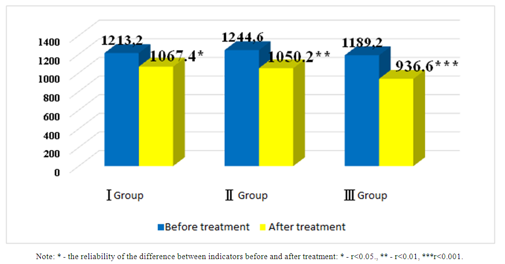
Figure 1. Comparative analysis of pre- and post-treatment N-pro brain sodium uretic peptide indicators in patients with chronic heart failure (pg/ml)
No reliable differences (R>0.05) were observed between the groups when the N-pro brain sodium uretic peptide indicator was compared between the groups before the treatments.
In the first group of patients, the level of N-pro brain sodium uretic peptide in the blood serum decreased by 1.14 times from 1213.2±40.2 pg/ml to 1067.4±36.4 pg/ml before and after treatments, and a reliable difference was noted (r<0.05). In the second group, its amount before treatment was 1244.6±47.2 pg/ml and after it was 1.2 times decreased to 1050.2±39.8 pg/ml, and a highly reliable (r<0.01) difference was found. N-pro brain sodium uretic peptide index was 1189.2±32.5 pg/ml to 936.6 in group III who received standard treatment consisting of β-blockers + sacubitril-valsartan (yuperio) + MRKA + glucose-sodium co-transporter type 2 inhibitors (dapagliflozin/forsiga). It decreased 1.3 times to ±29.3 pg/ml and a highly reliable (r<0.001) difference was observed.
During the six-month prospective follow-up of the patients after the treatment, the functional state of the heart was also studied and the obtained results were evaluated in comparison with the indicators before the treatments. Table 1 below presents a comparative analysis between groups of pre- and post-treatment cardiac functional status indicators in patients with CHF enrolled in the study.
Table 1. Comparative analysis of heart functional status indicators after standard medical treatments with different content in patients with chronic heart failure
     
As shown in the table, the size of the left lobe in the first group after the treatment was 42.2±1.4 mm before and after the treatment. from 39.5±1.5mm. decreased to, but no reliable difference was detected (r>0.05). 42.6±1.5 mm after treatment in the second group. from 38.1±1.2 mm. reliable (r<0.05) and 41.8±1.4 mm in the third group. from 36.2±1.3 mm. highly reliable (r<0.01) decrease. Left ventricular end-systolic size before treatment was 4.6±0.16 and 4.7±0.15 cm in the first and second groups, respectively. was equal to 4.1 ± 0.14 and 4.2 ± 0.13 cm after the treatments, and a reliable (r<0.05) difference was noted. In the third group, it improved by 1.23 times to 4.8±0.15 cm before treatment and 3.9±0.2 cm after treatment, and a highly reliable difference was observed (r<0.01). In the first group of patients who received β-blockers + AAFI or ARA + MRKA, the left ventricular end-diastolic size decreased from 5.8±0.13 cm to 5.5±0.12 cm, and no reliable difference was noted (r>0.05). In patients receiving the second β-blockers + sacubitril-valsartan (yuperio) + MRKA, its size improved by 10% from 6.1±0.2 cm to 5.5±0.15 cm and a reliable difference was found (r<0.05). In the third group of patients who were prescribed β-blockers + sacubitril-valsartan (yuperio) + MRKA + glucose-sodium co-transporter type 2 inhibitors (dapagliflozin/forsiga), the left ventricular end-diastolic size changed positively by 18% after treatments and a highly reliable difference was noted (from 6.3±0.16 to 5.2±0.2 cm, r<0.001).
Left ventricular end-diastolic volume improved from 186.6±5.8ml to 161.4±6.3ml and from 188.2±6.4ml to 158.8±6.6ml before and after treatment in the first and second groups, respectively. Differences in both groups were reliable (r<0.01). In the third group, a highly reliable difference (r<0.001) was found, which decreased by 1.27 times to 190.5±6.5 ml before treatment and 150.2±5.8 ml after treatment.
Left ventricular end-systolic volume improved by 15% from 90.4±5.2ml to 76.5±5.6ml in the first group before and after treatment, but no significant difference was noted (r>0.05). In the second group, a reliable difference of 20.5% was found, from 92.6±5.8 ml to 73.5±5.5 ml. In the third group, left ventricular end systolic volume changed from 89.4±5.6 ml to 68.2±5.2 ml by 23.7% and a reliable difference was observed (р<0.01). After treatment, the left ventricular ejection fraction increased from 40.6±1.8% to 47.8±2.0% in the first group (r<0.05), from 39.6±1.8% to 48.2±2.2% in the second group (r<0.01) and in the third group, a highly reliable (r<0.001) increase was noted from 38.2±1.6% to 51.5±1.4%. Left ventricular myocardium weight before and after treatment in the first group increased from 204.2±10.2 to 186.5±9.8 g, from 205.6±10.7 g to 182.5±11.3 g in the second group, and from 202.6±9.9 g in the third group. Although there was a significant decrease of 176.2±10.4 g, the changes in all groups were not reliable (r>0.05).
The correlation between N-pro-natriuretic peptide and left ventricular ejection fraction was studied in the patients included in the study, and it is presented in Figure 2 below.
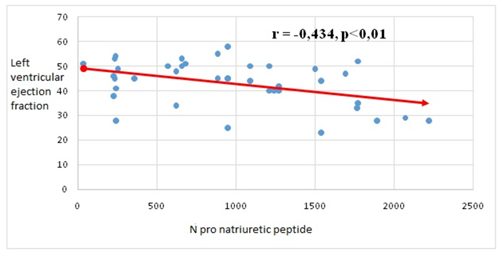
Figure 2. Correlation between N-pro natriuretic peptide and left ventricular ejection fraction in patients with chronic heart failure
As shown in the figure, a moderately strong negative (r = -0.434, p<0.01) correlation was found between the left ventricular ejection fraction and N pro-natriuretic peptide. This indicates a decrease in the ejection fraction of the left ventricle of the heart in parallel with the increase of the studied peptide.
In the analysis, highly reliable positive changes in intracardiac hemodynamics, in particular, end-systolic and diastolic volume and left ventricular ejection fraction, were found in the third group compared to the other two groups. The obtained results showed that the combined use of sacubitril-valsartan (yuperio) and glucose-sodium co-transporter type 2 inhibitors (dapagliflozin) as part of the standard treatment in patients with CHF is highly effective. It also has a positive effect on the restoration of the functional state of the heart by drugs belonging to the group of inhibitors of glucose sodium cotransporter type 2.

4. Conclusions

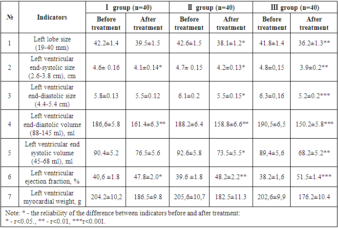
After treatments, positive changes in N-pro natriuretic peptide indicators were observed in all groups of patients. But in the third group, changes in its indicators at a highly reliable level confirm the effect of sacubitril + valsartan and dapagliflozin (Forsiga) on the functional state of the heart compared to the first two groups. Also, highly reliable positive changes in systolic and diastolic volume and left ventricular ejection fraction were observed in the group containing glucose-sodium co-transporter type 2 inhibitors (dapagliflozin), which showed that it is important in stabilizing cardiac hemodynamics.

References

[1]  Alzahrani, S., Alosaimi, M., Malibarey, W. M., Alhumaidi, A. A., Alhawaj, A. H., Alsulami, N. J., Alsharari, A. S., Alyami, A. A., Alkhateeb, Z. A., Alqarni, S. M. et al. (2019). Saudi Family Physicians' Knowledge of Secondary Prevention of Heart Disease: A National Assessment Survey. Archieves of Pharmacy Practice, 10(4), 54-60.
[2]  Anavekar NS, McMurray JJ, Velazquez EJ et al. Relation between renal dysfunction and cardiovascular outcomes after myocardial infarction. N Engl J Med 2004; 351: 1285-1295.
[3]  Bayliss E. A. [et al.] Description of barriers to self-care by per- sons with comorbid chronic diseases // Annals of Family Medicine. – 2003. – Vol. 1, № 1. – P. 15–21.
[4]  Benjamin EJ, Blaha MJ, Chiuve SE, Cushman M, Das SR, Deo R, de Ferranti SD, Floyd J, Fornage M, Gillespie C, Isasi CR, Jiménez MC, Jordan LC, Judd SE, Lackland D, Lichtman JH, Lisabeth L, Liu S, Longenecker CT, Mackey RH, Matsushita K, Mozaffarian D, Mussolino ME, Nasir K, Neumar RW, Palaniappan L, Pandey DK, Thiagarajan RR, Reeves MJ, Ritchey M, Rodriguez CJ, Roth GA, Rosamond WD, Sasson C, Towfighi A, Tsao CW, Turner MB, Virani SS, Voeks JH, Willey JZ, Wilkins JT, Wu JH, Alger HM, Wong SS, Muntner P; American Heart Association Statistics Committee and Stroke Statistics Subcommittee. Heart Disease and Stroke Statistics-2017 Update: A Report From the American Heart Association. Circulation. 2017 Mar 7; 135(10): e146-e603. doi: 10.1161/CIR.0000000000000485.
[5]  Bhatt A.S., Ambrosy A.P., Dunning A., DeVore A.D., Butler J., Reed S., Voors A., Starling R., Armstrong P.W., Ezekowitz J.A., Metra M., Hernandez A.F., O'Connor C.M., Mentz R.J. The burden of non-cardiac comorbidities and association with clinical outcomes in an acute heart failure trial - insights from ASCEND-HF. Eur J Heart Fail. 2020 Jun; 22(6): 1022-1031.
[6]  Bui, A. L., Horwich, T. B., & Fonarow, G. C. (2011). Epidemiology and risk profile of heart failure. Nature reviews. Cardiology, 8(1), 30-41.
[7]  Davis M.E., Richards A.M., Nicholls M.G., Yandle T.G., Frampton C.M., Troughton R.W. Introduction of metoprolol increases plasma B-type cardiac natriuretic peptides in mild, stable heart failure. Circulation 2006; 113(7): 977–985.
[8]  Eisenstein E.L., Nelson C.L., Simon T.A., Smitten A.L., Lapuerta P., Mark D.B. Vasopeptidase inhibitor reduces inhospital costs for patients with congestive heart failure: results from the IMPRESS trial. Inhibition of Metallo Protease by BMS-186716 in a Randomized Exercise and Symptoms Study in Subjects with Heart Failure. Am Heart J 2002; 143(6): 1112–1117.
[9]  Groenewegen, A., Rutten, F. H., Mosterd, A., & Hoes, A. W. (2020). Epidemiology of heart failure. European Journal of Heart Failure, 22(8), 1342-1356.
[10]  Lam CSP, Chandramouli C, Ahooja V, Verma S. SGLT-2 Inhibitors in Heart Failure: Current Management, Unmet Needs, and Therapeutic Prospects. J Am Heart Assoc. 2019 Oct 15; 8(20): e013389. doi: 10.1161/JAHA.119.013389.
[11]  Permadi, A. W., Hartono, S., Wahjuni, E. S., & Lestari, N. K. D. (2020). The Combination of Physical Exercise Programs in Patients with Heart Failure. International Journal of Pharmaceutical and Phytopharmacological Research, 10(1), 22-28.
[12]  Reibis R., Jannowitz C., Halle M., Pittrow D., Gitt A., Völler H. Management and outcomes of patients with reduced ejection fraction after acute myocardial infarction in cardiac rehabilitation centers. Curr Med Res Opin 2015; 31(2): 211–219.
[13]  Richards A.M. Variability of NT-proBNP levels in heart failure: implications for clinical application. Heart 2007; 93(8): 899–900.
[14]  Seferović PM, Petrie MC, Filippatos GS, Anker SD, Rosano G, Bauersachs J, Paulus WJ, Komajda M, Cosentino F, de Boer RA, Farmakis D, Doehner W, Lambrinou E, Lopatin Y, Piepoli MF, Theodorakis MJ, Wiggers H, Lekakis J, Mebazaa A, Mamas MA, Tschöpe C, Hoes AW, Seferović JP, Logue J, McDonagh T, Riley JP, Milinković I, Polovina M, van Veldhuisen DJ, Lainscak M, Maggioni AP, Ruschitzka F, McMurray JJV. Type 2 diabetes mellitus and heart failure: a position statement from the Heart Failure Association of the European Society of Cardiology. Eur J Heart Fail. 2018 May; 20(5): 853-872. doi: 10.1002/ejhf.1170. Epub 2018 Mar 8. PMID: 29520964.
[15]  Shiba N, Shimokawa H. Chronic kidney disease and heart failure--Bidirectional close link and common therapeutic goal. J Cardiol. 2011 Jan; 57(1): 8-17.
[16]  Tang S., Peng D., Hu Y., Chen J. Protective effects of valsartan and benazepril combined with atorvastatin on cardiorenal syndrome in rats. Eur Rev Med Pharmacol Sci 2015; 19(5): 759–766.
[17]  Virani, S. S., Alonso, A., Aparicio, H. J., Benjamin, E. J., Bittencourt, M. S., Callaway, C. W., Carson, A. P., Chamberlain, A. M., Cheng, S., Delling, F. N. et al. (2021). Heart Disease and Stroke Statistics-2021 Update: A Report from the American Heart Association. Circulation. 143(8): e254-e743.
[18]  Batyushin M.M., Vrublevskaya N.S. Clinical manifestations of kidney damage in chronic heart failure. Nephrology. 2010; 14(4): 27-30.
[19]  Berezikova Ekaterina Nikolaevna, Pustovetova Maria Gennadievna, Shilov Sergey Nikolaevich, Efremov Anatoly Vasilievich, Teplyakov Alexander Trofimovich, Safronov Igor Dmitrievich, & Samsonova Elena Nikolaevna (2014). Prognostic role of metabolic risk factor (hyperhomocysteinemia) in the development of chronic heart failure. Circulatory Pathology and Cardiac Surgery, 18 (1), 20-25.
[20]  Vasyuk YA. Possibilities and limitations of echocardiographic research in assessing left ventricular remodeling in CHF. Heart failure 2003; 4(2):107-1103.
[21]  Some unresolved issues of chronic heart failure / Ed. S.N. Tereshchenko. – M., 2007.
[22]  Ponikovich P., Voors A.A., Anker S.D., Bueno H., Cleland J.F., Kots A.J. et al. ESK recommendations for the diagnosis and treatment of acute and chronic heart failure 2016. Russia. cardiol. magazine 2017; 1(141): 7–81.
[23]  Preobrazhensky D.V. Anemia in chronic heart failure: a look at pathogenesis and ways of correction. Cardiology Therapy No. 2 (157), 2019.
[24]  Storozhakov G.I., Gendlin G.E., Reznik E.B. If the heart hurts, the kidneys suffer: cardiorenal syndrome in chronic heart failure // General Medicine. – 2009. – No. 1. – P. 27-36.