-
Paper Information
- Next Paper
- Previous Paper
- Paper Submission
-
Journal Information
- About This Journal
- Editorial Board
- Current Issue
- Archive
- Author Guidelines
- Contact Us
American Journal of Medicine and Medical Sciences
p-ISSN: 2165-901X e-ISSN: 2165-9036
2023; 13(12): 1946-1948
doi:10.5923/j.ajmms.20231312.27
Received: Nov. 26, 2023; Accepted: Dec. 16, 2023; Published: Dec. 18, 2023

Analysis of the Research Results: Comparative Morphology of the Liver Nervous System of Mammal Animals by Type of Food
Shodiyarova Dilfuza Saydullayevna1, Boykuziyev Xayitboy Xudoyberdiyevich2, Ortikova Yulduzxon Oldilxon Kizi3
1Phd Student, Department of Histology, Cytology and Embryology, Samarkand State Medical University, Samarkand, Uzbekistan
2Candidate of Medical Science, Associate Professor, Department of Histology, Cytology and Embryology, Samarkand State Medical University, Samarkand, Uzbekistan
3Student, Faculty of Preventive Medicine, Samarkand State Medical University, Samarkand, Uzbekistan
Copyright © 2023 The Author(s). Published by Scientific & Academic Publishing.
This work is licensed under the Creative Commons Attribution International License (CC BY).
http://creativecommons.org/licenses/by/4.0/

In this article, the comparative morphology of the nervous system of the liver of rabbits, representing herbivorous mammals, and dogs, representatives of carnivorous mammals, was studied, and the results of the study were analyzed. Livers of 10 adult rabbits and 10 dogs were studied for the study. Research results show that the nervous system of the liver of mammals with different types of feed has specific morphological and morphometric characteristics depending on the type of feed [1,3,6,8,11,12,13,14,15].
Keywords: Food type, Liver of mammals, Adrenergic and cholinergic nervous system of the liver
Cite this paper: Shodiyarova Dilfuza Saydullayevna, Boykuziyev Xayitboy Xudoyberdiyevich, Ortikova Yulduzxon Oldilxon Kizi, Analysis of the Research Results: Comparative Morphology of the Liver Nervous System of Mammal Animals by Type of Food, American Journal of Medicine and Medical Sciences, Vol. 13 No. 12, 2023, pp. 1946-1948. doi: 10.5923/j.ajmms.20231312.27.
Article Outline
1. Introduction
- The role of the liver in the process of digestion is very large. Because dozens of complex biochemical reactions take place in it at the same time, and these processes do not interfere with each other. The management system is important in the implementation of such complex biochemical processes [2,4,5,7,9,10]. Studying the morphological and functional bases that show the importance of the nervous system in managing such complex and systematic processes is one of the urgent problems of our medicine today. Therefore, we set out to study the unexplored aspects of this problem.
2. Methods
- 10 adult rabbit and 10 dog livers were studied. The obtained material was frozen (fixed) in 12% formalin. Sections taken from the prepared paraffin blocks were studied by staining using Bilshovsky-Gross, Karnovsky-Rutz methods. To identify adrenergic nerve fibers, unhardened cryostat sections were processed in a 2% solution of glyoxylic acid according to the method of V.N. Shvalev and N.I. Juchkova (1979) and studied under a luminescent LUMAM-I2 microscope. The obtained morphological and morphometric data were analyzed and relevant conclusions were drawn.
3. Analysis and Result
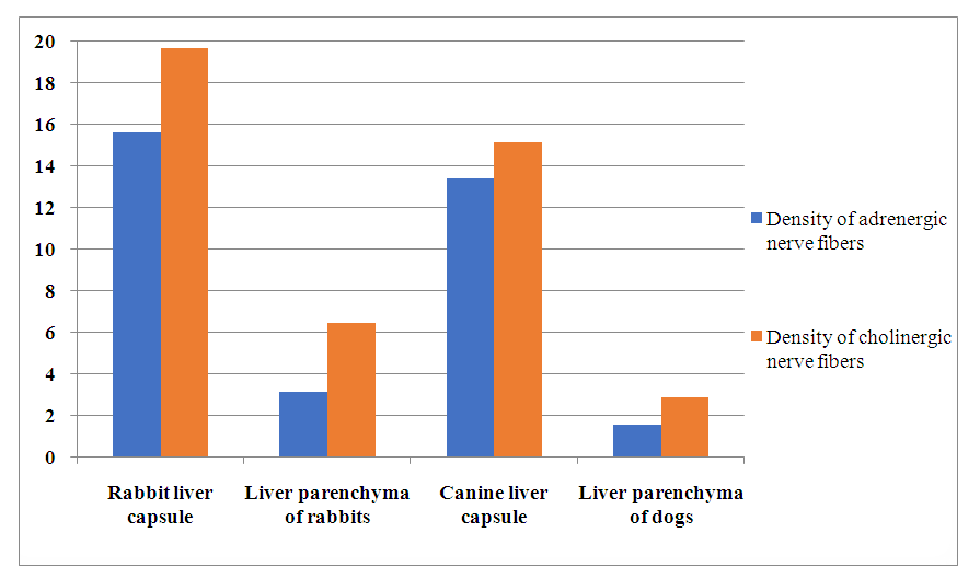
- Cryostat sections of rabbit liver treated with a 2% solution of glyoxylic acid, all elements of adrenergic nerve fibers can be seen under a fluorescent microscope. We find adrenergic nerve fibers in all parts of the liver of rabbits.However, in Glisson’s capsule of the liver, adrenergic nerve fibers are very dense compared to its blood vessels, bile ducts and parenchyma. In the capsule of the liver of rabbits, adrenergic nerve fibers are located in the form of large, medium, small bundles or in the form of individual fibers. These bundles are located along the wall of blood vessels and form different tangles of different sizes. During the division of Glisson’s capsule and blood vessels into smaller blood vessels, adrenergic nerve fibers are also divided into smaller bundles along the direction of the divided blood vessels and form a thick mesh along the wall of these blood vessels. Because adrenergic nerve fibers contain fluorogenic amines (catecholamines), they emit a bright blue-green light. Sometimes, these colors remind of the northern rain. In cases where fibers in large bundles are very close to each other or overlap, these light rays merge and appear in the form of long light-emitting corridors. Separate fibers of the adrenergic nervous system coming out of large bundles penetrate into the surrounding tissue or inside the wall of blood vessels, forming a smaller mesh in its muscular layer.Smaller bundles of adrenergic nerve fibers branch off from larger bundles of interstitial connective tissue and form a thick mesh along the walls of interstitial arteries and veins and bile ducts. The individual fibers coming out of this mesh go inside the lobes and go towards the central vein between the liver plates and sinusoidal capillaries. In some cases, these fibers end by forming different-shaped expansions near the cells of the liver plate or the wall of sinusoidal capillaries. It is certainly the terminals of adrenergic nerve fibers, that is, connections (synapses) formed with capillaries or liver cells.The location density of adrenergic nerve fibers In the rabbit liver capsule is 15.6±2.40 (relative to the field of view of the microscope). The density of adrenergic nerve fibers in the parenchyma of the liver is 3.15±0.41. Like adrenergic nerve fibers, cholinergic nerve fibers are found in all parts of the rabbit liver. Most of them are located in the liver capsule and the wall of blood vessels.There are relatively fewer cholinergic nerve fibers in the wall of bile ducts and parenchyma of the liver. In the capsule of the liver and in the wall of the large blood vessels, cholinergic nerve fibers are located in the form of large bundles and thick net tangles formed by these bundles. These large tufts and tangles penetrate the liver capsule and form smaller tufts and tangles along the walls of blood vessels and around the vessels. Within the interstitial connective tissue, the interstitial and interstitial blood vessels and bile duct walls form small meshes. Individual fibers coming out of these entanglements go inside the lobes and go to the central vein around the liver plates and sinusoidal capillaries. In some cases, the individual fibers penetrating the liver parenchyma divide and end up forming different expansions near the capillary wall or around the liver cells. Such expansions can also be found in individual fibers coming out of large bundles, near the wall of the liver capsule, or around the wall of blood vessels. These extensions are the synapses formed by the terminals of cholinergic nerve fibers, i.e. nerve endings or target organs. The average density of cholinergic nerve fibers in the liver capsule of rabbits is 19.64±2.12. This index is equal to 6.45±0.71 in rabbit liver parenchyma (inside).Thus, all these data obtained as a result of the study are morphological and morphometric characteristics of the nervous system of the liver of rabbits.
4. Morphology of the Canine Liver Nervous System
- The capsule of the liver of dogs is much richer in adrenergic nerve fibers than other parts. They are mainly located along the wall of blood vessels in the liver capsule. In the wall of large blood vessels, large bundles of adrenergic nerve fibers form thick net tangles around the blood vessels. When large blood vessels divide into smaller blood vessels, large bundles of adrenergic nerve fibers also divide into smaller bundles, forming a thick mesh around the small blood vessels. Separate fibers from large and medium bundles penetrate the wall of blood vessels and form an intravascular network in its muscular layer. In some cases, such individual fibers end up forming expansions next to the muscle fibers. Such adrenergic nerve fibers emit a bright blue-green light. Because they contain catecholamines, that is, fluorogenic amines. The level of light transmission may not be uniform across fibers or individual parts. This is due to the fact that mediators are not uniformly distributed in adrenergic nerve fibers, or in other words, the functional state of neurons. In large fibers, sometimes the rays radiating from adjacent or overlapping fibers combine to form bright light-emitting corridors. Smaller bundles separated from large bundles located in the wall of large blood vessels form a small network in the interlobular connective tissue, interlobular, in the wall of blood vessels and bile ducts. Separate fibers from the network of these small bundles enter the liver lobe, form a very sparse network between the liver plate and sinusoidal capillaries, and are directed towards the central vein. Some individual fibers end by forming expansions near the capillary wall or near the liver cells. These extensions have a higher level of light transmission. Because these expansions are nerve endings and mediators accumulate in such nerve endings. Especially if these terminals are synapses formed with working organs, i.e. terminals of efferent nerve fibers, they emit a brighter light.The average density of adrenergic nerve fibers In the liver capsule of dogs is 13.38±1.12, and this indicator is 1.56±0.18 in the liver parenchyma. Along with adrenergic nerve fibers, cholinergic nerve fibers can be seen in the liver of dogs. For this purpose, cholinergic nerve fibers are clearly identified when preparations made from the liver of dogs are stained by the Kornovsky-Rutz method. Looking at such preparations, it can be noted that the main part of cholinergic nerve fibers and nerve endings is located in the capsule of the liver of dogs. These fibers are located along the wall of the large blood vessels of the liver capsule and sometimes away from the blood vessels, forming individual large bundles. In the wall of large blood vessels, cholinergic nerve fibers form thick reticular tangles around large bundles and vessels. Individual fibers from these tangles penetrate into the surrounding tissues or into the walls of blood vessels and form small meshes in the middle layer of the muscle. Large bundles and small bundles separated from tangles form a loose mesh in interlobular connective tissue, in the walls of veins and arteries around the lobes, as well as in the wall of bile ducts. Individual fibers coming out of this network enter the lobe and go along the wall of sinusoidal capillaries towards the central vein [table 1, histogram 1].
|
 | Histogram 1 |
 Abstract
 Reference
 Full-Text PDF
 Full-text HTML