-
Paper Information
- Next Paper
- Paper Submission
-
Journal Information
- About This Journal
- Editorial Board
- Current Issue
- Archive
- Author Guidelines
- Contact Us
American Journal of Medicine and Medical Sciences
p-ISSN: 2165-901X e-ISSN: 2165-9036
2023; 13(11): 1726-1730
doi:10.5923/j.ajmms.20231311.29
Received: Oct. 10, 2023; Accepted: Nov. 12, 2023; Published: Nov. 21, 2023

Sonographic Criteria for Ultrasound Diagnosis of Hip Dysplasia
Shirov B. F., Mardieva G. M., Yanova E. On., Mansurov D. G.
Samarkand Generally Accepted Medical University, Samarkand, Republic of Uzbekistan
Copyright © 2023 The Author(s). Published by Scientific & Academic Publishing.
This work is licensed under the Creative Commons Attribution International License (CC BY).
http://creativecommons.org/licenses/by/4.0/

Introduction. The analysis of the effectiveness of ultrasound diagnostics in the seroscale two-dimensional B-mode, in children under the age of 6 months with suspected hip dysplasia of varying severity. Purpose of the research. To evaluate the effectiveness of ultrasound sonography in the diagnosis of various hip dysplasia in children under 6 months of age, as well as to determine the relationship of pathology with risk factors. Materials and methods. A screening examination of 300 children undergoing inpatient treatment at the Samarkand Regional Multidisciplinary Children's Medical Center was conducted. Of these, 70 had hip dysplasia of varying severity, as well as 20 healthy children as a control group. Sonography according to the method of R. Graf was carried out using a linear sensor with a frequency of 5-7 MHz on the device Toshiba XARIO-200 (Japan). Results. Of the risk factors in our study, the most significant were pelvic or leg presentation of the fetus, female sex and tight swaddling. After ultrasound sonography using the R.Graf technique, all patients, depending on the parameters of angle α (development of the acetabulum bone dome) and angle β (development of the acetabulum cartilage zone), were divided into 7 groups. The first group consisted of healthy children with normal hip joint indicators from the control group. The second and third groups included children with the so-called immature hip joint, but differed in age. In the second group – children under 3 months, in the third - from 3 to 6. Children from these groups require only observation by an orthopedist. The fourth group of children already had instability of the hip joint and requires the initiation of therapeutic treatment. We identified children from the fifth group as patients with subluxation, which requires active treatment and monitoring of the condition in dynamics. The sixth and seventh groups consisted of children already with a complete dislocation of the hip, one differing in the orientation of the acetabulum lip. In group 6, it was above the femoral head, and in group 7 – below it, which in turn has a greater chance of developing complications after treatment and a longer recovery stage. According to the results of the study, it was revealed that the smaller the angle α, the greater the degree of underdevelopment of the joint. Conclusion. Ultrasonographic assessment of qualitative and quantitative parameters of hip joints allows differentiating different stages of their dysplasia. From the group of patients with identified hip dysplasia, patients with early immature joint changes that do not manifest clinically prevailed. Based on this, it is necessary to include ultrasound of the hip joint in the seroscale two-dimensional sonography mode in the standard examination of newborns to objectify the pathological process.
Keywords: Ultrasound, Sonography, Hip joint, TBS, Dysplasia, R.Graf
Cite this paper: Shirov B. F., Mardieva G. M., Yanova E. On., Mansurov D. G., Sonographic Criteria for Ultrasound Diagnosis of Hip Dysplasia, American Journal of Medicine and Medical Sciences, Vol. 13 No. 11, 2023, pp. 1726-1730. doi: 10.5923/j.ajmms.20231311.29.
Article Outline
1. Introduction
- Dysplasia of the development of the hip joint includes a wide range of abnormalities of the development of the hip joint of varying severity. In addition to physical examination, ultrasound is the preferred imaging method for screening for hip dysplasia in children under six months of age. The Graph method is the most widely used method of ultrasound examination of the hips of infants; a step-by-step approach will be shown in this article [1]. Hip dysplasia of varying degrees in newborns According to various authors, this group of pathologies occurs in 3-5% of newborns, and in some countries, such as Italy, Czechoslovakia, Hungary, Georgia, 5-6 times more often [2].The methods and goals of treatment have not changed dramatically over the past 20 years, although recent developments over the past 5-10 years have focused on optimal observation methods, imaging methods to guide treatment, evaluating the results of treatment methods and clarifying indications for treatment. It is important for both doctors and families to understand that the treatment of hip dysplasty can be complicated and accompanied by complications [3].According to 2013 studies, this pathology occurs on average 3 times more often in girls than in boys.In the most well-known and frequently used Tonnis classification, two degrees of subluxation and dislocation of the hip are distinguished. These systems allow, first of all, to establish indications for performing extra-articular and intra-articular interventions. Hartofilakidis et al. proposed a classification based on the allocation of joints by the nature of underdevelopment of the acetabulum and the ratio between the true and false articular fossa [4].From population studies, it was concluded that about 75-85% of newborns have morphologically normal hips, 13-25% are immature, while 2-4% have dysplastic hips. In most cases, dysplasia is observed, and only 10% of patients have complete dislocation, i.e. approximately 1:1000. The incidence of hip dysplasia varies depending on geographical, genetic and cultural factors, but generally ranges from 0.006 in Africans to 7.6% in Native Americans. Hip dysplasia should be checked in all newborns, but especially in those who have risk factors, including pelvic presentation, in those with a positive family history of first-line relatives, in female children, primogeniture (the mother's first child), lack of water, birth weight more than 4 kg in full-term children. The only postpartum risk factor is tight swaddling, in which the baby is wrapped so that the hips and knees are fully stretched and brought together, which is a common practice.Early diagnosis and treatment of such pathologies are important to ensure the best possible clinical outcome. Hip dysplasia includes a whole range of physical and visualization data that allows diagnosing pathology of varying degrees, from mild instability and developmental abnormalities to outright dislocation. Hip dysplasia is asymptomatic in infancy and early childhood, and, therefore, screening of infants is carried out to identify this unusual condition. Traditional screening methods include newborn examination and periodic physical examination, as well as selective use of radiographic studies. In recent years, ultrasound has become increasingly popular, allowing infants with varying degrees of dysplasia to undergo early diagnosis and treatment, thereby reducing the frequency of manifestations at a later stage, especially conducting a general examination of infants with a high risk of developing pathology.
2. Purpose of the Research
- To study monographic aspects of hip dysplasia in children under 6 months of age and to determine the relationship of pathology with risk factors.
3. Material and Methods of the Research
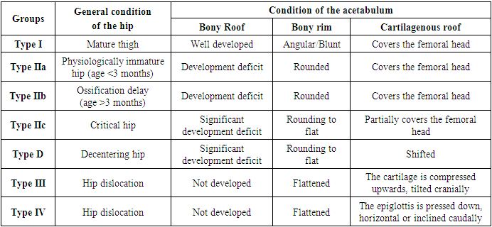
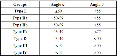
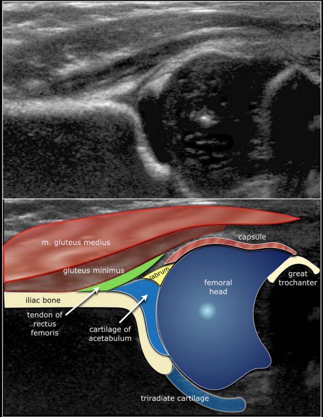
- The analysis of ultrasound of the hip of 300 children who were on inpatient treatment at the Samarkand Regional Multidisciplinary Children's Medical Center was carried out, of which 70 patients were found to have various degrees of severity of the corresponding symptoms of dysplasia. All patients were examined by sonography using an ultrasound device "Toshiba XARIO-200 with a linear sensor with a frequency of 5-7 MHz. The quantitative and qualitative indicators of hip joints performed according to the method of R. Graf (tab.1, tab.2) were studied in the work. The essence of this technique is to assess the development of the bone dome of the acetabulum (angle α), as well as the development of the cartilaginous zone of the acetabulum (angle β) (Fig.1).
|
|
4. Results of the Research
- At the first stage of the study, risk factors for the development of hip dysplasia in the examined children were determined. According to our research, the most significant was the pelvic or leg presentation of the fetus, noted in 28 children (40%). This is due to the fact that obstetric errors could have been made during childbirth. Next in frequency of occurrence is the female sex, identified in 23 girls, which was 32.8%. The world community has not yet reached a consensus on this risk factor. The most likely reason for this ratio of boys and girls is the influence of the mother's female sex hormones on the pelvic muscles in girls. The third most important factor was the regional factor of incorrect too tight swaddling, established in 12 children (17.1%). The so-called "beshiki" (national cradles with too tight swaddling) are the reason for frequent visits to pediatric orthopedists in our region. In the remaining 7 children, namely in 10.1% of cases, a combination of several of the above factors was observed (Fig.2).
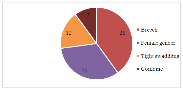 | Figure 2. Distribution of examined children with hip dysplasia by risk factors |
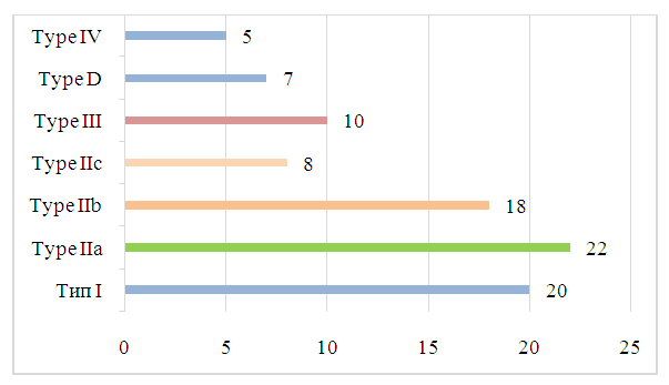 | Figure 3. Distribution of examined children depending on Type a of TBS dysplasia according to the results of the study |
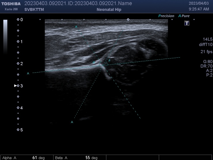 | Figure 4. Healthy child K., 3 months old. Normal hip joint (Type I – α =60°, β=55°) |
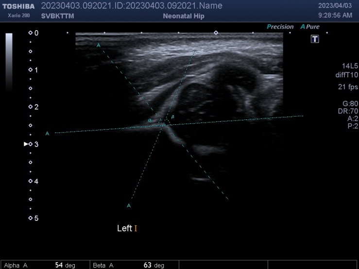 | Figure 5. Patient N., 5 months of age. "Immature" hip joint (Type IIa – α =54°, β =>63°) |
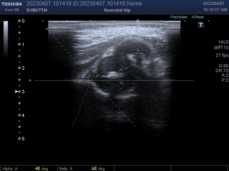 | Figure 6. Patient M., 2 months of age. "Subluxation" of the hip joint (Type III. α =48°, β = 68°) |
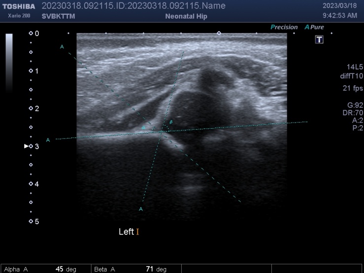 | Figure 7. Patient I., 3 months of age."Complete hip dislocation" (Type IV. α=<45°, β >71) |
5. Discussion of the Results of the Study
- Thus, ultrasound examination of the hip joints is advisable to be carried out in young children to identify signs of hip dysplasia of varying severity. Ultrasound examination should begin with B-mode. The smaller the angle α and the larger the angle β, the greater the degree of underdevelopment of the joint. If minor changes in the hip joints are detected, it is recommended to consult a traumatologist or a pediatric orthopedist.Early diagnosis of hip dysplasia in children is the key to effective therapy. In case of hip dysplasia of Turea II, follow-up up to a year is recommended. If the pathology does not resolve itself, then physiotherapy is recommended. With Turei III, conservative treatment should be carried out, and Type IV is a direct indication for surgical treatment. Ultrasonographic assessment of qualitative and quantitative parameters of hip joints allows differentiating different stages of their dysplasia. From the group of patients with identified dysplasia, patients with early changes in the immature joint prevailed (57.5%), which do not manifest clinically, based on this, it is necessary to include ultrasound of the hip joint in the sonography mode in the standard examination of newborns to objectify the pathological process.Assessment of the hip joints in B-mode does not require significant time and the results of ultrasound examination make it possible to prescribe pathogenetically based treatment, monitor the effectiveness of the therapy in dynamics, without exposing the child to radiation.Conclusion. Ultrasound sonography of the hip joint is an effective method of rapid non-invasive detection of hip dysplasia of varying severity in young children. The main advantages of the methods are the absence of radiation exposure, speed, non-invasiveness, the possibility of repeated studies, as well as higher efficiency compared to classical radiography in the case of children aged 1-6 months. This, in turn, increases the chances of early detection and the appointment of effective treatment without surgery.
Conflict of Interest
- The authors declare that there is no conflict of interest.
Funding Source
- The was conducted without sponsorship.
 Abstract
 Reference
 Full-Text PDF
 Full-text HTML