-
Paper Information
- Previous Paper
- Paper Submission
-
Journal Information
- About This Journal
- Editorial Board
- Current Issue
- Archive
- Author Guidelines
- Contact Us
American Journal of Medicine and Medical Sciences
p-ISSN: 2165-901X e-ISSN: 2165-9036
2023; 13(11): 1711-1714
doi:10.5923/j.ajmms.20231311.26
Received: Oct. 18, 2023; Accepted: Oct. 30, 2023; Published: Nov. 11, 2023

Morphology of Lymphopoietic Function of Regional Lymph Nodes of Bacterial Infection in Purulent Pyelonephritis
 Abstract
 Reference
 Full-Text PDF
 Full-text HTML
Jakhongir A. Shadmanov1, Rajabboy I. Isroilov2
1Department of Urology, Tashkent Medical Academy, Tashkent, Uzbekistan
2Department of Pathological Anatomy, Tashkent Medical Academy, Tashkent, Uzbekistan
Correspondence to: Jakhongir A. Shadmanov, Department of Urology, Tashkent Medical Academy, Tashkent, Uzbekistan.
| Email: |  |
Copyright © 2023 The Author(s). Published by Scientific & Academic Publishing.
This work is licensed under the Creative Commons Attribution International License (CC BY).
http://creativecommons.org/licenses/by/4.0/

In this scientific research, morphological changes characteristic of the lymphocytopoietic function of regional lymph nodes in cystitis and pyelonephritis were studied. If in the initial period of the disease the barrier and drainage function of lymphatic vessels and regional lymph was increased, over time it is observed that B-lymphocytic lymphocytopoietic function prevails in regional lymph nodes. In the morphological aspect, the lymphocytopoietic function is confirmed by activation and increase by proliferation of lymphoid and macrophage cells in the cortical layer of lymph nodes.
Keywords: Pyelonephritis, Cystitis, Drainage, Lymphocytopoiesis, Lymph vessel, Lymph node, Sinusoid, Morphofunctional zone, Proliferation
Cite this paper: Jakhongir A. Shadmanov, Rajabboy I. Isroilov, Morphology of Lymphopoietic Function of Regional Lymph Nodes of Bacterial Infection in Purulent Pyelonephritis, American Journal of Medicine and Medical Sciences, Vol. 13 No. 11, 2023, pp. 1711-1714. doi: 10.5923/j.ajmms.20231311.26.
Article Outline
1. Introduction
- Pyelonephritis in 80% of cases is caused by E. coli, this bacillus normally lives in the intestine, enters the urinary tract in violation of the hygiene of the pelvis and calyx, as well as the interstitial tissues [8]. As a result, starting from the renal tissue, it causes stagnation in the tissue of venous and lymphatic vessels. When the development of this process begins, due to the development of compensatory processes, pyelo-renal reflux occurs, the calyx and pelvis expand. As a result, lymphangitis and lymphadenitis develop from the spread of infection to the lymphatic capillaries of the kidneys and ureters. Lymphadenitis can acquire a lymphoproliferative character in response to antigens of microorganisms. As humoral immunity to bacterial infection develops, lymphocytes proliferate in the lymph node, mainly in the B-lymphocytic zone, and lymphocytopoiesis develops [6].Lymphocytopoiesis, which is the process of formation, proliferation and differentiation of lymphoid cells during the formation of lymphocytes, is the development of all lymphoid cells synchronously with each other [1,2]. Lymphocytopoiesis proceeds in stages both in the embryonic and postpartum periods. Lymphocytopoiesis, in turn, consists of T-lymphocytopoiesis and B-lymphocytopoiesis. Each of them consists of three stages: the bone marrow stage, which occurs in antigen-independent central organs, and antigen-dependent stages, which occur in peripheral lymphoid organs.In B-lymphocytopoiesis, B-lymphocytes first mature in the bone marrow, then migrate to the spleen and lymph nodes, differentiate in them and accumulate in a zone specific to B-lymphocytes, that is, in lymphoid follicles, germinal, perilymphoid ring and marginal areas. B-lymphocytes detects antigens with the participation of a macrophage and a T helper, identifies the antigen and synthesizes immunoglobulin in response to the antigen [3,4]. For the synthesis of immunoglobulin, the B-lymphocyte turns into a plasma cell, and this process is called “B-lymphocyte plasmatization". In this process, macrophages, monocytes, histiocytes and a dendritic cell phagocytose, process and determine antigens that have entered the tissue, and help lymphocytes detect them using histomoskeletal antigens on their outer surface.Immature T-lymphocytes in T-lymphocytopoiesis are formed in the bone marrow, then migrate to the cortical layer of the thymus, where, in a sterile state, they turn into cortical thymocytes on average within a week. After a week, 2-4% of cortical thymocytes survive, the rest (96-98%) undergo apoptosis and phagocytosis by thymus macrophages. Macrophages determine the products of phagocytized thymocytes, distribute them to the surviving thymocytes, and they differentiate. These surviving thymocytes should be able to identify their own histomoskeletal antigen on the surface of other cells and perform a state of innate immunological tolerance.The selection of surviving thymocytes with such high requirements prevents their transition to a state of autoaggression in relation to private cells in the body, in violation of which autoimmune diseases develop [5,7]. When angigen enters the body, it is determined by macrophages and is presented to the T-lymphocyte, T-lymphocytes undergo a “blastotransformation” reaction, turning into an activated lymphocyte, that is, an immunocyte, and exhibit cytotoxic action against the antigen, destroying it.
2. The Purpose of the Study
- This scientific research was aimed at studying morphological changes characteristic of lymphocytopoietic function in regional lymph nodes in patients with purulent pyelonephritis.
3. Material and Methods
- To achieve this goal in 2021-2022 during the autopsy of corpses of patients with purulent pyelonephritis, lymph nodes located in the pelvis were taken for histological examination in the department of adult Pathology at the Center of Republican Pathological Anatomy of the Ministry of Health of the Republic of Uzbekistan. The lymph nodes were divided into two equal parts and hardened within 72 hours in 10% neutralized formalin dissolved in a phosphate buffer. Then it was washed in running water for 3-4 hours, dehydrated in a series of alcohols with an increased concentration and poured paraffin with wax, prepared blocks. Histological sections of 5-6 microns were made from paraffin blocks, stained with hematoxylin-eosin dyes, examined in the lenses of a light microscope 20, 40, studied, and took computer pictures of the desired areas.
4. Results and Discussion
- Examination of the lymph nodes of the inguinal region at the microscopic level in chronic purulent cystitis and pyelonephritis showed that, since this disease is a bacterial infectious disease, lymphocytopoietic function prevails in the lymph nodes, that is, lymphoproliferative inflammation. Macroscopically, it was found that the lymph nodes of the inguinal region are enlarged to varying degrees, that is, the smallest is 854 mg, and the largest is 1760 mg.It was found that the lymph nodes have a bean-shaped and rounded appearance, the outer surface is smooth, but the color has changed due to inflammation, i.e. it has acquired a brownish hue due to the presence of red, bluish and grayish foci. When cut, the texture is soft, with a rotten appearance, the color is spotty.In histological preparations treated with hematoxylin and eosin dyes by the general morphological method, in the initial period of the disease with purulent pyelonephritis, there is a sharp swelling of the interstitial substance in all morphofunctional areas of the lymph node, expansion of the subcapsular sinusoid, loosening of the tissue structures of the fibrous membrane, rarefaction of cells. It is claimed that the predominance of dyscirculatory and edematous processes coincides with the exudative period of inflammatory disease.Primary follicles located in the mucous membrane of the lymph node are loosened and are defined as enlarged in area, with a sparse arrangement of lymphocytes, macrophages and reticular cells. The presence in the composition of lymphocytes of cells with different sizes of the nucleus and dark and light color indicates increased lymphocytopoietic activity in them (Fig. 1). It is known that from the location of B-lymphocytes in the lymphoid follicles of the mucous membrane of the lymph nodes, it is revealed that the plasmatization reaction of B-lymphocytes develops to develop humoral immunity in response to bacterial infection.
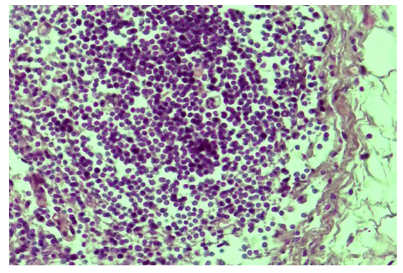 | Figure 1. Primary lymphoid follicle of the cortical layer of the lymph node, the interstitial substance of which is strongly swollen, lymphocytes are located in a loosened and hyperchromasized form. Staining: G-Е. Increased: 10х40 |
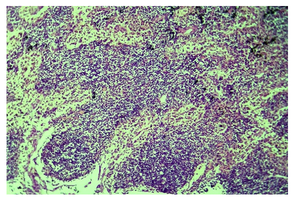 | Figure 2. General view of the lymph node tissue, at the initial stage of purulent pyelonephritis, the sinusoids are dilated, the cortex and paracortical layer are hyperplasized.Staining: G-Е. Increased: 10х10 |
 | Figure 3. View of the lymph node in a semi-thin section, in a state of activation of large and medium lymphocytes. Staining: toluidine blue. Increased: 10х100 |
 | Figure 4. Proliferation and increase of B-lymphocytes in the lymph node of the inguinal region in chronic purulent pyelonephritis. Staining: G-Е. Increased: 10х10 |
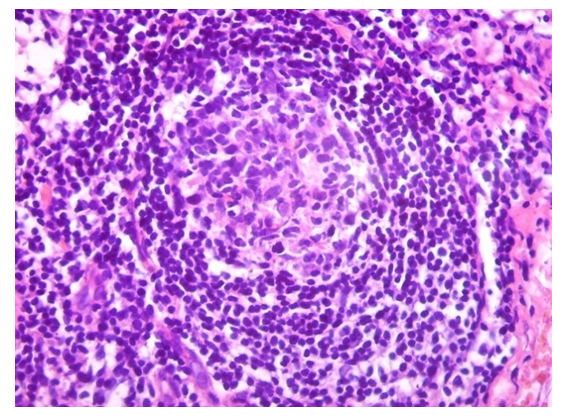 | Figure 5. Secondary follicle of the inguinal lymph node in chronic purulent pyelonephritis, an increase in the number of reticulocytes, lymphoblasts in the germinal center. Staining: G-Е. Increased: 10х40 |
 | Figure 6. Expansion of all morphofunctional zones of secondary lymphoid follicles. Staining: G-Е. Increased: 10х40 |
5. Conclusions
- While at the initial stage of the development of bacterial infectious purulent pyelonephritis, the barrier and drainage function in the lymphatic vessels and regional lymph nodes increases, over time there is a predominance of the development of B-lymphocytic lymphocytopoietic function in the lymph nodes.It was determined that in the initial period of the disease with purulent pyelonephritis, the processes of dyscirculation and edema prevail in the lymph node, the sinusoids of both the cortex and the cerebral layer are sharply expanded, fragments of tissues and cells, microorganisms are present in the cavity, and later in the parenchyma of the lymph node, a lymphocytopoietic reaction begins to develop intensively, both in the lymphoid follicles of the cortical layer and in the paracortical layer the proliferation of lymphoid and monocyte-macrophage cells begins.At the beginning of the lymphocytopoietic reaction, the primary follicles first loosen and expand in area, the lymphocytes, macrophages and reticular cells contained in them are activated and sparsely located, hyperplasia of the lymph node mucosa in response to bacterial infection indicates the predominance of B-lymphocytopoiesis.Over time, the zone of secondary lymphoid follicles expands and increases; the increased lymphocytopoietic function in it is evidenced by the observation that in the germinative zone, reticular cells with dendrites acquired the appearance of a “stellate sky”due to transformation into whitish cells.Small lymphocytes in the lymphocytic layer are fully developed, formed lymphocytes that, as a result of migration to the medullar layer, undergo an immunological response, the so-called “plasmatization” inside the sinusoid of the medullar layer and in the soft bands between them, turning into plasma cells and synthesizing immunoglobulins, forming antibodies corresponding to the antigens of existing microorganisms, and humoral immune response.