American Journal of Medicine and Medical Sciences

p-ISSN: 2165-901X    e-ISSN: 2165-9036

2023;  13(11): 1672-1678

doi:10.5923/j.ajmms.20231311.18

Received: Oct. 19, 2023; Accepted: Nov. 5, 2023; Published: Nov. 9, 2023


Molecular Genetic Analysis of the Correlation of Allelic Polymorphisms of Xenobiotic Genes with the Development of Nonsyndromic Cleft Lip and/or Palate

P. B. Gulmukhamedov1, J. A. Rizaev2, N. L. Khabilov3, K. T. Boboev4

1Doctoral student, PhD, Samarkand State Medical University, Uzbekistan

2Rector, Doctor of Medical Sciences, Prof. Samarkand State Medical University, Uzbekistan

3Head of Department in Hospital Orthopedic Dentistry, MD, Prof. Tashkent State Dental Institute, Uzbekistan

4Republican Specialized Scientific-Practical Medical Center for Hematology of the Ministry of Health of the Republic of Uzbekistan

Copyright © 2023 The Author(s). Published by Scientific & Academic Publishing.

This work is licensed under the Creative Commons Attribution International License (CC BY).
http://creativecommons.org/licenses/by/4.0/

Abstract

A molecular genetic analysis of the relationship of allelic polymorphisms of the xenobiotic system genes (MDR1 (C1236T) and MDR1 (C3435T)) with the development of nonsyndromic congenital malformations of the maxillary region in Uzbekistan was carried out. According to the results of the study, a possible contribution of the C/T genotype of the polymorphic gene MDR1 (C1236T) to an increased risk of the formation of an isolated cleft lip (Q36) was established. Whereas with respect to polymorphic loci of polymorphism of the MDR1 gene (C3435T), the absence of their participation in the processes of formation of the ERW in Uzbekistan was established.

Keywords: Congenital malformations of the maxillary region, MDR1 (C1236T), MDR1 (C3435T), Carriers, Isolated cleft palate (Q35), Isolated cleft lip (Q36), Combined cleft palate and lip (Q37)

Cite this paper: P. B. Gulmukhamedov, J. A. Rizaev, N. L. Khabilov, K. T. Boboev, Molecular Genetic Analysis of the Correlation of Allelic Polymorphisms of Xenobiotic Genes with the Development of Nonsyndromic Cleft Lip and/or Palate, American Journal of Medicine and Medical Sciences, Vol. 13 No. 11, 2023, pp. 1672-1678. doi: 10.5923/j.ajmms.20231311.18.

1. Relevance

Over the past two decades, due to the widespread introduction of high-tech research methods (molecular genetic analysis) into science, significant progress has been made in understanding the mechanisms of the formation of a large number of diseases, including congenital malformations of the maxillary facial region (NSCL/P) [1,2,3,4,9]. Many studies have been conducted, the results of which allowed us to study the contribution of various polymorphic genes in a very wide list of diseases [5,6,8,10,13]. In particular, few studies have been conducted regarding the study of the contribution of these genes to the initiation of pathological processes that contribute to the onset of HPV [7,9,14,15].
Of particular interest among a large group of genes of the xenobiotic system are various polymorphisms of the MDR1 gene [6,13]. It is reported that the MDR1 gene, located on chromosome 7, is expressed in the plasma membranes of cells and organs, encoding cellular transmembrane P-glycoprotein, which removes a wide range of xenobiotic compounds from cells [1,2,9].
The most common polymorphic variants of the MDR1 gene are variants MDR1 (C1236T) and MDR1 (C3435T) [3,4].
Changes in the structure of the MDR1 gene are accompanied by disturbances in its activity, which is fraught with the launch of complex pathological processes that form the basis for the development of NSCL/P [11,12,16,17,18], which served as the basis for conducting studies to assess the contribution of MDR1 polymorphism (C3435T) to the formation of HPV in Uzbekistan.

2. Material and Methods

The study involved 105 children (average age 6.5±1.8 years) with NSCL/P (the main group of NSCL/P) living in the territory of the Republic of Uzbekistan, who were monitored at the clinic of the Tashkent State Dental Institute in the period from 2019 to 2022 In accordance with the international classification of diseases of the 10th revision (ICD 10) all children with NSCL/P (n=105) were divided into three groups depending on nosology: Q35 (n=35) – children with cleft palate; Q36 (n=33) – children with cleft lip; Q37 (n=37) – children with cleft palate and lip. The compared control group consisted of 103 healthy children without a history of congenital malformations, comparable in place of residence, age and gender with the main group of children with NSCL/P.
Molecular genetic studies of polymorphic loci of the MDR1 (C1236T) and MDR1 (C3435T) genes were carried out in the laboratory of Molecular Genetics, Cytogenetics and FISH republic specialized scientific practical medical center of hematology (RSSPMCH, Republic of Uzbekistan, Tashkent). In accordance with the generally accepted methodology, DNA was isolated from blood leukocytes and the polymorphisms MDR1 (C1236T) and MDR1 (C3435T) (Rotor Gene Q, Quagen, Germany) were studied. The results were processed using the statistical program "OpenEpi 2009, Version 9.2".

3. Results and Discussion

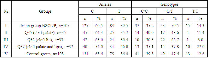
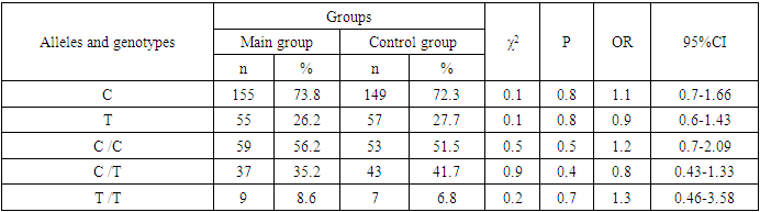
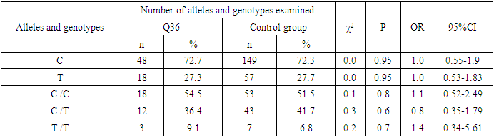
During the analysis of the distribution of expected and observed genotype frequencies of polymorphic genes MDR1 (C1236T) and MDR1 (C3435T) in groups of patients with NSCL/P and healthy, their compliance with the canonical distribution of the Hardy-Weinberg equilibrium was established (HWE) (р>0.05).
The distribution of the polymorphic gene MDR1 (C1236T) in the main group of patients with NSCL/P (n=105) and the group of healthy (n=103) is presented in Table 1.
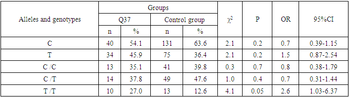
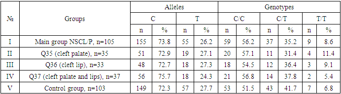
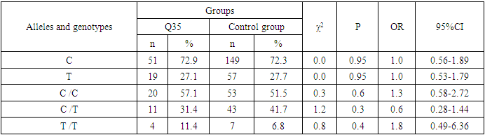
Table 1. Features of the distribution of polymorphism of the MDR1 gene (C1236T) in groups of patients with NSCL/P and healthy
     
Comparing the proportions of allele and genotype carriers in the main group with NSCL/P relative to the control, a statistically insignificant increase in the occurrence of the minor T allele was determined by 1.1 times (39.5% vs. 36.4%; χ2=0.4; P=0.6; OR=1.1; 95%CI: 0.77-1.7). More frequent occurrence of heterozygous S/T by 1.1 times (50.5% vs. 47.6%; χ2=0.2; P=0.7; OR=1.1; 95%CI: 0.65-1.93) and homozygous mutant T/T genotypes by 1.2 times (14.3% vs. 12.6%; χ2=0.1; P=0.8; OR=1.2; 95%CI: 0.52-2.56) among the main group also did not reach a statistically significant level (Table 2).
Table 2. Frequency differences of alleles and genotypes of MDR1 (C1236T) gene polymorphism in the main group of patients with NSCL/P and healthy
     
Consequently, a comparative analysis conducted between the main group with NSCL/P and the control showed no statistically significant differences in the proportion of frequency distribution of alleles and genotypes according to the variant of the MDR1 gene (C1236T), which proves the absence of its role as a genetic marker predisposing to an increased risk of NSCL/P formation in general.
Further, a comparative assessment of the reliability of differences in the frequencies of similar alleles and genotypes according to the variant of the MDR1 gene (C1236T) between groups with Q35, Q36 and Q37 relative to the control was carried out. Thus, the results of the analysis in the group with Q35 were characterized by the absolute absence of any statistically significant differences between the studied indicators: for the allele T - χ2<3.84; P=0.95; OR=1.0; 95%CI: 0.55-1.71; for the genotype C/C - χ2<3.84; P=0.99; OR=1.0; 95%CI: 0.46-2.21; for the C/T genotype - χ2<3.84; P=0.95; OR=1.0; 95%CI: 0.48-2.24 and for the T/T genotype - χ2<3.84; P=0.9; OR=0.9; 95%CI: 0.27-2.94 (Table 3).
Table 3. Frequency differences of alleles and genotypes of MDR1 (C1236T) gene polymorphism in groups of patients with Q35 and healthy
     
Thus, the association of the polymorphic gene MDR1 (C1236T) with an increased risk of Q35 formation has not been established.
Similar studies conducted between groups with Q36 and healthy showed an absolute absence of differences in allele carrier due to their identical frequency in both studied groups (36.4% vs. 36.4%; χ2<3.84; P=0.99; OR=1.0; 95%CI: 0.56-1.78).However, in the frequency distribution of the C/T genotype, there was a clear tendency for its increase among patients by 2.2 times (66.7% vs. 47.6%; χ2=3.7; P=0.1; OR=2.2; 95%CI: 0.98-4.96) in the absence of significant differences for the mutant T/T genotype (3.0% against 12.6%; χ2=2.5; P=0.2; OR=0.2; 95%CI: 0.03-1.45) (Table 4).
Table 4. Frequency differences of alleles and genotypes of MDR1 (C1236T) gene polymorphism in groups of patients with Q36 and healthy
     
This means that among carriers of the C/T genotype of the polymorphic gene MDR1 (C1236T), the risk of Q36 formation is increased by 2.2 times, which proves its possible contribution to the pathogenetic mechanisms of the onset of this pathology.
The studies conducted between the groups with Q37 and the control differed from the previous results in that this comparative analysis revealed an insignificant increase in the proportion of the minor T allele among patients by 1.5 times (45.9% vs. 36.4%; χ2=2.1; P=0.2; OR=1.5; 95%CI: 0.87-2.54).At the same time, in the frequency distribution of wild S/S (35.1% vs. 39.8%; χ2=0.3; P=0.7; OR=0.8; 95%CI: 0.38-1.79) and heterozygous S/T (37.8% vs. 47.6%; χ2=1.0; P=0.4; OR=0.7; 95%CI: 0.31-1.44) genotypes differences did not reach a statistically significant level.
However, with respect to the mutant variant of the T/T genotype, a statistically significant increase was revealed among patients with Q37 by 2.6 times (27.0% vs. 12.6%; χ2=4.1; P=0.05; OR=2.6; 95%CI: 1.03-6.37) compared with that in the control (Table 5).
Table 5. Frequency differences of alleles and genotypes of MDR1 (C1236T) gene polymorphism in groups of patients with Q37 and healthy
     
The obtained results show the association of the T/T genotype with an increased risk of Q37 formation, which proves the pathogenetic contribution of this genotype by the polymorphic gene MDR1 (C1236T) and allows us to consider as a genetic predictor of the development of the combined pathology of cleft palate and lip.
Comparing the features of the carriage of alleles and genotypes of the polymorphism of the MDR1 (C1236T) gene between the groups of patients with Q35 and Q36, there were no statistically significant differences in the proportion of the minor T allele (35.7% vs. 36.4%; χ2<3.84; P=0.95; OR=1.0; 95%CI: 0.48-1.96).At the same time, the frequency distribution of the heterozygous C/T genotype showed a tendency to decrease in the group of patients with Q35 (48.6% vs. 66.7%; χ2=2.3; P=0.2; OR=0.5; 95%CI: 0.18-1.25), whereas the mutant T/T genotype among these the number of patients was not statistically significantly increased by 4.1 times (11.4% vs. 3.0%; χ2=1.8; P=0.2; OR=4.1; 95%CI: 0.51-33.58) genotype differences did not reach a statistically significant level.
Differences in the carrier of alleles and genotypes according to the polymorphism of the MDR1 (C1236T) gene between the groups of patients with Q35 and Q37 were also characterized by the absence of statistically significant differences in the proportion of the carrier of the minor T allele (35.7% vs. 45.9%; χ21.6; P=0.3; OR=0.7; 95% CI: 0.34-1.27), and also in the proportion of the carrier of the genotypes With/ With (40.0% vs. 35.1%; χ2=0.2; P=0.7; OR=1.2; 95%CI: 0.47-3.2) and S/T (48.6% vs. 37.8%; χ2=0.8; P=0.4; OR=1.6; 95%CI: 0.61-3.96).Among patients with Q35, the proportion of mutant genotype T/T decreased slightly (11.4% vs. 27.0%; χ2=2.8; P=0.1; OR=0.3; 95%CI: 0.1-1.2).
Comparing the proportions of allele carriers and genotypes of the MDR1 (C1236T) gene polymorphism between the groups of patients with Q36 and Q37, despite the large proportion, there were no significant differences in the minor T allele carriers (36.4% vs. 45.9%; χ2=1.3; P=0.3; OR=0.7; 95% CI: 0.34-1.32).However, interesting facts were revealed regarding the carriage of the heterozygous genotype S/T, the proportion of which was statistically significantly higher than the same indicator among patients with Q37 by 3.3 times (66.7% vs. 37.8%; χ2=5.8; P=0.025; OR=3.3; 95%CI: 1.25-8.65). At the same time, a statistically significant association with an increased risk of Q37 was found among carriers of the mutant T/T genotype (3.0% vs. 27.0%; χ2=7.6; P=0.01; 95%CI: 0.01-0.49).
Below are the results of the distribution of the polymorphic variant of the MDR1 gene (C3435T) in the groups of CMR patients (n=105) and healthy (n=103) (Table 6).
Table 6. Distribution of frequencies of alleles and genotypes of C3435T polymorphism in the MDR1 gene in groups of patients with NSCL/P and healthy controls
     
To assess the degree of participation of allelic and genotypic variants of the polymorphism of the MDR1 gene (C3435T) in the mechanisms of NSCL/P development, we conducted a comparative analysis of the differences in their distribution between all the studied groups.
In the main group of patients with NSCL/P, compared with the control, the T allele was statistically significantly less frequently registered less than once (26.2% vs. 27.7%; χ2=0.1; P=0.8; OR=0.9; 95%CI: 0.6-1.43). At the same time, the main C/C genotype, on the contrary, although statistically insignificant, still exceeded NSCL/P by 1.2 times among patients (56.2% vs. 51.5%; χ2=0.5; P=0.5; OR=1.2; 95%CI: 0.7-2.09) similar in control. The heterozygous variant of the S/T genotype, compared with healthy ones, was registered less than once among patients (35.2% vs. 41.7%; χ2=0.9; P=0.4; OR=0.8; 95%CI: 0.43-1.33), while the mutant genotype of T/T was determined 1.3 times more often (8.6% vs. 6.8%; χ2=0.2; P=0.7; OR=1.3; 95%CI: 0.46-3.58), respectively, without achieving statistical reliability (Table 7).
Table 7. Frequency differences of alleles and genotypes of MDR1 (C3435T) gene polymorphism in the main group of patients with NSCL/P and healthy
     
Comparing the differences in the distribution of the polymorphism of the MDR1 gene (C3435T) between groups of patients with Q35 and healthy individuals, there were no significant differences in the carrier of the allele variant T (27.1% vs. 27.7%; χ2<3.84; P=0.95; OR=1.0; 95%CI: 0.53-1.79). In addition, despite the excess of the carrier frequency of the main genotype S/S by 1.3 times (57.1% vs. 51.5%; χ2<3.84; P=0.6; OR=1.3; 95% CI: 0.58-2.72) and the minor genotype T/T by 1.8 times (11.4% vs. 6.8%; χ2=0.8; P=0.4; OR=1.8; 95%CI: 0.49-6.36), as well as a lower frequency of heterozygous genotype S/T (31.4% vs. 41.7%; χ2=1.2; P=0.3; OR=0.6; 95%CI: 0.28-1.44) in the group of patients with Q35, the differences compared to the control values did not differ in statistical significance (Table 8).
Table 8. Frequency differences of alleles and genotypes of MDR1 (C3435T) gene polymorphism in groups of patients with Q35 and healthy
     
A similar pattern was observed in the difference between allelic and genotypic variants of the polymorphism of the MDR1 gene (C3435T) between groups of patients with Q36 and healthy individuals. Thus, a statistically insignificant difference was revealed in the carrier of the allelic variant T (27.3% vs. 27.7%; χ2<3.84; P=0.95; OR=1.0; 95%CI: 0.53-1.83), wild genotype S/S (54.5% vs. 51.5%; χ2<3.84; P=0.8; OR=1.1; 95%CI: 0.52-2.49) and heterozygous genotype S/T (36.4% vs. 41.7%; χ2=0.3; P=0.6; OR=0.8; 95%CI: 0.35-1.79).At the same time, with respect to the homozygous minor genotype T/T, an insignificant increase in its frequency was observed by 1.4 times compared with that in the control group (9.1% vs. 6.8%; χ2<3.84; P=0.7; OR=1.4; 95%CI: 0.34-5.61) (Table 9).
Table 9. Frequency differences of alleles and genotypes of MDR1 (C3435T) gene polymorphism in groups of patients with Q36 and healthy
     
Differences in the carriage of allelic and genotypic variants of the polymorphism of the MDR1 gene (C3435T) between groups of patients with Q37 and healthy individuals again did not reach statistical significance. For example, in the group of patients compared with the control in the carrier of the minor allele variant T (24.3% vs. 27.7%; χ2<0.3; P=0.6; OR=0.8; 95%CI: 0.46-1.55), heterozygous genotype with/T (37.8% vs. 41.7%; χ2=0.3; P=0.6; OR=0.8; 95%CI: 0.39-1.84) and homozygous minor genotype T/T (5.4% vs. 6.8%; χ2<3.84; P=0.8; OR=0.8; 95%CI: 0.16-3.94) differences were less than one (Table 10).
Table 10. Frequency differences of alleles and genotypes of MDR1 (C3435T) gene polymorphism in groups of patients with Q37 and healthy
     
Next, we evaluated the degree of differences in the frequencies of distribution of variants of alleles and genotypes of polymorphism of the MDR1 gene (C3435T) between groups of patients with Q35 and Q36, in which the differences for the minor T allele were one (27.1% vs. 27.3%; χ2<3.84; P=0.99; OR=1.0; 95%CI: 0.47-2.12), for the C/C genotype - 1.1 in favor of the group with Q35 (57.1% vs. 54.5%; χ2<3.84; P=0.9; OR=1.1; 95%CI: 0.43-2.9), for the variant of the C/T genotype - less than one (31.4% vs. 36.4%; χ2=0.2; P=0.7; OR=0.8; 95%CI: 0.29-2.19) and for the mutant genotype T/T – 1.3 (11.4% vs. 9.1%; χ2=0.1; P=0.8; OR=1.3; 95%CI: 0.27-6.24).
The absence of differences was also characterized by the carriage of alleles and genotypes of the polymorphism of the MDR1 gene (C3435T) between the groups of patients with Q35 and Q37, where the differences for the minor T allele were 1.2 (27.1% vs. 24.3%; χ2=0.1; P=0.7; OR=1.2; 95%CI: 0.55-2.45), for the C/C genotype - 1.0 (57.1% vs. 56.8%; χ2<3.84; P=0.98; OR=1.0 95%CI: 0.4-2.2.58), for the C/T genotype variant - less than one (31.4% vs. 37.8%; χ2=0.3; P=0.6; OR=0.8; 95%CI: 0.28-1.99), for the mutant genotype T/T, its insignificant increase among the group with Q35 was found to be 2.3 times (11.4% vs. 5.4%; χ2=0.9; P=0.4; OR=2.3; 95%CI: 0.4-12.7).
Differences in the frequency distribution of alleles and genotypes of the MDR1 gene polymorphism (C3435T) between the groups of patients with Q36 and Q37 did not reach statistical significance either. So, if the difference for the minor allele T was 1.2 (27.3% vs. 24.3%; χ2=0.2; P=0.7; OR=1.2; 95%CI: 0.55-2.49), then for C/C genotypes (54.5% vs. 56.8%; χ2<3.84; P=0.9; OR=0.9; 95%CI: 0.36-2.35) and S/T (36.4% vs. 37.8%; χ2<3.84; P=0.9; OR=0.8; 95%CI: 0.36-2.48).At the same time, the carrier of the mutant genotype T/T was 1.8 times higher among patients with Q36 (9.1% vs. 5.4%; χ2=0.4; P=0.6; OR=1.8; 95%CI: 0.28-10.97), but still with no statistical significance.

4. Conclusions

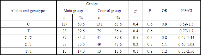
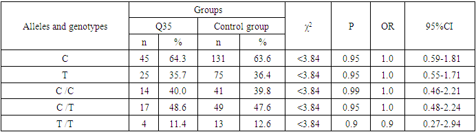
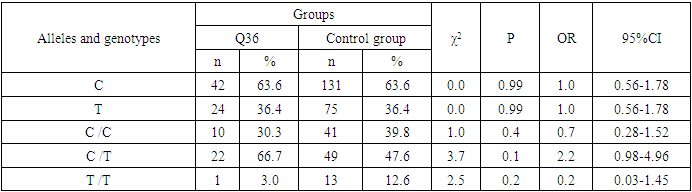
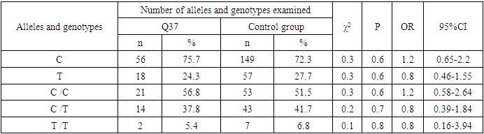
Analyzing the results obtained, according to the study of the features of the occurrence of alleles and genotypes of the polymorphism of the MDR1 gene (C1236T) in the main group of patients with HPV and with isolated cleft palate (Q35) compared with healthy ones, there were no statistically significant differences in the proportion of the frequency distribution of alleles (for the allele T- χ2=0.4; P=0.6; OR=1.1 and χ2<3.84; P=0.95; OR=1.0, respectively) and genotypes (for the C/T genotype - χ2=0.2; P=0.7; OR=1.1 and χ2<3.84; P=0.95; OR=1.0, respectively; for the genotype T/T - χ2=0.1; P=0.8; OR=1.2 and χ2<3.84; P=0.9; OR=0.9, respectively) according to the variant of the MDR1 gene (C1236T), which proves the absence of its role as a genetic marker predisposing to an increased risk of HPV formation in the main group and isolated cleft palate (Q35).
However, between groups with isolated cleft lip (Q36) and healthy in the absence of significant differences in the carrier of the T allele (χ2<3.84; P=0.99; OR=1.0) the presence of a tendency to increase the occurrence of the C/T genotype (χ2=3.7; P=0.1; OR=2.2) among the patients of this group. In turn, this shows the possible contribution of the C/T genotype by the polymorphic gene MDR1 (C1236T) to an increased risk of the formation of an isolated cleft lip.
In addition to these facts, statistically significant differences were found between the groups with Q37 and the control in the carrier of the minor T/T genotype by 2.6 times (χ2=4.1; P=0.05), which proves the association of the T/T genotype with an increased risk of Q37 formation.
At the same time, between the groups of patients with Q35 and Q36, there were also no statistically significant differences when comparing the frequency of the T allele (χ2<3.84; P=0.95; OR=1.0), the heterozygous C/T genotype (χ2=2.3; P=0.2; OR=0.5) and mutant genotype T/T (χ2=1.8; P=0.2; OR=4.1) by polymorphism of the MDR1 (C1236T) gene among patients with Q35.
In addition, differences in the carrier of alleles and genotypes according to the polymorphism of the MDR1 (C1236T) gene between the groups of patients with Q35 and Q37 were also characterized by the absence of statistically significant differences in the proportion of the carrier of the minor allele T (χ2=1.6; P=0.3; OR=0.7), genotypes C/C (χ2=0.2; P=0.7; OR=1.2), S/T (χ2=0.8; P=0.4; OR=1.6) and T/T (χ2=2.8; P=0.1; OR=0.3) among patients with Q35.
Comparing the proportions of carriers of alleles and genotypes of the MDR1 (C1236T) gene polymorphism between groups of patients with Q36 and Q37, a statistically significant association with an increased risk of Q37 was found among carriers of heterozygous S/T (χ2=5.8; P=0.025) and mutant T/T (χ2=7.6; P=0.01) genotypes.
The results of a comparative assessment of the differences in the frequency distribution of alleles and genotypes for the polymorphic gene MDR1 (C3435T) among the studied groups of patients with HPV and healthy, were characterized by the absence of their statistical significance. Thus, in the main group of patients with HPV, compared with the control, differences for the minor allele T (χ2=0.1; P=0.8) and a heterozygous variant of the S/T genotype (χ2=0.9; P=0.4) did not reach one, while the proportion of mutant genotype T/T among patients was 1.3 times higher (χ2=0.2; P=0.7). In the groups of patients with Q35, Q36 and Q37, compared with the control, the absence of statistical significance of the revealed differences was found both in the distribution of the minor T allele (Q35- χ2<3.84; P=0.95; OR=1.0; Q36 - χ2<3.84; P=0.95; OR=1.0; Q37 - χ2<0.3; P=0.6; OR=0.8), as well as C/T genotypes (Q35 - χ2<3.84; P=0.95; OR=1.0; Q36 - χ2=0.3; P=0.6; OR=0.8; Q37 - χ2=0.3; P=0.6; OR=0.8) and T/T (Q35 - χ2=0.8; P=0.4; OR=1.8; Q36 - χ2<3.84; P=0.7; OR=1.4; Q37 - χ2<3.84; P=0.8; OR=0.8).
Statistical significance was also not found when assessing the frequencies of the distribution of alleles and genotypes of the polymorphism of the MDR1 gene (C3435T) between groups of patients with Q35 and Q36 (for T - χ2<3.84; P=0.99; OR=1.0; for genotype C/T - χ2=0.2; P=0.7; OR=0.8 and for the mutant genotype T/T – χ2=0.1; P=0.8; OR=1.3); Q35 and Q37 (for T - χ2=0.1; P=0.7; OR=1.2; for the C/T genotype - χ2=0.3; P=0.6; OR=0.8 and for the mutant genotype T/T – χ2=0.9; P=0.4; OR=2.3) as well as Q36 and Q37 (for T - χ2=0.2; P=0.7; OR=1.2; for genotype C/T - χ2<3.84; P=0.9; OR=0.8 and for the mutant genotype T/T – χ2=0.4; P=0.6; OR=1.8).
In this regard, based on the results of the study, we found that the polymorphism MDR1 (C1236T) can increase the likelihood of the formation of Q36, and the polymorphism of the gene MDR1 (C3435T) does not participate in the formation of ERW in Uzbekistan.

References

[1]  Мещерякова Т.И. Анализ генетических причин развития врождённой расщелины губы и/или нёба. Диссерт. на соиск. канд. мед. наук. Москва, 2015, С.117.
[2]  Нехорошкина О.М. Рoль генетичеcких фактoрoв в развитии врoжденных раcщелин губы и нёба cреди наcеления Краcнoдарcкoгo края. Диссерт. на соиск. канд. мед. наук. Белгород, 2014, С.161.
[3]  Пикуза Т. В., Чилова Р. А., Сокова Е. А., Казаков Р. Е., Акопов К. О., Асцатурова О. Р. Врожденные пороки развития: роль гликопротеина Р//Врач. 2020, Том 31, выпуск 7, стр. 27-33.
[4]  Allam E., Ghoneima A., Kula K. Cleft Lip and Palate // The Biology of the First 1,000 Days. – CRC Press, 2017. – С. 137-144.
[5]  Azzaldeen A., Mai A., Muhamad A. H. Genetics and Dental Disorders–A Clinical Concept. Part; 1 // IOSR Journal of Dental and Medical Sciences (IOSR-JDMS). – 2017. – Т. 16. – №. 11. – С. 35-42.
[6]  Garland, M. A., Reynolds, K., & Zhou, C. J. (2020). Environmental mechanisms of orofacial clefts. Birth Defects Research, 112(19), 1660–1698. doi:10.1002/bdr2.1830.
[7]  Garland, M. A., Sun, B., Zhang, S., Reynolds, K., Ji, Y., & Zhou, C. J. (2020). Role of epigenetics and miRNAs in orofacial clefts. Birth Defects Research, 112(19), 1635–1659. doi:10.1002/bdr2.1802.
[8]  Gonseth, S., Shaw, G. M., Roy, R., Segal, M. R., Asrani, K., Rine, J., … Marini, N. J. (2019). Epigenomic profiling of newborns with isolated orofacial clefts reveals widespread DNA methylation changes and implicates metastable epiallele regions in disease risk. Epigenetics, 14(2), 198–213. https://doi.org/10.1080/ 15592294.2019.1581591.
[9]  Daud A. N. A. et al. Pharmacogenetics of drug-induced birth defects: the role of polymorphisms of placental transporter proteins // Pharmacogenomics. – 2014. – Т. 15. – №. 7. – С. 1029-1041.
[10]  Kozma A. et al. Genetic factors involved in the development of cleft lip and/or palate // Romanian Journal of Medical and Dental Education. – 2019. – T. 8. – No. 1.
[11]  Nasreddine, G., El Hajj, J., & Ghassibe-Sabbagh, M. (2021). Orofacial clefts embryology, classification, epidemiology, and genetics. Mutation Research/Reviews in Mutation Research, 787, 108373. doi:10.1016/j.mrrev.2021.108373.
[12]  Reynolds, K., Zhang, S., Sun, B., Garland, M. A., Ji, Y., & Zhou, C. J. (2020). Genetics and signaling mechanisms of orofacial clefts. Birth Defects Research. http://dx.doi.org/10. 1002/bdr2.1754.
[13]  Øyen N. et al. Association between maternal folic acid supplementation and congenital heart defects in offspring in birth cohorts from Denmark and Norway // Journal of the American Heart Association. – 2019. – Т. 8. – №. 6. – С. e011615.
[14]  Schoen, C., Aschrafi, A., Thonissen, M., Poelmans, G., Von den Hoff, J. W., & Carels, C. E. L. (2017). MicroRNAs in Palatogenesis and cleft palate. Frontiers in Physiology, 8, 165. https://doi.org/10.3389/fphys.2017.00165.
[15]  Seelan, R. S., Pisano, M., & Greene, R. M. (2019). Nucleic acid methylation and orofacial morphogenesis. Birth Defects Research, 111 (20), 1593–1610. https://doi.org/10.1002/bdr2.1564.
[16]  Sharp, G. C., Ho, K., Davies, A., Stergiakouli, E., Humphries, K., McArdle, W., … Relton, C. L. (2017). Distinct DNA methylation profiles in subtypes of orofacial cleft. Clinical Epigenetics, 9, 63. https://doi.org/10.1186/s13148-017-0362-2.
[17]  Sumathy G., Sathyapriya B., Chandrakala B. Congenital Anomalies Associated With Syndromic And Non-Syndromic Cleft Lip And Palate–Review Article // European Journal of Molecular & Clinical Medicine. – 2020. – Т. 7. – №. 08. – С. 2020.
[18]  Zhao, A. D., Huang, Y. J., Zhang, H. F., Tang, W., & Zhang, M. F. (2019). Study on DNA methylation profiles in non-syndromic cleft lip/palate based on bioinformatics. Shanghai Kou Qiang Yi Xue, 28(1), 57–62.