Odilov K. K. 1, Nishanov D. A. 2, Madaliev A. A. 2, Abdikarimov Kh. G. 2
1Bukhara Regional Branch of the Republican Specialized Scientific and Practical Medical Center of Oncology and Radiology, MH RUz
2RSNPMTSOiR MH RUz
Copyright © 2023 The Author(s). Published by Scientific & Academic Publishing.
This work is licensed under the Creative Commons Attribution International License (CC BY).
http://creativecommons.org/licenses/by/4.0/

Abstract
Relevance of the topic: In the structure of human malignant tumors, soft tissue sarcomas (STS) occupy a very modest place, accounting for about 1%. In synovial sarcoma, an analysis was made of the relationship between histological and immunohistochemical characteristics and the number of cells carrying the translocation. The presence of p53 mutations and its accumulation is being actively studied in soft tissue sarcomas. Compared with other types of soft tissue sarcomas, increased levels of p53 expression are found in synovial sarcoma. Overexpression of the Bc1-2 gene prevents the characteristic morphological signs of apoptosis. Bcl-2 is the leading gene that determines the mechanism of cell death by suppressing apoptosis [4]. High expression of this oncogene may be an independent indicator of overall and disease-free survival in soft tissue sarcomas. VEGF is the most effective direct angiogenic factor known. One of the features of VEGF is increased vascular permeability, which is one of the additional mechanisms of neoangiogenesis and can lead to the accumulation of plasma fibrin in tissues. Antigen KI-67 is a nuclear protein associated with cell proliferation. Changing Ki-67 expression levels had no significant effect on cell proliferation in vivo.
Keywords:
Soft tissue sarcoma, Proliferative activity, Apoptosis, Vascular endothelial growth factor, Histological structure, Immunohistochemistry, Recurrence, Metastasis, Survival
Cite this paper: Odilov K. K. , Nishanov D. A. , Madaliev A. A. , Abdikarimov Kh. G. , Immunohistochemical Markers in the Diagnosis and Treatment of High-Grade Soft Tissue Sarcomas, American Journal of Medicine and Medical Sciences, Vol. 13 No. 11, 2023, pp. 1646-1650. doi: 10.5923/j.ajmms.20231311.11.
1. The Purpose of the Study
To analyze immunohistochemical markers in high-grade soft tissue sarcomas.
2. Material and Research Methods
The material of our The message was 88 patients with high-grade soft tissue sarcomas of various localizations. A total of 88 patients who underwent biopsy and surgery in 2016-2022 were selected. in RSNPMTSOiR and its Bukhara, Tashkent regional branches and the city of Tashkent branch. Case histories of patients, as well as slides and cassettes of patients from these institutions, were studied and an immunohistochemical study was carried out. At the same time, out of 88 sick men, there were -50 (56.7%), and women - 38 (43.3%). The age of the patients ranged from 10 to 77 years, with an average of -47.1 years. Of the 88 patients, in 32 (36.5%) the tumor was localized in the soft tissues of the thigh, in 6 (6.8%) - in the lower leg, in 5 (5.6%) - in the shoulder, in 7 (7.9%) - in the forearm, in 9 (10.2%) - in the retroperitoneal space, in 24 (27.4%) - in the trunk, in 5 (5.6%) - in the head and neck. In most cases, the size of the tumor was more than 5 cm (70.4%).The extent of tumor spread was assessed by TNM classification. At the same time, in 18 (20.5%), T1N0M0-1 was detected, in 57 (64.8%), T2bN0M0, and 13 (14.7%) had T2N0M1.All patients were examined comprehensively, according to the accepted standard of diagnosis and treatment. The patients underwent clinical and laboratory studies, ultrasound and X-ray studies, MSCT, MRI and, if necessary, PET/CT. In all 88 patients, the diagnosis was verified histologically and an immunohistochemical study was performed. Histological examination determined the histological type and degree of tumor differentiation.During the immunohistochemical study, we analyzed the molecular structures in cells, cell arrangement, histogenesis of tumor cells, angiogenesis parameters (VEGF), proliferative and mitotic activity of cells (Ki -67), expression of the suppressor gene (p53) and apoptosis index in tumor cells x (Sun1-2).Of the 88 patients, 20 (25%) had malignant fibrous histiocytoma, 20 (25%) had angiogenic sarcoma, 20 (25%) had synovial sarcoma, 20 (25%) had rhabdomyosarcoma, and 8 (9.1%) had leiomyosarcoma. Immunohistochemical study was carried out in the laboratory of pathomorphology RSNPMTSOiR wet archival materials were provided by the scientific director of the department of pathomorphology R SNPMTSOiR Nishanov D.A. reviewed and concluded. Sections are taken from wet archives from the surgical removal of tumors. Materials were processed for 12 hours in a histoprocessor Thermo _ Fisher Scientific” for histological examination of biopsy and surgical materials according to the instructions. Sections 4 μm thick were prepared from paraffin blocks, stained with hematoxylin-eosin, and microscopic examinations were carried out under a Leica microscope. Microsystems Germany".
3. Results and Discussion Research
In 20 patients with malignant fibrous histiocytoma, when studying the VEGF index, it was found that the assessment was given by the density of vessels in one area. The results obtained show that in all 20 patients, from 5 to 10 vessels were detected in one area and had a positive reaction for endothelial tissue. This indicates that patients with this type of sarcoma have a low gland reserve.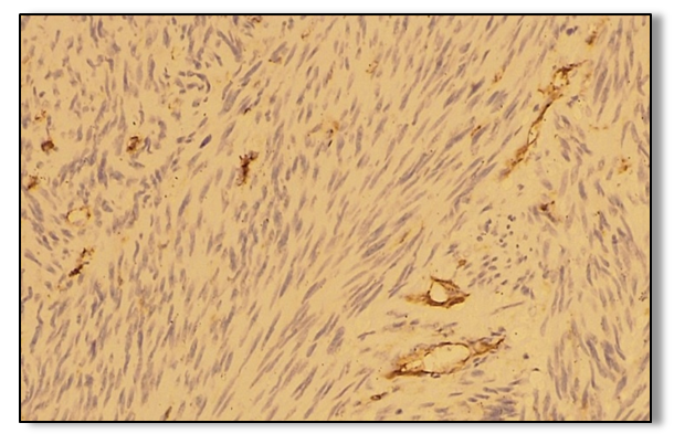 | Rice 1.1. Positive vascular endothelial response to the VEGF marker in malignant fibrous histocytoma. IGX - Dub Chromagen. About. 10 x 40 |
When immunohistochemical analysis of the gene Ki -67 found that the results were evaluated as a mild, moderate and severe positive reaction. Of the 20 patients observed, 2 (10%) had a weak positive response, 4 (20%) had a moderate positive response, and 14 (70%) had a high positive response.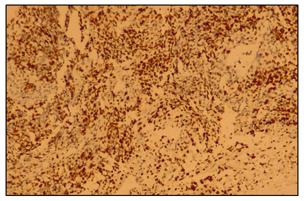 | Rice 1.2. Malignant fibrous histiocytoma - highly positive reaction to the Ki-67 gene. IGX - Dub Chromogen. About. 10 x 40 |
The weakly positive reaction was characterized by 10% with low activity, the average activity was 10-20% and the high proliferative activity was more than 20%.Table 1. The level of proliferative activity of the Ki-67 gene in malignant fibrous histocytoma (n= 20)
 |
| |
|
According to the immunohistochemical analysis, it was found that highly proliferative active cells were found in 70% of the nuclei in MFH, which indicates a high aggressive course of this tumor.In our observations with MFH, the Bcl-2 marker was used to determine tumor apoptosis, which regulates cell death by controlling the permeability of the mitochondrial membrane in patients. The results obtained were evaluated as mild, moderate and severe positive reactions. Of the 20 patients, 3 (15%) had a mild positive response, 7 (35%) had a moderate positive response, and 10 (50%) had a high positive response.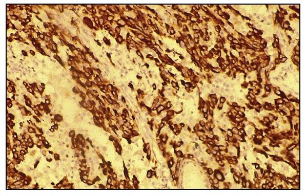 | Rice 1.3. Positive high-level Bcl-2 gene response in malignant fibrous histiocytoma. IGX - Dub Chromogen. About. 10 x 40 |
Brown staining of the mitochondral membrane, endoplasmic the reticulum and nuclear membrane at MFH indicates the presence of Bcl-2 oxidation and apoptosis. It was found that the Bcl-2 protein is present in abundance in the cytoplasm of DFG and helps to determine tumor apoptosis.According to the literature, it is known that the p53 protein, which is a tumor suppressor gene, that is, a nuclear transcription protein that helps cause damage to the genome. In addition, the suppressor gene helps the introduction of the cell cycle, as well as the further development of pathology, controls its existence. The results obtained were assessed as mild, moderate and pronounced positive reactions. Of the 20 patients, 3 (15%) had a weakly positive reaction, 7 (35%) had a moderate positive reaction, and 10 (50%) had a high positive reaction.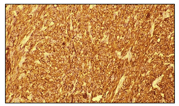 | Rice 1.4. Malignant fibrous histiocytoma p53 high positive protein. IGX - Dub Chromogen. About. 10 x 40 |
With angiosarcoma of soft tissues, we studied the density of blood vessels in 20 patients in the field of view and identified 30-40 blood vessels were positive reactions. This indicates the presence of aggressiveness and increased tumor metastasis. With angiogenic soft tissue sarcoma, out of 20 patients, 3 (15%) had a mild degree of positive reaction, 4 (20%) had an average degree of positive reaction, and 13 (65%) had a high degree of positive reaction. The results of the study show that in 4 (20%) cases, the proliferative activity of tumor cells was moderate, in 13 (65%) - high activity and 3 (15%) - mild activity. The highest degree of proliferative activity indicates the high aggressiveness of the tumor in the course of the disease.With angiogenic soft tissue sarcoma, out of 20 patients, 2 (10%) had a mild degree of positive reaction and 14 (70%) had a high degree of positive reaction in the determination of bcl -2 protein. The results of the study show that in 70% of cases there is a high activity of apoptosis in tumor cells, the average and light activity of bcl -2 was in 20% and 10% of cases, respectively. The bcl -2 protein is most commonly found in the cell cytoplasm and shows apoptosis in tumor cells. When analyzing the expression of the p53 suppressor gene in soft tissue angiosarcoma, it was shown that out of 20 patients studied, 3 (15%) had a mild degree of positive reaction, 7 (35%) had an average degree of positive reaction, and 10 (50%) had a high degree of positive reaction. reactions. A high degree of positive reaction indicates an aggressive course of the tumor process.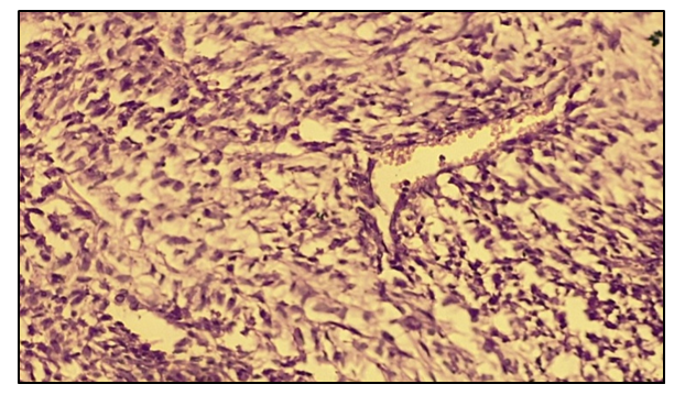 | Rice 2. Microscopic view of angiosarcoma average differentiated type. Stained with heme -eosin. About. 10 x 40 |
Patient O.M., 58 years old. Diagnosis: Angiogenic soft tissue sarcoma of the lumbar spine. The macroscopic dimensions of the tumor are 10.0x7.0x6.5 cm. Inside, the capsule oozes spots of burning and decay. In appearance, the tumor resembles fish meat. The tumor capsule was preserved. In synovial sarcoma, a positive reaction in the determination of VEGF was characterized by the presence of 35-45 vascular endothelial cells in the field of view. This is to say about the high expression of VEGF and the aggressive course of the tumor process.Indicators Ki 67 in synovial sarcoma out of 20 patients, 2 (10%) had a mild degree of positive reaction, 3 (15%) had a moderate degree of positive reaction, and 15 (75%) had a high degree of positive reaction. There was no negative reaction. In this case, the presence of high expression of Ki 67 is characterized by an aggressive course of the disease. When studying the rate of apoptosis bcl -2 in high-grade soft tissue sarcomas, synovial sarcoma, it was found that out of 20 patients, 2 (10%) had a mild degree of positive reaction, 4 (20%) had an average degree of positive reaction and 14 (70%) had a high degree of positive reaction.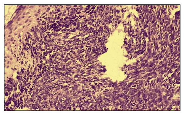 | Rice 3. Microscopic view of synovial sarcoma of a high differentiated type. Stained with heme -eosin. About. 10 x 40 |
With synovial sarcoma, brown staining of the endoplasmic reticulum and nuclear membrane is determined, which indicates the presence of the bcl -2 protein and apoptosis of tumor cells. At the same time, in 70% of cases, apoptosis is determined in proliferatively active cells.With synovial soft tissue sarcoma, p53 indicators in 2 (10%) patients had a mild positive reaction, 5 (25%) had a moderate positive reaction, and 13 (65%) had a high positive reaction.When analyzing the VEGF index among patients with soft tissue rhabdomyosarcoma, it was found that in 20 patients with microscopic examination in the field of view, the density of vessels corresponded to 10-15 vessels of endothelial cells, which was considered a positive reaction. This indicates that this form of soft tissue sarcoma was characterized by an average degree of aggressiveness due to the low supply of tumor cells with blood vessels compared to other forms of tumors. In the study of negative reactions were not observed.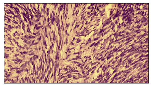 | Rice 4. Microscopic view of low-grade rhabdomyosarcoma. Heme-eosin staining. About. 10 x 40 |
When studying Ki -67 indicators, it was found that out of 20 patients, 6 (30%) had a mild degree of positive reaction, 10 (50%) had an average degree of positive reaction, and 4 (20%) had a high degree of positive reaction. There were no negative reactions. With rhabdomyosarcoma, the presence of a low and medium degree of expression (80%) indicates a less aggressive course of the tumor process and the proliferative activity of tumor cells.The study of p53 indices in soft tissue rhabdomyosarcoma showed that out of 20 patients, 6 (30%) had a mild positive reaction, 8 (40%) had a moderate positive reaction, and 6 (30%) had a high positive reaction. A high positive reaction was characteristic of the moderate degree of aggressiveness of the tumor process in rhabdomyosarcoma. There were no negative reactions.When analyzing the VEGF indices among patients with soft tissue leiomyosarcoma, it was found that in 8 patients, microscopic examination in the field of view of the density of the vessels of endothelial cells corresponded to 10-15 vessels, which was considered a positive reaction. This indicates that patients with this type of sarcoma have a low iron supply, a low ability to metastasize to other organs, and a moderately aggressive course of the disease. In the study of negative reactions were not observed.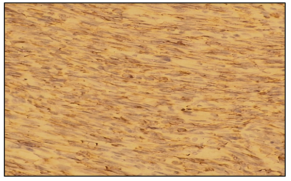 | Rice 5. Microscopic view of a leiomyosarcoma of a moderately differentiated type. Heme-eosin staining. About. 10 x 40 |
When studying Ki 67 indicators, it was found that out of 8 patients, 2 (25%) had a mild degree of positive reaction, 5 (62.5%) had an average degree of positive reaction, and 1 (12.5%) had a high degree of positive reaction. There were no negative reactions.When studying the bcl -2 indicators, it was found that out of 8 patients, 2 (25%) had a mild degree of positive reaction, 4 (50%) had an average degree of positive reaction, and 2 (25%) had a high degree of positive reaction. There were no negative reactions.The study of p53 parameters in soft tissue leiomyosarcoma found that out of 8 patients, 1 (12.5%) had a mild degree of positive reaction, 6 (75%) had an average degree of positive reaction, and 1 (12.5%) had a high degree of positive reaction. positive reaction.The average positive reaction was typical for the average degree of aggressiveness of the tumor process in leiomyosarcoma. There were no negative reactions. Table 2. VEGF in high-grade soft tissue sarcomas
 |
| |
|
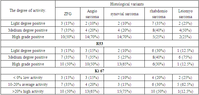
Table 3. Parameters of immunohistochemical markers in high-grade soft tissue sarcomas bcl -2
 |
| |
|
4. Conclusions
The analysis of the immunohistochemical study showed that in high-grade soft tissue sarcomas there is a certain profile for each histological variant, its severity largely determines the aggressiveness of the tumor process. This must be taken into account in determining the tactics of treatment, in the choice of chemotherapy, targeted therapy, and the prognosis of the disease. Studied immunohistochemical markers VEGF, Ki -67, bcl -2 and p 53 each of them has its own characteristics in determining tumor aggressiveness and can be recommended as a prognostic factor in high-grade soft tissue sarcomas.
References
| [1] | Abdullaev R.Ya., Golovko T.S., Khvistyuk A.N. and other Ultrasonic diagnosis of tumors of the musculoskeletal system. - Kharkov: New word, 2008.- 128 p. |
| [2] | Gorbunova V.A. New directions of drug treatment of soft tissue sarcomas // Medical and organizational problems in generalized sarcomas, 2009 RONTS im. Blokhin. Report at the 2009 meeting. |
| [3] | Gorbunova V.A. New approaches in the drug treatment of soft tissue sarcomas // Sarcomas of bones, soft tissues and skin tumors. - 2009. - No. 1. - S. 38-45. |
| [4] | Adamyan A.A., Romashov Yu.V., Adzhieva Z.A. Surgical correction of soft tissue deformities of the lower extremities // Analyzes of plastic, reconstructive surgery. - 2006. - No. 3. - S. 30-32. |
| [5] | Bosch A.JI., Fos S.N. The study of SMT: from immunohistochemistry to molecular biology. Practical approach // Sarcomas of bones, soft tissues and skin tumors. - 2010. -№1. - S. 24-30. |
| [6] | Aliev M.D. Introduction to oncoorthopedics // Sarcomas of bones, soft tissues and skin tumors. - 2009. - Т1. - S. 14-17. |
| [7] | Babichenko I.I., Kovyazin V.A. New methods of immunohistochemical diagnosis of tumor growth: Textbook. - M.: RUDN, 2008. - 109 p. |
| [8] | Bullwinkel, J., Baron-Luhr, B., Ludemann, A., Wolenberg, S., Gerdes, J., and Scholzen, T. (March 2006). "The Ki-67 protein is associated with ribosomal RNA transcription in resting and proliferating cells". J. Sell. Physiol. 206(3): 624–35. |
| [9] | Rahmanzadeh R, Hutmann G., Gerdes J., Scholzen T. (June 2007). "Light inactivation of pKi-67 by a chromophore results in inhibition of ribosomal RNA synthesis". Cell Profile. 40(3): 422–30. |


 Abstract
 Reference
 Full-Text PDF
 Full-text HTML

