-
Paper Information
- Paper Submission
-
Journal Information
- About This Journal
- Editorial Board
- Current Issue
- Archive
- Author Guidelines
- Contact Us
American Journal of Medicine and Medical Sciences
p-ISSN: 2165-901X e-ISSN: 2165-9036
2023; 13(10): 1545-1549
doi:10.5923/j.ajmms.20231310.37
Received: Oct. 3, 2023; Accepted: Oct. 17, 2023; Published: Oct. 20, 2023

Comparative Morphological and Morphometric Changes in the Gallbladder Wall of Dogs and Rabbits in Conditions of Experimental Calculus and Inflammatory Cholecystitis
Boboev Askar Ibodullaevich, Oripov Firdavs Suratovich
Samarkand State Medical University, Siab College of Public Health Named after Abu Ali Ibn Sina, Samarkand, Uzbekistan
Correspondence to: Boboev Askar Ibodullaevich, Samarkand State Medical University, Siab College of Public Health Named after Abu Ali Ibn Sina, Samarkand, Uzbekistan.
| Email: |  |
Copyright © 2023 The Author(s). Published by Scientific & Academic Publishing.
This work is licensed under the Creative Commons Attribution International License (CC BY).
http://creativecommons.org/licenses/by/4.0/

This article presents a model of experimental calculous cholecystitis in mammals. The morphology and morphometric data of different layers of the gallbladder wall are described. The results of the study showed that the morphometric parameters of the mucous, muscular-fibrous layers in rabbits and dogs of the experimental group were significantly higher than in animals of the control group. It was found that the gallbladder in the experimental group was significantly thickened compared to the control group of animals. This is a reaction of the gallbladder wall in response to calculous cholecystitis, as a result of which inflammatory processes develop, caused by the mechanical action of stones on the gallbladder wall. The data given in the article can serve as the necessary information for doctors to make a timely diagnosis and choose the right tactics for treating patients with such pathological processes. At the same time, it helps to prevent postoperative complications and properly conduct the rehabilitation process.
Keywords: Experimental animals, Hepatobiliary system, Calculous cholecystitis, Gallbladder, Morphological changes
Cite this paper: Boboev Askar Ibodullaevich, Oripov Firdavs Suratovich, Comparative Morphological and Morphometric Changes in the Gallbladder Wall of Dogs and Rabbits in Conditions of Experimental Calculus and Inflammatory Cholecystitis, American Journal of Medicine and Medical Sciences, Vol. 13 No. 10, 2023, pp. 1545-1549. doi: 10.5923/j.ajmms.20231310.37.
1. Introduction
- According to statistics from the World Health Organization, various pathological conditions of the hepatobiliary system occur in people aged 15 to 95 years, and gallstones occur in one in ten thousand of the world's population. This pathological process is more common in men over forty years old, but the results of research by scientists show that this disease is getting younger every year. Changes and problems that occur in the liver and gallbladder as a result of this pathology attract the attention of many researchers. Scientists studied the structure and function of the liver in experimental hepatitis in dogs [3], morphological changes in the liver in acute cholecystitis [20], obstructive jaundice, acute cholangitis, biliary sepsis and their pathogenetic relationship, principles of differential diagnosis [9], morphology of nervous structures of the common bile duct [11], age-related features of morphological changes in the membranes of the gallbladder in acute cholecystitis [19], comparative morphology of the liver and gallbladder of humans and laboratory animals [17] were studied. Some researchers studied the bacterial flora and histological structure of choledocholithiasis in patients with choledocholithiasis and cholangitis [5], the morphological structure and correction of the liver in cholelithiasis [14], morphological changes in cholelithiasis in chronic cholecystitis [6], the formation of cholelithiasis in children, observed the frequency incidence of anomalies of the gallbladder in Gilbert's syndrome [15], differences in the conservative or surgical treatment of gallstone disease in young children [16], morphological changes in gallstones [1]. Investigators analyzed the morphological features of benign gallbladder tumors [12], the sonographic assessment of cholesterol gallstones in the general population [10], age-related anatomical features of the liver and gallbladder in adolescents and adults [22], and morphological features were determined. esophagogastroduodenal region in individuals with cholecystectomy [8,18]. Other scientists studied the specific effect of laser light on the morphology of the liver and biliary tract in cholelithiasis [23], morphological changes in the liver in calculous cholecystitis in overweight people [21], the interaction of inflammatory mediators and morphological changes in the gallbladder in destructive form of cholecystitis. Researchers [4,7] noticed that each type (form) of calculous cholecystitis has its own morphological features. Scientists have established complications in the biliary tract after cholecystectomy [13], functional and morphological changes in the liver in experimental acute obstructive cholestasis [2,18]. The results of the study of experimental studies conducted in the clinic and on mammals show that diseases of the biliary system have not been studied enough, and this, in turn, remains an urgent problem for practical and theoretical medicine. These data show how common the pathology of the liver, gallbladder and biliary tract is among the population, and it remains one of the unresolved urgent problems of medicine. Various internal and external factors can cause morphological changes in the morph functional structure of the gallbladder. The scientific research that we set ourselves is aimed at studying the little-studied aspects of this problem.
2. Material and Methods
- The gallbladder of dogs and rabbits was taken as material. Experimental animals were studied in two groups. The first group consisted of [7] dogs and [8] rabbits in the control group. The second group of animals was an experimental group of [14] dogs and [16] rabbits and was called a model of calculous cholecystitis. To do this, the animals of the experimental group were surgically opened the gallbladder under anesthesia and poured 4-6 non-sterile stones into it. Animals in the control group were surgically opened and again sutured under anesthesia. Animals of both the control and experimental groups were kept under the same vivarium conditions. Animals of both groups were euthanized under anesthesia one month after the operation. When looking for gallstones in the animals of the experimental group, stones were found in the common bile duct, and in some animals even in the intestines. Gallbladder material obtained from slaughtered animals was fixed in 12% formalin and embedded in paraffin to prepare histological sections. The resulting sections were stained with hematoxylin-eosin and the Van Gieson method. The thickness of the layers of the gallbladder wall was measured using an optical ruler, and the obtained digital data were subjected to statistical processing.
3. Rsults Research
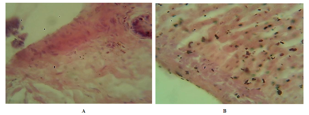
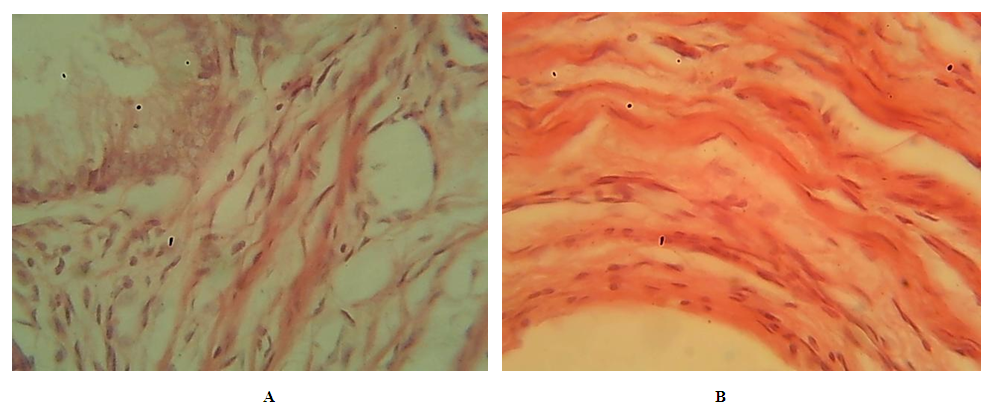
- The gallbladder (vesica fe ll ea) is elongated pear-shaped. It has a base, body, funnel and neck. The length of the gallbladder is about 10 cm, and its base reaches the anterior edge of the liver. The gallbladder is one of the important organs of the digestive system, it collects and condenses the bile produced by the liver. When food is eaten, the fluid secreted from the gallbladder enters the duodenum and activates the enzymes involved in the digestion of food, breaking down the fats contained in it. The body contains about 50-80 ml of fluid and fluid. If there are no problems with digestion, the gallbladder is also actively working. Otherwise, bile, i.e. serum, can accumulate in it, stone formation and other diseases can occur. Mucous, muscular-fibrous, adventitial (serous, covering only the lower surface) membranes are isolated on the wall of the gallbladder. The mucous membrane of the gallbladder consists of a special plate, consisting of epithelium and connective tissue, formed by multi-branched folds. The mucous membrane of the bile ducts located outside the gallbladder and liver is covered with a single-layer prismatic epithelium with a cuticular border in the apical part, and the nuclei of these cells are located in the basal part. Among the prismatic cells of the epithelium of the mucous membrane there are goblet cells, and in the region of the neck of the bladder there are mucous glands. The proper layer consists of loose fibrous irregular connective tissue rich in blood vessels. The muscular-fibrous membrane of the gallbladder consists of bundles of smooth muscles directed in different directions. In the region of the body of the gallbladder, the muscles are located longitudinally, and in the cervical part - along the circumference. Between the muscle fibers are layers of connective tissue. The gallbladder is surrounded by an adventitial membrane, consisting of loose fibrous irregular connective tissue that contains large blood vessels and nerves.The gallbladder walls of rabbits and dogs consist of mucosal, musculoskeletal, and adventitial layers lined with a single layer of prismatic epithelium, like the human gallbladder. In dogs and rabbits of the experimental group, swelling and thickening of the gallbladder wall, foci of leukocyte infiltration can be observed. Animals of the experimental group are characterized by the presence in the walls of the gallbladder, fibrosis and foci of inflammatory infiltration, and therefore there is a significant thickening of the wall of the gallbladder compared to the animals of the control group. (Fig. 1.2).
 | Figure 1. The structure of the gallbladder wall of control animals. A-with both; B rabbit. Hematoxylin-eosin staining. Ok. 7, vol. 40 |
 | Figure 2. The structure of the gallbladder wall of animals of the experimental group. A-with both; B rabbit. Hematoxylin-eosin staining. Ok. 7, vol. 40 |
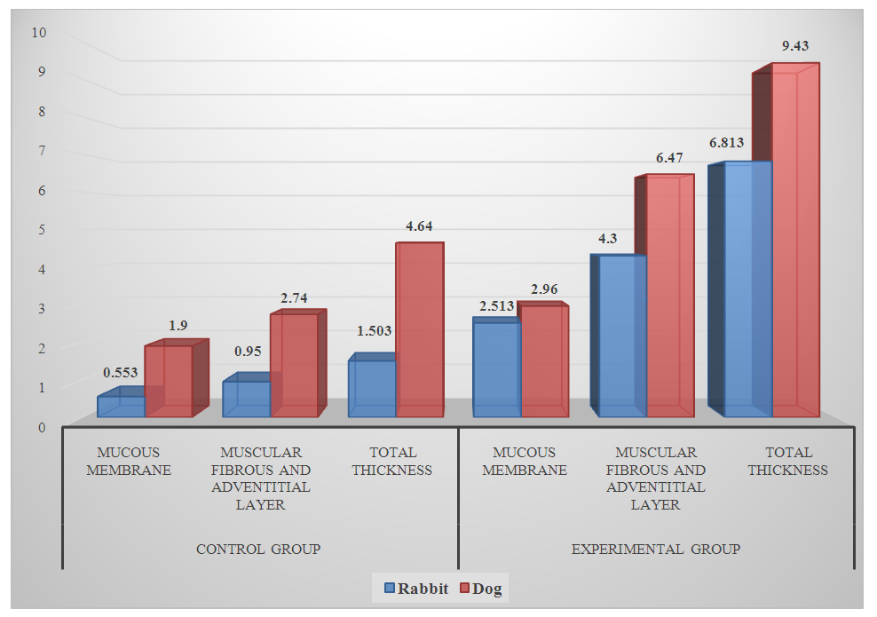
 | Figure 3. Comparative morphometric parameters of the gallbladder wall of mammals in the control and experimental groups |
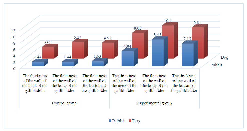
 | Figure 4. Comparative morphometric parameters of different parts of the gallbladder wall in mammals |
4. Conclusions
- With experimental calculous cholecystitis in experimental rabbits, the morphometric parameters of the mucous and muscular-fibrous layers, as shown by statistical data, in rabbits of the experimental group, the thickness increases by 4-6 times, and in dogs only twice. When comparing the thickness of different sections of the gallbladder separately in rabbits of the experimental group, a thickening of 3-6 times is also noted, but in dogs it is twice as compared with the control group of animals. The results of the study showed that the morphology of the gallbladder wall changes to varying degrees in experimental cholelithiasis in rabbits and dogs, which is most likely due to the nature of the nutrition of the above animals.
 Abstract
 Reference
 Full-Text PDF
 Full-text HTML