American Journal of Medicine and Medical Sciences

p-ISSN: 2165-901X    e-ISSN: 2165-9036

2023;  13(10): 1477-1481

doi:10.5923/j.ajmms.20231310.25

Received: Aug. 20, 2023; Accepted: Sep. 25, 2023; Published: Oct. 10, 2023


Features of the Clinical Course and Diagnosis of Hyperandrogenia in Mayer-Rokitanski-Kuster-Hauser Syndrome

Adilova M. N.1, Negmadjanov B. B.2, Rabbimova G. T.3

1Applicant of the Department of Obstetrics and Gynecology No. 2, Samarkand Medical University, Uzbekistan

2Doctor of Medical Sciences, Professor, Samarkand Medical University, Uzbekistan

3Candidate of Medical Sciences, Associate Professor, Samarkand Medical University, Uzbekistan

Copyright © 2023 The Author(s). Published by Scientific & Academic Publishing.

This work is licensed under the Creative Commons Attribution International License (CC BY).
http://creativecommons.org/licenses/by/4.0/

Abstract

Congenital utero-vaginal aplasia or aplasia Mayer-Rokitansky-Küster-Hauser syndrome (MRKH) is characterized by unfused uterine buds, aplasia of the cervix and vagina, but normal or hypoplastic bilateral adnexa and is clinically manifested by primary amenorrhea. Patients with MRKH have a normal developmental female phenotype and karyotype (46,XX) and an incidence of 1 in 4000 or 5000 births (Cheroki et al. 2006). MRKH syndrome is considered to be a malformation of the Müllerian (paramesonephric) ducts, occurring in utero between 4 and 12 weeks of gestation, in the oviducts, uterus, cervix and upper vagina, while the Wolffian (mesonephric) ducts regress without fusing with the Müllerian ducts ( Ludwig 1998).

Keywords: Clinical, Course, Mayer-rokitanski

Cite this paper: Adilova M. N., Negmadjanov B. B., Rabbimova G. T., Features of the Clinical Course and Diagnosis of Hyperandrogenia in Mayer-Rokitanski-Kuster-Hauser Syndrome, American Journal of Medicine and Medical Sciences, Vol. 13 No. 10, 2023, pp. 1477-1481. doi: 10.5923/j.ajmms.20231310.25.

1. Introduction

Mayer-Rokitansky-Küster-Hauser syndrome (MRKH) is a rare congenital disorder of the female reproductive system that is characterized by the absence or underdevelopment of the uterus and vagina. However, women with MRKH may experience hyperandrogenism, which is associated with increased levels of male hormones in the blood. Hyperandrogenism (HA), on the other hand, is a condition in which the levels of male hormones such as testosterone, dihydrotestosterone and dehydroepiandrosterone (DHEA) are elevated in a woman. According to the literature, 10–20% of women exhibit certain signs; HA is not only a medical, but also a social problem, since it leads to various cosmetic defects (excessive oily skin and hair, the development of acne), as well as psycho-emotional reactions that reduce the quality of life of women, as well as social problems associated with restrictions in choice professions and employment. In women with MRKH, hyperandrogenism may occur due to dysfunction of the ovaries, which continue to secrete male hormones into the blood despite the absence of the uterus and vagina. Symptoms of hyperandrogenism may include a rough voice, increased muscle mass, increased hair growth on the face, chest, and abdomen, and menstrual irregularities. In some cases, hyperandrogenism can lead to impaired fertility, as well as an increased risk of developing cardiovascular disease and diabetes. To diagnose hyperandrogenism in patients with MRKH, it is necessary to conduct a comprehensive examination, including analysis of the level of male hormones in the blood, assessment of the clinical manifestations of hyperandrogenism, ultrasound examination of the ovaries, assessment of insulin and blood sugar levels, as well as genetic studies. Treatment of hyperandrogenism in MRKH syndrome may include the use of hormonal medications, such as contraceptives or androgen antagonists, which reduce the level of male hormones in the blood and improve the clinical symptoms of hyperandrogenism. There is little literary data on the endocrinological features of the condition of the ovaries with aplasia of the uterus and vagina, and they are contradictory. There are isolated publications on data on hyperandrogenism syndrome in patients with aplasia of the vagina and uterus. Therefore, the study of patients with aplasia of the vagina and uterus in combination with hyperandrogenism of ovarian origin, the characteristics of clinical and diagnostic criteria are relevant.
The purpose of the study was to study the features of the clinical course and diagnosis of hyperandrogenism syndrome with aplasia of the vagina and uterus.

2. Material and Research Methods

The studies were carried out in the department of pediatric gynecology of a multidisciplinary medical center and the private company “Doctor” in the period from 2009-2023. Clinical data and results of diagnostic criteria of 110 patients with Rokitansky-Küster-Hauser-Mayer syndrome were studied. Criteria for inclusion in the main group: patients with aplasia of the vagina and uterus in combination with one or more clinical (acne, alopecia, hirsutism) manifestations of hyperandrogenism (clinical and laboratory signs) aged 16-35 years; Exclusion criteria: patients with aplasia of the vagina and uterus aged less than 16 and more than 35 years; androgen-producing tumors, hypothyroidism, type 1 diabetes mellitus, hyperprolactinemia of organic origin, hypercortisolism, taking medications with side effects of hyperandrogenism. Determination of hormone levels was carried out in all patients at the first visit and over time using enzyme immunoassay. Hormones such as testosterone, TSH, LH, FSH, DHEAS, and prolactin were determined using an enzyme immunoassay in the laboratory. Assessment of the severity of acne and hirsutism in hyperandrogenism of ovarian origin can be carried out using various scales and indices. One of the most common indices of hirsutism is the Ferriman-Gallwey index, which evaluates the level of hairiness on the face, chest, abdomen, back and upper extremities on a 9-point scale. According to the Acne Qualification Group scale, the severity of acne was assessed based on the number of inflammatory elements on the face, as well as the number of comedones.

3. Research Results

We assessed the severity of acne and hirsutism in patients of the study groups. There is an opinion that the clinical symptoms of hyperandrogenism develop in direct dependence on the degree of androgen metabolism disorders. When assessing the severity of acne, the most commonly used classification of the American Academy of Dermatology was used: grade I is characterized by the presence of comedones and up to 10 papules; II degree – comedones, papules, up to 10 pustules; III degree – comedones, papules, pustules, up to 5 nodes; IV degree is characterized by a pronounced inflammatory reaction in the deep layers with the formation of painful nodes and cysts. When examining patients, 30.8% of subjects were diagnosed with acne of varying severity. When assessing the severity of acne, the most commonly used classification of the American Academy of Dermatology was used: grade I is characterized by the presence of comedones and up to 10 papules; II degree – comedones, papules, up to 10 pustules; III degree – comedones, papules, pustules, up to 5 nodes; IV degree is characterized by a pronounced inflammatory reaction in the deep layers with the formation of painful nodes and cysts. Based on the accepted classification, the vast majority of patients in the main clinical group had grade I acne in 46.6% of cases, and grade II acne was noted in 25.6% of patients. Grades III and IV (the most severe) were recorded in 11.5% and 16.2% of cases, respectively. Despite the lack of a clear relationship between the severity of acne and the presence of biochemical markers of hyperandrogenism, it should be noted that the subjects most often had II and III severity of acne (p = 0.001). In the control group, there were no cases of acne in accordance with the accepted classification. The most significant symptom in the diagnosis of hyperandrogenism is hirsutism. In contrast, acne or seborrhea is more often the result of changes in androgen metabolism. In this regard, in all patients, the hirsute number was determined by the sum of the indifferent (the degree of hair growth of the forearms and legs) and hormonal numbers (the degree of hair growth of the remaining 9 areas of the body), using the classic 4-point Ferriman-Gallwey scale by D. Ferriman and J. Gallwey [3].
The total number of points (hirsut number) from 1 to 7 characterizes normal hair growth, from 8 to 11 points – borderline, more than 12 points – hirsutism. Hair growth is considered excessive when the hirsute number is more than 12 points. In the control group, hirsutism was not detected in any case. In the main group, a hirsute number of more than 12 points on the Ferriman-Gallway scale was detected in 33.3%, in 53.3% of cases the hirsute number was within the border zone from 8 to 11 points (Table 1).
Table 1. Assessment of the degree of hirsutism in patients of the study groups
     
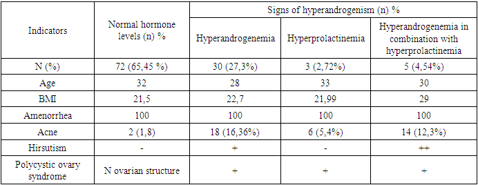
Depending on the level of hormones, patients of the main group are divided into subgroups: patients with hyperandrogenemia, hyperprolactinemia, hyperandrogenemia in combination with hyperaprolactinemia (Table 2).
Тable 2. Clinical signs of hyperandrogenism in patients with aplasia of the vagina and uterus
     
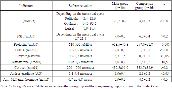
As a result of the study, a high incidence of various complaints in patients with HA syndrome was established, and features of the dynamics of hormone levels were determined. In MRKH syndrome in combination with hyperandrogenism, there were statistically significant differences in the hormonal background, namely, the level of LH, FSH, testosterone, prolactin, as well as the level of AMH (anti-Mullerian hormone) were statistically significantly lower than those of the comparison group. The endocrine status of patients with MRKH syndrome was characterized by the presence of hyperandrogenism of ovarian origin with significantly lower AMH values. Thus, patients with aplasia of the uterus and vagina are at risk for the development of various endocrine disorders and therefore require more thorough examination and continuity of management to determine the timing and methods of reproductive function.
Тable 3. Hormonal background of patients with SMRCC
     
Table 4. Indicators of lipid metabolism in patients with SMRKH combined with hyperandrogenism of ovarian origin
     
As can be seen from the table, the values of low-density lipoproteins and high-density lipoproteins are statistically significantly different from each other. Thus, the level of LDL is 3.8 times higher in patients with aplasia of the vagina and uterus in combination with hyperandrogenism, and HDL is 3.3 times lower than normal values, this indicates a violation of lipid metabolism in patients of the main group.
Considering that one of the mechanisms of the pathogenesis of PCOS is insulin resistance, we examined the level of fasting insulin, the level of glucose in the blood of patients of both groups and the HOMA index (Table 5).
Table 5. Data from a survey of carbohydrate metabolism in obese women
     
The table shows that blood glucose levels and impaired glucose tolerance differ statistically significantly between the main group and the comparison group. In addition, insulin levels and the presence of hyperinsulinemia were also statistically significantly different between the two groups. However, the HOMA index normally does not have statistically significant differences.
Thus, indicators of carbohydrate and lipid metabolism in patients with aplasia of the vagina and uterus in combination with hyperandrogenism of ovarian origin indicate a violation of carbohydrate metabolism, characterized by an increase in the level of glucose and insulin in the blood, as well as a decrease in sensitivity to insulin, which can lead to the development of diabetes. And levels of lipids, such as cholesterol and triglycerides, are elevated in the blood of patients with this condition, while HDL levels are decreased. This may increase the risk of developing cardiovascular diseases such as atherosclerosis and myocardial infarction.
Thus, based on the results of the analysis, we can conclude that MRKH syndrome type II (complex type) was observed in 30% of patients with aplasia of the uterus and vagina, most often combined with an abnormality of the urinary system. The most common clinical manifestations are a combination of hyperandrogenemia, hyperprolactinemia, and hyperandrogenemia in combination with hyperprolactinemia. During an ultrasound examination of MRKH syndrome, every 2nd girl had an abnormally high location of the ovaries, which had an elongated shape and a small-caliber follicular structure. The hormonal status of patients with MRKH syndrome was characterized by the presence of signs of ovarian hyperandrogenism with significantly lower AMH values compared to healthy peers. Patients with aplasia of the uterus and vagina are at risk for the development of various endocrine disorders and therefore require more thorough examination and continuity of management to determine the timing and methods of reproductive function.

References

[1]  Adamyan L.V. et al. Aplasia of the uterus and vagina (Mayer-Rokitansky-Küster-Hauser syndrome) in combination with endometriosis: unresolved aspects of etiology and pathogenesis (literature review) // Problemy Reproduktsii. – 2016. – T. 22. – No. 3.
[2]  Boyarsky K.Yu. Ovarian stimulation and folliculogenesis in the late 90s: on the threshold of the future (literature review) // Problems of reproduction - 1997. - No. 4. - pp. 11-15. Вихляева Е.М. Болезнь поликистозных яичников // Азиатский вестник акушеров-гинекологов. - 1999. - Т.5. - № 1-2. - С. 60-73.
[3]  Gasparov A.S., Kulakov V.I., Bogdanova E.D. Features of the clinical course and effectiveness of treatment of polycystic ovary disease in adolescence and mature reproductive ages // Problems of reproduction. - 1995. - No. 4. - pp. 11-17.
[4]  Demidov V.N., Alieva E.A. Strukov A.V. Possibilities of echography in the diagnosis of polycystic ovary syndrome // Obstetrics and gynecology. - 1991. - No. 1. - P. 40-42.
[5]  Demidov V.N., Zykin B.I. Ultrasound diagnostics in gynecology. - Medicine: M., 1990. - P. 123-125.
[6]  Clinical guide to ultrasound diagnostics / Ed. Mitkova V.V., Medvedeva M.V. - M., 1997. - T.3. - pp. 132-174.
[7]  Kruglyak D. A., Buralkina N. A., Ipatova M. V., Uvarova E. V. Mayer–Rokitansky–Küster–Hauser syndrome: modern treatment methods, psychological and social aspects (analytical review) // Reproductive Health children and teenagers. 2018. No. 3. URL: https://cyberleninka.ru/article/n/sindrom-mayera-rokitanskogo-kyustera-hauzera-sovremennye-metodiki-lecheniya-psihologicheskie-i-sotsialnye-aspekty-analiticheskiy (access date: 09.14.2022).
[8]  Manukhin L.B., Gevorkyan M.A. Polycystic ovary syndrome // Problems of reproduction. - 1999. - No. 6. - pp. 21-26.
[9]  Pishchulin A.A., Butov A.V., Udovichenko O.V. Ovarian hyperandrogenism syndrome of non-tumor origin (literature review) // Problems of reproduction. - 1999. - N3. - pp. 24-28.
[10]  Raisova A.T. Current problems of hyperandrogenism // Clinician. - 1995. - N3. - pp. 54-59.
[11]  Strizhakov A.N., Davydov A.I. Clinical transvaginal echography. - M., 1994. - P. 121-144.
[12]  Czech E. Ultrasound diagnostics in obstetrics and gynecology (translated from Czech). - M., 1982.
[13]  Ai Z, Zhu X, Chen H, Chen R. Precocious puberty or growth hormone deficiency as initial presentation in Mayer-Rokitansky-kuster-Hauser syndrome: a clinical report of 5 cases. BMC Pediatr. 2022 Jul 14; 22(1): 418. doi: 10.1186/s12887-022-03474-0. PMID: 35836205; PMCID: PMC9281080.
[14]  Botsis D., Kassanos D., Pyrgiotis E., Zourlas P.A. Sonographic incidenсe of polycystic ovaries in a gynecological population // Ultrasound-Obstet. Gynec, 1995, Sept. 6(3): 182-5.
[15]  Campbell S., Goessens L., Goswany R. Real-time ultrasonography for determination of ovarian morphology and volume // Lancet, 1982, 1, 8269, p. 425-426.
[16]  Dewailly D., Duhamel A., Robert Y. et al. Interrelationship between ultrasonography and biology in the diagnosis of polycystic ovarian syndrome // Ann. N.Y. Acad. Sci., May 28, N 687, p. 206-216.
[17]  Dobson M.G. Transvaginal ultrasound // Churchill Livingstone, 1995.
[18]  Filicori M., Flamigni C. The Ovary: Regulation, Dysfunction and Treatment // Bologna (Italy), 1996.
[19]  Franks S. Ultrasound diagnosis of PCO. In Аdvances in Polycystic Ovary Disease, Mayo Clinic, Ro chester, April 1996.
[20]  Goldstein S.R., TimorATritsch I.E. Ultrasound in Gynecology // Churchill Livingstone Inc., 1995.
[21]  Henes M, Jurow L, Peter A, Schoenfisch B, Taran FA, Huebner M, Seeger H, Brucker SY, Rall KK. Hyperandrogenemia and ovarian reserve in patients with Mayer-Rokitansky-Küster-Hauser syndrome type 1 and 2: potential influences on ovarian stimulation. Arch Gynecol Obstet. 2018 Feb; 297(2): 513-520. doi: 10.1007/s00404-017-4596-1. Epub 2017 Nov 24. PMID: 29177592.
[22]  Odhaib SA, Mohammed MJ, Al-Ali AJH, Mansour AA. Clinical and Radiological Findings in Mayer-Rokitansky-Küster-Hauser Syndrome Type 2: Case report. Sultan Qaboos Univ Med J. 2021 Nov; 21(4): 635-638. doi: 10.18295/squmj.4.2021.036. Epub 2021 Nov 25. PMID: 34888086; PMCID: PMC8631212.
[23]  Peters HE, Laeven CHC, Trimbos CJMA, van de Ven PM, Verhoeven MO, Schats R, Mijatovic V, Lambalk CB. Anthropometric biomarkers for abnormal prenatal reproductive hormone exposure in women with Mayer-Rokitanksy-Küster-Hauser syndrome, polycystic ovary syndrome, and endometriosis. Fertil Steril. 2020 Dec; 114(6): 1297-1305. doi: 10.1016/j.fertnstert.2020.06.029. Epub 2020 Oct 6. PMID: 33036791.
[24]  Rall K, Conzelmann G, Schäffeler N, Henes M, Wallwiener D, Möhrle M, Brucker SY. Acne and PCOS are less frequent in women with Mayer-Rokitansky-Küster-Hauser syndrome despite a high rate of hyperandrogenemia: a cross-sectional study. Reprod Biol Endocrinol. 2014 Mar 18; 12:23. doi: 10.1186/1477-7827-12-23. PMID: 24641817; PMCID: PMC4003801.
[25]  Rosenberg C., Pardo J., Kaplan B. et al. A modified transvaginal sonographic technique for better ovary evaluаtion // Ultras. Obstetr. Gynec., 1992, v. 2, N 1, p. 153.
[26]  Tsarna E, Eleftheriades A, Eleftheriades M, Kalampokas E, Liakopoulou MK, Christopoulos P. The impact of Mayer-Rokitansky-Küster-Hauser Syndrome on Psychology, Quality of Life, and Sexual Life of Patients: A Systematic Review. Children (Basel). 2022 Apr 1; 9(4): 484. doi: 10.3390/children9040484. PMID: 35455528; PMCID: PMC9025055.
[27]  Wang Y, He YL, Yuan L, Yu JC, Xue HD, Jin ZY. Typical and atypical pelvic MRI characteristics of Mayer-Rokitansky-Küster-Hauser syndrome: a comprehensive analysis of 201 patients. Eur Radiol. 2020 Jul; 30(7): 4014-4022. doi: 10.1007/s00330-020-06681-4. Epub 2020 Mar 5. PMID: 32140817.