-
Paper Information
- Previous Paper
- Paper Submission
-
Journal Information
- About This Journal
- Editorial Board
- Current Issue
- Archive
- Author Guidelines
- Contact Us
American Journal of Medicine and Medical Sciences
p-ISSN: 2165-901X e-ISSN: 2165-9036
2023; 13(10): 1444-1447
doi:10.5923/j.ajmms.20231310.18
Received: Sep. 12, 2023; Accepted: Sep. 28, 2023; Published: Oct. 7, 2023

Pathomorphological Changes in the Brain Cortex of Rats under Different Times of Exposure to Energy Drink
 Abstract
 Reference
 Full-Text PDF
 Full-text HTML
Oripov Firdavs Suratovich, Eshkabilova Surayyo Turaevna
Samarkand State Medical University, Samarkand, Republic of Uzbekistan
Copyright © 2023 The Author(s). Published by Scientific & Academic Publishing.
This work is licensed under the Creative Commons Attribution International License (CC BY).
http://creativecommons.org/licenses/by/4.0/

In the 21st century, an urgent problem is the excessive popularity of energy drinks, which have a stimulating and tonic effect on the human nervous system. Energy drinks are non-alcoholic or low-alcohol highly carbonated drinks containing a large amount of caffeine and other biologically active substances. Currently, schoolchildren, students and ordinary people engaged in mental work in large numbers use them during a period of special workload at work or in school. There are quite a few reports in the available literature about the harmful effects of energy drinks on the body. This article presents the results of a study of the cellular and tissue structures of the brain of rats that were orally administered the Iguana energy drink for 30, 60 and 90 days. We have established, to varying degrees, alterative-dystrophic and necrobiotic changes in glial and ganglion cells and the vascular system of the brain of rats, which significantly depend on the timing of the intake of energy drinks.
Keywords: Energy drinks, Caffeine, Biologically active substances a, Morphology, Nervous system, Cerebral cortex, Vascular system of the brain
Cite this paper: Oripov Firdavs Suratovich, Eshkabilova Surayyo Turaevna, Pathomorphological Changes in the Brain Cortex of Rats under Different Times of Exposure to Energy Drink, American Journal of Medicine and Medical Sciences, Vol. 13 No. 10, 2023, pp. 1444-1447. doi: 10.5923/j.ajmms.20231310.18.
Article Outline
1. Purpose and Objectives of the Study
- Energy drinks are soft drinks that are typically high in caffeine and other stimulants, and may also be fortified with plant extracts and nutrients (such as vitamin B, taurine, and amino acids). These drinks are becoming more and more popular among teens and young adults in the US and around the world. [1]. The attraction and consumption of energy drinks among young people, especially students, is related to both the pleasant taste and the promise of being a stimulant that keeps them awake [2].These drinks appeared on the market in the 1960s in Europe and Asia, and since the end of the last century they have spread throughout the world [3]. There are reports in the literature about the short- and long-term effects of energy drink consumption on the cardiovascular and central nervous systems. Energy drinks have been shown to cause a number of side effects, especially in young adults, including high blood pressure, serious cardiovascular disease, kidney disease, metabolic disturbances, poor sleep, seizures, and neuropsychiatric side effects [4,5,6,8]. The power engineer does not give energy, it only opens the energy channels of the organism itself. A person does not receive energy, but uses his internal resources.Excessive consumption of energy drinks containing caffeine and taurine, which are potent psychoactive substances, can modify neurotransmission and inevitably affect the functioning of the nervous systems. Disorders of the emotional sphere, the appearance of unmotivated fear, the development depression, sleep disorders, appetite, an increase in the frequency of committing antisocial acts. Toblin RL et al noted the appearance aggressive behavior, disobedience to orders and insomnia in conscripts after taking energy drinks [7].Today, the opinions of scientists and specialists are divided: someone considers “energy drinks” to be completely harmless, like ordinary soda; others, on the contrary, argue that they can act like drugs, and are certainly addictive and addictive. Energy drinks have a negative impact not only on the cardiovascular system, but also cause disorders of the nervous system.In order to identify the morphological aspects of the negative impact of this kind of drinks, we decided to study in an experiment in rats structural changes in the cerebral cortex during prolonged administration of the widely used Iguana drink.
2. Materials and Research Methods
- This report presents the results of a study of structural changes in the cerebral cortex of 26 outbred white male rats weighing 220–250 g. The animals were kept under standard vivarium conditions, in ordinary cages with wire mesh at room temperature, regulated at 21°C±10°C, humidity from 45% to 50% and light/dark cycles (24/12 hours). They were fed a standard rat diet and given free access to water. The animals were allowed to acclimatize to the experimental conditions by observing them for 10 days prior to the experiment. Animals _ was divided into 4 groups:• Group I (control) included six rats.• Group II (short-term group energy drink - Iguana), included seven rats given an energy drink at a dose of 7.5 ml / kg / day orally through a gastric tube for 1 month.• Group III: included six rats treated with Iguana energy drink at a dose of 7.5 ml / kg / day orally through a gastric tube for 2 months.• Group IV (long-term energy drink group): consisted of seven rats treated with an energy drink at a dose of 7.5 ml/kg/day orally via gavage for 3 months.During the last days of the experiment, the rats were not given food at night, and then in the morning they were anesthetized with light inhalations of ether, and blood samples were taken directly from the heart for laboratory tests. Rats were slaughtered by decapitation, observing all the provisions of bioethics, the brain was removed, which was fixed in 10% neutral formalin and the cut pieces were embedded in paraffin. Histological sections were stained with hematoxylin-eosin, according to van Giesson and Nissl, who examined on a light microscope using lenses of various magnifications and microphotographed the necessary areas.
3. Research Results and Their Discussion
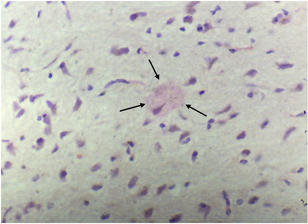
- Microscopic examination of the cerebral cortex of rats treated with Iguana for 30 days showed uniform loosening in all parts of the brain, but most pronounced in the upper layers. The perivascular areas appeared to be moderately dilated, and the pericellular areas were significantly more dilated (photo 1). There were focal extravasates, that is, diapedetic petechial hemorrhages. Single lymphocytes were found in the walls of uneven blood-filled intracerebral capillaries. When staining the sections according to Nissl, there was not a rough and not a relief violation of the cytoarchitectonics of the cerebral cortex, which was expressed by a slight increase in the size of the cells with their hypochromia (photo 2). The tigroid substance looked slightly swollen and dull.
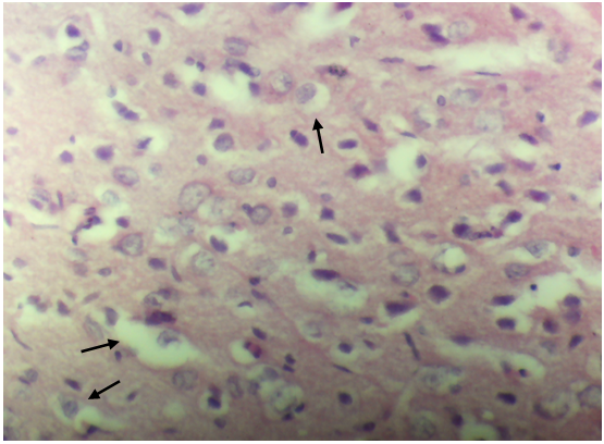 | Photo 1. The cerebral cortex. Hematoxylin-eosin staining. Ob.40. Severe pericellular edema |
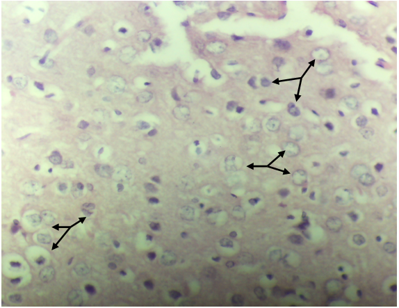 | Photo 2. The cerebral cortex. Hematoxylin-eosin staining. Ob.40. Dystrophic swelling of cells with hypochromia of their nuclei |
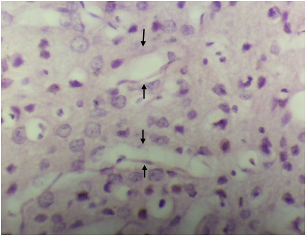 | Photo 3. The cerebral cortex. Hematoxylin-eosin staining. Ob.40. Dystonia of the walls of intracerebral capillaries |
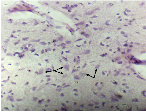 | Photo 4. The cerebral cortex. Nissl coloring. Ob.40. Swelling of the nuclei of neurons and uneven staining of the tigroid substance |
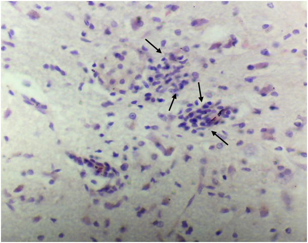 | Photo 5. Cortex. Hematoxylin-eosin staining. Ob.40. Lymphoid cell infiltrates in the walls of blood vessels |
 | Photo 6. The cerebral cortex. Nissl coloring. Ob.40. Hypochromia of the nucleus and fine-grained change in the tigroid substance |
4. Conclusions
- Based on the obtained results of histological examination, it can be concluded that with prolonged use of an energy drink a number of significant alterative-reactive changes can occur in vessels and layers cerebral cortex. The adverse side effects of this common energy drink increase with the duration of exposure. Significantly significant, widespread and relief changes in experimental rats and their absence in control animals demonstratively show their damaging side effect on the brain structures of animals.The data obtained from the results of the conducted studies can be used in the implementation of solving important social problems related to the regulation of energy drinks in society.