-
Paper Information
- Next Paper
- Previous Paper
- Paper Submission
-
Journal Information
- About This Journal
- Editorial Board
- Current Issue
- Archive
- Author Guidelines
- Contact Us
American Journal of Medicine and Medical Sciences
p-ISSN: 2165-901X e-ISSN: 2165-9036
2023; 13(9): 1336-1339
doi:10.5923/j.ajmms.20231309.35
Received: Aug. 21, 2023; Accepted: Sep. 13, 2023; Published: Sep. 28, 2023

Histological Study of Adrenal Gland in Cases of Traumatic Brain Injury
 Abstract
 Reference
 Full-Text PDF
 Full-text HTML
Sukhrob Rayimberdiev1, Ibragim Bakhriev2
1Andijan State Medical Institute, Andijan, Uzbekistan
2Tashkent Medical Academy, Tashkent, Uzbekistan
Copyright © 2023 The Author(s). Published by Scientific & Academic Publishing.
This work is licensed under the Creative Commons Attribution International License (CC BY).
http://creativecommons.org/licenses/by/4.0/

The article presents the results of a comparative study of the morphofunctional reaction of the human adrenal glands in cases of violent death caused by traumatic brain injury. The pathomorphological characteristics of the adrenal glands in people who died of a brain variant of thanatogenesis of people who died from mechanical trauma were compared. Morphological differences in the adrenal response were revealed. It is proposed to use the assessment of the morphofunctional state of the adrenal glands as additional forensic criteria for diagnosing death from traumatic brain injury.
Keywords: Traumatic brain injury, Adrenal glands, Morphology, Forensic diagnostics
Cite this paper: Sukhrob Rayimberdiev, Ibragim Bakhriev, Histological Study of Adrenal Gland in Cases of Traumatic Brain Injury, American Journal of Medicine and Medical Sciences, Vol. 13 No. 9, 2023, pp. 1336-1339. doi: 10.5923/j.ajmms.20231309.35.
1. Introduction
- Currently, there is increasing interest in studying the mechanisms of damage and structural restructuring of the organs of the endocrine system caused by various pathological factors [2,4,5], affecting both physiological and emotional nature, which cause stress reactions in the body, which can become the pathogenetic basis of various conditions [3]. The harmful effect of a stressor depends on its strength, duration or repeatability, as well as on the reactivity of the organism itself, which has been subjected to excessive stress. Therefore, the same stressor in different people can cause different consequences and manifestations.On the one hand, a stress reaction can be considered as a way to achieve resistance of the body under the action of excessive factors and be adaptive, with subsequent restructuring of the body's defense mechanisms. On the other hand, stress can be a determinant that has a damaging effect on organs and their systems, which ultimately leads to the development of pathologies. Constant stress effects on the body cause changes in the mass of stress marker organs, changes in the concentration of stress response hormones and general changes in the physical condition of a person in an unfavorable direction.The endocrine system, having a wide range of hormonal influences on various organs and systems, plays a key role in the body's response to environmental factors and its adaptation processes to changing external conditions.The main effector gland of internal secretion during the development of a stress reaction is the adrenal gland, the cells of the cortical layer of which secrete the hormone cortisol, responsible for adaptation, resistance and stress resistance of the body. The study of the histological reaction of the adrenal gland [1], taking into account its multifaceted functions, can be widely used to develop methods for correcting the pathology that has arisen, and the possibility of comparing the differences in the histological picture reflecting the functional state of the organ allows it to be used as diagnostic criteria [6].
2. Materials and Methods
- The object of the study was the adrenal glands of 43 men who committed suicide (a fact established by the investigation) by hanging and died of mechanical asphyxia. As a control, the adrenal glands of 39 men who died from a life-incompatible mechanical injury without the development of the agonal period were used.The adrenal glands (AG) were extracted and fixed in calcium formol for 24 hours. For histological examination, fragments from the middle part of the AG were taken, on which all zones of cortical (CZ) and cerebral (CS) substance were present.On a telemetry unit consisting of a Micros light microscope, a Celeron-2000 personal computer, and a Sony camera, a karyometric study of at least 30 endocrinocytes was performed using Adobe Photo Shop 6.0 for Windows [Lakin G.F., 1980.]. The average area of endocrinocyte nuclei and its coefficient of variability were calculated.
3. Results and Discussion
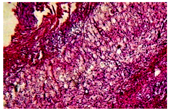
- The results of the morphological study showed that against the background of severe traumatic brain injury in the adrenal glands, the development of some discirculatory, dystrophic-necrotic and dysregenerative changes is noted. At the same time, these pathomorphological changes are more pronounced in the glomerular and bundle zones of the cortical layer and manifested by loosening, vacuolization, sometimes complete destruction of parenchymal cells. The glomerular zone destroys its typical structure due to pronounced vacuolar dystrophy and necrosis of adrenocorticocytes (Fig. 1).
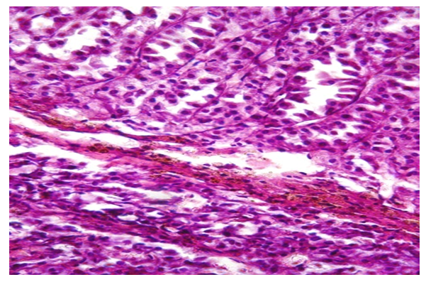 | Figure 1. Glomerular zone, stroma edema, vacuolization adrenocorticocytes. Color: G-E. X: 10x20 |
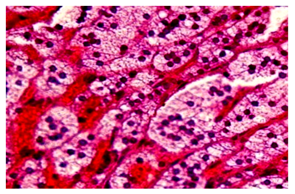
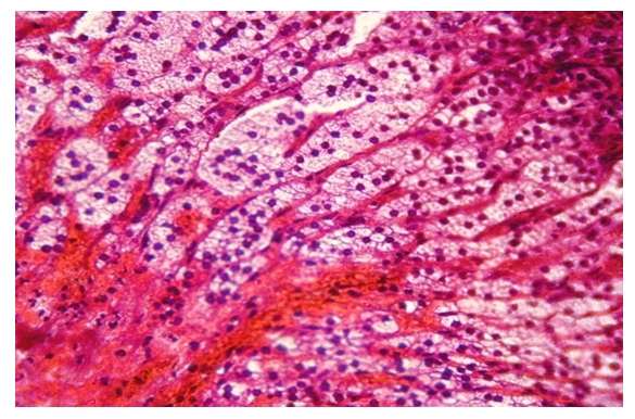 | Figure 2. Foci of parenchymal decay and foci of hemorrhage in the bundle zone. Color: G-E. X: 10x20 |
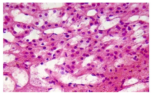 | Figure 3. Increased edematous phenomena, the formation of foci of regenerates in the glomerular zone of the adrenal cortex. Color: G-E. X: 10x20 |
 | Figure 4. Bundle zone, focal interstitial edema, preservation of the bundle arrangement of adrenocorticocytes. Color: G-E. X: 10x20 |
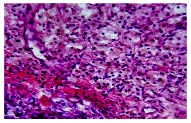
 | Figure 5. Pronounced destruction of adrenocorticocytes of the bundle zone of the cortex. Color: G-E. X:10x40 |
 | Figure 6. Bundle zone, edema, loosening of cells in the form of the formation of destructive adrenocorticocytes. Color: G-E. X: 10x20 |
4. Conclusions
- Thus, against the background of severe traumatic brain injury, pronounced dyscirculatory, dystrophic-destructive changes develop in the adrenal glands, and these changes were more pronounced in the glomerular and bundle zones of the cortical layer, and in the cerebral layer, along with edematous changes, some activation of pheochromacytoma cells was noted. With combined trauma, some increases in discirculatory and edematous phenomena were noted separately, but foci of vacuolar rearrangements of adrenocorticocytes remained in the cortical layer. At the same time, there was an increase in dyscirculatory and edematous phenomena, active destruction of parenchymal cells was also observed, especially in the glomerular zone of the cortex in the form of the formation of foci of necrosis, destruction and hemorrhage.The obtained data can be used to increase the validity of a forensic medical conclusion on the thanatogenesis of trauma, which indicates the expediency of referral to a microscopic examination of the adrenal glands from the corpses of persons who died as a result of traumatic brain injury or suspected of such.