-
Paper Information
- Next Paper
- Previous Paper
- Paper Submission
-
Journal Information
- About This Journal
- Editorial Board
- Current Issue
- Archive
- Author Guidelines
- Contact Us
American Journal of Medicine and Medical Sciences
p-ISSN: 2165-901X e-ISSN: 2165-9036
2023; 13(9): 1274-1276
doi:10.5923/j.ajmms.20231309.21
Received: Sep. 1, 2023; Accepted: Sep. 17, 2023; Published: Sep. 23, 2023

Assessing the Factors of Endothelial Dysfunction in Patients with Anterior Uveitis Who Underwent COVID-19
Rizaeva Manzurakhon Asrorovna1, Kamilov Khalidbek Mukhamedjanovich2, Khamraeva Gavhar Khusanovna2
1Republican Clinical Ophthalmological Hospital, Tashkent, Uzbekistan
2Department of Ophthalmology, Center for the Development of Professional Qualification of Medical Workers, Tashkent, Uzbekistan
Copyright © 2023 The Author(s). Published by Scientific & Academic Publishing.
This work is licensed under the Creative Commons Attribution International License (CC BY).
http://creativecommons.org/licenses/by/4.0/

To analyze the assessment of endothelial dysfunction in patients with anterior uveitis who have undergone COVID-19. Endothelial dysfunction (ED) is characterized by an imbalance between factors of relaxation and contraction, procoagulant and anticoagulant substances, and pro-inflammatory and anti-inflammatory mediators. It plays a vital role in the pathogenesis of anterior uveitis in COVID-19 patients.The study included 64 patients (64 eyes) with anterior uveitis who had undergone COVID-19 treatment at the Republican Clinical Ophthalmological Hospital. The patients' ages ranged from 20 to 61 years. A control group of 20 healthy individuals (20 eyes) was also included. All patients underwent various ophthalmic tests, including visometry, pneumotonometry, perimetry, biomicroscopy, B-scanning, and ophthalmoscopy. A blood test was conducted using enzyme immunoassay to measure fibronectin, endothelin-1, and Von Willebrand factor in blood plasma. a large selection of HUMAN ELISA kits and IFA-Fn kits manufactured by “NVO Immunoteks” closed joint stock company (Russia) were applied using a Mindray semi-automatic analyzer for enzyme-linked immunosorbent assay (ELISA) of endothelial growth factor. The research showed a significant increase in vascular endothelial growth factor in the examined patients by an average of 41% compared to the control group. The level of endothelin-1 in patients with anterior uveitis who underwent COVID-19 was increased by 2.7 times relative to the comparison group. Such an increase in the level of endothelin-1 indicates an endothelial dysfunction that worsens as the disease progresses and is due to the state of ischemia that occurs when blood circulation is disturbed. In conclusion, endothelial dysfunction plays a crucial role in the development of anterior uveitis in COVID-19 patients. Therefore, finding methods to treat it will reduce the risk of vascular complications. Keywords for this study include endothelial vascular dysfunction, anterior uveitis, post-COVID, vascular endothelial growth factor (VEGF), endothelin-1, fibronectin, and Von Willebrand factor.
Keywords: Endothelial vascular dysfunction, Anterior uveitis, Post-COVID, Vascular endothelial growth factor (VEGF), Endothelin-1, Fibronectin, Von Willebrand factor
Cite this paper: Rizaeva Manzurakhon Asrorovna, Kamilov Khalidbek Mukhamedjanovich, Khamraeva Gavhar Khusanovna, Assessing the Factors of Endothelial Dysfunction in Patients with Anterior Uveitis Who Underwent COVID-19, American Journal of Medicine and Medical Sciences, Vol. 13 No. 9, 2023, pp. 1274-1276. doi: 10.5923/j.ajmms.20231309.21.
1. Introduction
- At present, it has become apparent that the endothelium plays a major role in regulating vascular tone, modulating hemostasis, affecting vascular permeability, and controlling the growth of blood vessels [1]. However, endothelial dysfunction can activate the secretion of vasoconstrictor factors, among which endothelin-1 (ET-1) has an important role. Tissue hypoxia and increased levels of cellular hormones and cytokines actively contribute to the development of endothelial dysfunction. In response to endogenous factors, endothelial cells are activated, promoting the synthesis of factors such as ET-1 and intercellular adhesive molecules. Additionally, endothelial cells interact with cells in the bloodstream, releasing ET-1 and other factors into the blood, where their chemotaxis can induce leukocytes and platelets to migrate into the vascular wall. Endothelial cells also evoke adhesion by expressing specific adhesion molecules (selectins, integrins, and the immunoglobulin supergene family) that can interact with ligands on leukocytes and platelets.Based on the analysis of current literature on endothelial dysfunction, we can state that this problem remains relevant and has not been fully studied regarding the mechanism of development of ED in anterior uveitis in patients with COVID-19 [3-7]. Accordingly, the results of our research with patients with anterior uveitis who underwent COVID-19 indicate the damaging effect of the studied parameters on the functional state of vascular endothelial cells. Therefore, we decided to assess the state of the endothelium in patients with anterior uveitis who have undergone COVID-19. A holistic view of endothelial dysfunction as a significant link in the pathogenesis of COVID-19 enables us to prevent the risk of developing pathology in patients with uveitis, including developing pathogenetically substantiated directions of pharmacotherapy.The objective of the research is to analyze the methods of research in patients with anterior uveitis who have undergone COVID-19.
2. Subjects and Methods
- Sixty-four patients (64 eyes) with anterior uveitis were treated under our medical supervision. The patients' ages ranged from 20 to 61 years. The control group comprised 20 healthy individuals. All patients underwent visometry, pneumotonometry, perimetry, biomicroscopy, B-scan, and ophthalmoscopy. As for special investigative methods, a blood test was carried out by enzyme immunoassay. Fibronectin, endothelin-1, and von Willebrand factor in blood plasma were analyzed using HUMAN kits and IFA-Fn kits manufactured by PJSC "NVO Immunoteks" (Russian Federation) for enzyme-linked immunosorbent assay (ELISA) of endothelial growth factor using a Mindray semi-automatic analyzer.
3. Results
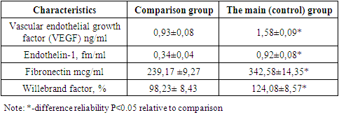
- In endothelial dysfunction, proliferation, and maturation of endothelial cells, emphasis is given to factors formed in the endothelium, specifically vascular endothelial growth factor (VEGF). Numerous studies have shown that the main stimulus of angiogenesis is oxygen deficiency, which causes hypoxia or ischemia, while HIF-1 promotes the intensity of vascular factors, namely vascular endothelial growth factor. Physiological angiogenesis is introduced by an adaptation response to oxygen deficiency since VEGF is considered a stress-induced protein regulated by glucose and oxygen. It has a strong influence on vascular permeability, is a powerful angiogenic protein, and is involved in neovascularization processes in pathological situations.Analysis of the research results presented in Table 1 showed a significant increase in vascular endothelial growth factor in patients by an average of 41% compared to other groups. Considering that VEGF is a stress-induced protein, its regulation is compared to other oxygen- and glucose-regulated proteins; therefore, an increase in its level can be considered as an adaptive response to hypoxia which leads to an increase in the pro-angiogenic factor for VEGF. It is commonly known that the main activators of the synthesis of endothelin-1 in the body are hypoxia and oxidative stress. These factors activate mRNA transcription, synthesis of endothelin precursors, their conversion into endothelin-1, and their secretion in a few minutes. As well, vascular endothelial growth factor activates intracellular mechanisms for the synthesis of endothelin-1.
|
4. Discussion
- Damage to endothelial cells is accompanied by a range of reactions that provide activation of the kallikrein-kinin system, the internal mechanism for the formation of prothrombinase activity, the fibrinolysis system, complement and a violation of the complex of functions performed by endothelial cells in the norm. Along with biosynthesis in the focus of inflammation, collagen catabolism occurs which is provided by collagenases of fibroblasts, macrophages, neutrophils, etc. Regulating influence on the proliferative, collagen-synthetic and collagenolytic functions of fibroblasts in the focus of inflammation is shown by lymphocytes, neutrophils and mast cells. As described above, the extracellular connective tissue of various organs contains a certain amount of macrophages belonging to the system of mononuclear phagocytes. Secretory products of macrophages include factors with intense opsonization (fibronectin).The main function of fibronectin involves attaching cells to matrices containing fibrillar collagen. Fibronectin is capable of binding to collagen at the stage of fibrillogenesis acting as an inhibitor of the growth of collagen fibers and thus regulating the density of the collagen framework. These fibronectin fibers can be mixed with other components of the extracellular matrix turning it into a solid supporting structure. An analysis of the research results showed an increase in soluble fibronectin by 31% when compared with the indicators of the control group.The endothelium in the physiological state inactivates the coagulation processes with the help of other mechanisms one of which being the synthesis of the von Willebrand factor. The latter is involved in the process of the internal cascade of fibrin formation, and stimulates the onset of thrombosis: it enables the attachment of platelet receptors to vascular collagen and fibronectin and to each other, i.e. strengthens adhesion and aggregation of platelets.In conclusion, the percentage of the von Willebrand factor increased by 21% when compared with the group of healthy individuals.Thus, it is worth mentioning that endothelial dysfunction plays a vital role in developing anterior uveitis in patients who have undergone COVID-19, and therefore the search for methods of its treatment will inevitably reduce the risk of vascular complications.Damage to endothelial cells leads to a range of reactions that activate the kallikrein-kinin system, the internal mechanism for the formation of prothrombinase activity, the fibrinolysis system, complement, and a disruption of the complex functions performed by endothelial cells in a healthy state. Collagen catabolism also occurs in the focus of inflammation, which is provided by collagenases from fibroblasts, macrophages, neutrophils, and other cells. Lymphocytes, neutrophils, and mast cells regulate the proliferative, collagen-synthetic, and collagenolytic functions of fibroblasts in the focus of inflammation. As mentioned earlier, the extracellular connective tissue of various organs contains a certain number of macrophages belonging to the system of mononuclear phagocytes. Secretory products of macrophages include factors with intense opsonization (fibronectin).The main function of fibronectin involves attaching cells to matrices containing fibrillar collagen. Fibronectin binds to collagen at the stage of fibrillogenesis, acting as an inhibitor of the growth of collagen fibers and thus regulating the density of the collagen framework. These fibronectin fibers can be mixed with other components of the extracellular matrix, turning it into a solid supporting structure. An analysis of the research results showed a 31% increase in soluble fibronectin compared to the control group.In a healthy state, the endothelium inactivates coagulation processes with the help of other mechanisms, one of which is the synthesis of the von Willebrand factor. The latter is involved in the process of the internal cascade of fibrin formation and stimulates the onset of thrombosis by enabling the attachment of platelet receptors to vascular collagen, fibronectin, and each other, strengthening adhesion and aggregation of platelets.In conclusion, the percentage of von Willebrand factor increased by 21% compared to the group of healthy individuals. Therefore, it is worth mentioning that endothelial dysfunction plays a vital role in developing anterior uveitis in patients who have had COVID-19, and the search for methods of treatment will inevitably reduce the risk of vascular complications.
 Abstract
 Reference
 Full-Text PDF
 Full-text HTML