-
Paper Information
- Next Paper
- Paper Submission
-
Journal Information
- About This Journal
- Editorial Board
- Current Issue
- Archive
- Author Guidelines
- Contact Us
American Journal of Medicine and Medical Sciences
p-ISSN: 2165-901X e-ISSN: 2165-9036
2023; 13(7): 1011-1015
doi:10.5923/j.ajmms.20231307.35
Received: Jul. 13, 2023; Accepted: Jul. 26, 2023; Published: Jul. 27, 2023

The Morphological State of the Kidneys of the Offspring in Conditions of Chronic Toxic Hepatitis in the Mother
Zulfiya Yusupovna Chorieva1, Dilorom Bakhtiyarovna Adilbekova2
1Termez Branch of Tashkent Medical Academy, Termez, Uzbekistan
2Doctor of Medical Sciences, Tashkent Medical Academy, Tashkent, Uzbekistan
Copyright © 2023 The Author(s). Published by Scientific & Academic Publishing.
This work is licensed under the Creative Commons Attribution International License (CC BY).
http://creativecommons.org/licenses/by/4.0/

Chronic toxic hepatitis in the mother negatively affects the postnatal growth and development of the kidneys of the offspring. A deep understanding of the pathogenesis of this morphological condition determines the future conduct of targeted, evidence–based, highly effective complex therapy in order to prevent pathology in children born and fed by mothers with chronic liver pathology.
Keywords: Kidney, Chronic toxic hepatitis, Mother, Offspring, Morphological state, Liver, Pathogenesis, Effect, Pathology, Therapy, Blood vessels, Tissues
Cite this paper: Zulfiya Yusupovna Chorieva, Dilorom Bakhtiyarovna Adilbekova, The Morphological State of the Kidneys of the Offspring in Conditions of Chronic Toxic Hepatitis in the Mother, American Journal of Medicine and Medical Sciences, Vol. 13 No. 7, 2023, pp. 1011-1015. doi: 10.5923/j.ajmms.20231307.35.
Article Outline
- The purpose of the study was to study the effect of chronic toxic hepatitis in the mother on the postnatal kidneys morphogenesis of the offspring.
1. Materials and Methods
- The experiments were carried out on outbred Wistar rats. The animals were divided into 2 groups: the 1st group (control)–intact animals (30 individuals), and the 2nd group–experimental (30 individuals)–rats, which were injected weekly for 6 weeks with heliotrin from a calculation of 0.5 mg/100 g of animal weight. The pathology of the mother’s liver in the early periods of postnatal ontogenesis in the vascular tissue structures of the kidneys of the offspring causes inflammatory – reactive changes. These pathomorphological changes in the kidneys of the offspring, in the early periods of postnatal ontogenesis, subsequently lead to a delay and lag in the processes of formation and development of the vascular tissue structures of the kidneys of the offspring.
2. The Relevance of the Study
- It is known that the birth and upbringing of healthy children depends primarily on the state of health of the mother. In this regard, it is relevant to study the effect of maternal pathology on the offspring. Recently, there has been a need to study relationships not only in the norm but also in diseases of the mother and (or) the father that complicate the pregnancy and childbirth process [3,5,6,7]. The question of the effect of maternal liver pathology on pregnancy and offspring has attracted the attention of researchers for a long time, because it is often one of the causes of death of young children, and often leads to various severe injuries of the internal organs of the offspring. The problem of the impact of various negative factors on the offspring is not only medical but also of great social importance. This is due to the fact that in recent decades, a demographic crisis has been observed all over the world–the birth rate is decreasing, and despite the development of technologies in medicine, the mortality of newborns is high. Scientists also warn about the effect of many drugs, unfavorable environmental factors, stress, and viral and infectious diseases that have an embryotoxic, fetotoxic and teratogenic effect, depending on the periods of embryo formation and how long they are exposed. Despite these factors, the influence of the mother’s liver pathology on the development, formation, and formation processes of the internal organs of the offspring has not been sufficiently studied.The aim of our study was to study the effect of chronic toxic hepatitis in the mother on the postnatal morphogenesis of the offspring's kidney. All animal experiments were carried out in accordance with the humane principles of the European Community (86/609/EEC) and the Declaration of Helsinki.Experiments were performed on Wistar rats. The rats were divided into 2 groups, the first group was called the control group and the second group was called the experimental group. Both groups of rats were injected with heliothrine alkaloid for 6 weeks to create a chronic form. 10 days after the last injection, men joined them and women in the control group. Rats born and fed from mothers with chronic toxic hepatitis were decapitated on days 3, 7, 21, and 30 of postnatal development, and pieces of kidney tissue were collected for histological examination. The material was subjected to general morphological, morphometric, and electron microscopic studies.
3. Results and Discussions
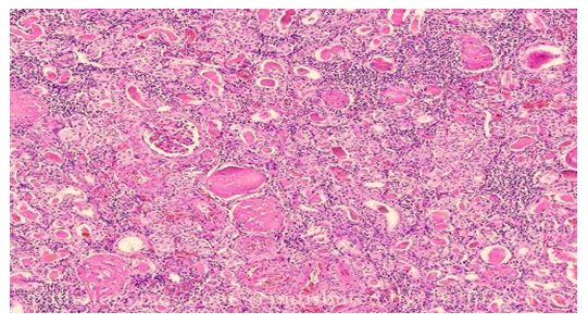
- The following picture was observed in the vascular tissue structures of the kidney in the 3–7 days of postnatal development of rat pups born and raised from mothers with chronic toxic hepatitis: microstructure of kidneys in newborn rat pups (3–7 days) was characterized by a low level of morphological differentiation. On the contrary, tubulointerstitial nephritis, lymphocytic infiltration and interstitial edema were found in the kidney spaces. A large number of emerging nephrons are detected, which, unlike the animals in the control group, are located in three rows instead of two. In this regard, the width of the nephrogenic layer is greater than that of animals in the control group. Most renal corpuscles are in the lower stages of development. The outer layer of the nephron capsules consists of a low prismatic epithelium, not squamous as in control animals. In places, there are clusters of prismatic cells without clear division into glomeruli and capsules. Compared to control animals, there are less formed glomeruli in the field of vision. This picture is combined with the expansion and abundance of capillary rings located here. Other parts of the nephron are also characterized by a low level of maturity. Other divisions of nephrons are also characterized by less clear maturity. Proximal convoluted tubules, which are lined with higher epithelium than controls, lack brush borders. The medulla contains significant layers of connective tissue and a small number of collecting ducts.The study of the histomorphological state of the kidneys of rat pups on the 14th day of postnatal life showed that the morphofunctional formation of the kidneys slows down. Thus, there are still individual forming nephrons, while in control rat pups such morphological formations are no longer present at these times. Against the background of such morphological immaturity, moderate dystrophic changes are noted. Some renal tubules are dilated and filled with desquamated epithelial cells. In other parts of the nephron, hydropic degeneration of the cytoplasm and pycnosis of the nuclei occur. On preparations stained with hematoxylin and eosin, changes in hemodynamics were found in the form of congestion, which manifested itself as a sharp expansion of interlobular arterioles and capillaries, and aggregation of the erythrocytes contained in them. Focal infiltrates represented by lymphocytes and macrophages were found in the interstitium of the cortical and medulla (Figure 1).
|
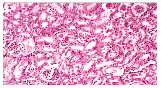 | Figure 2. The kidney of rat pups on the 30th day of postnatal life. Degenerative changes in the interstitial tissue of the cortical and medulla are revealed. Coloring G.–E. SW. 10x20 |
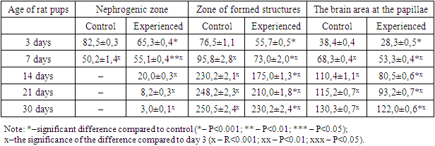
 | Figure 3. Morphometric parameters of kidneys from control female rats and offspring born with chronic toxic hepatitis |
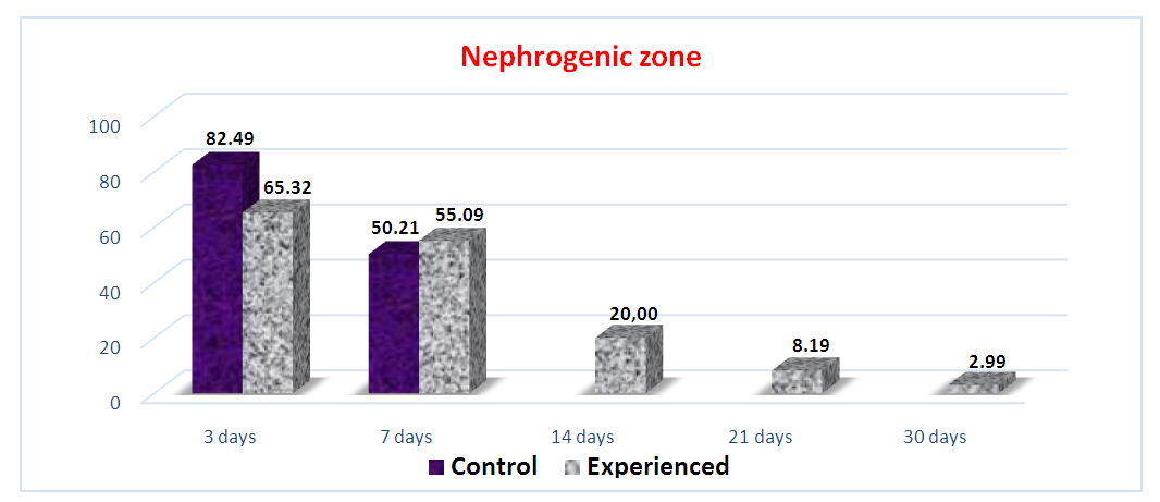
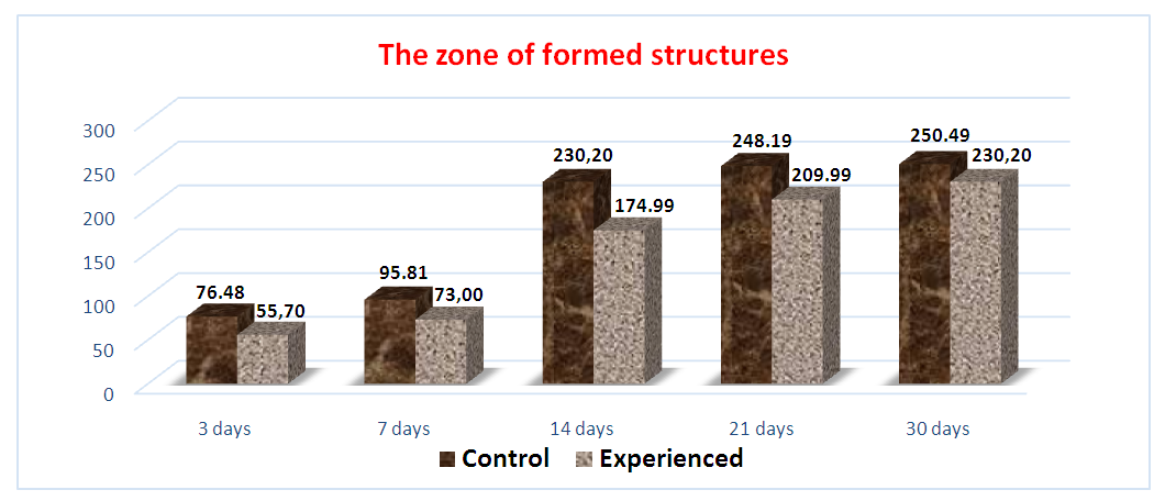 | Figure 4. Morphometric indicators of the kidneys of offspring born from female control rats and with chronic toxic hepatitis |
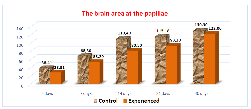 | Figure 5. Morphometric indicators of the kidneys of offspring born from female control rats and with chronic toxic hepatitis |
4. Conclusions
- 1. Chronic toxic damage to the mother’s liver negatively affects the processes of postnatal growth, development and formation of tissue structures of the offspring’s kidneys;2. Hepatotoxins introduced into the mother’s body before pregnancy and formed in her during hepatitis enter the bloodstream and then breast milk, contributing to the development of inflammatory, reactive and degenerative changes in the vascular tissue structures of the offspring’s kidneys. In the first periods after birth, it has a negative effect on the growth and development of offspring;3. Pathological changes in the vascular tissue structures of the offspring’s kidneys, which later lead to a delay in postnatal development processes and the formation of the entire organ and organ system;4. All this requires the development of evidence–based treatment and preventive measures to prevent pathology in children born and fed to mothers with chronic liver pathology.
 Abstract
 Reference
 Full-Text PDF
 Full-text HTML